Introduction
Diabetes mellitus (DM) is a severe metabolic disease
prevalent in a large number of patients worldwide (~5.38%) and is
characterized by high concentrations of blood sugar (1). The challenges in DM therapy arise from
complications that affect several organs, such as the heart, kidney
and skin: Many patients suffer from a delay in skin wound and ulcer
repair (2); and higher sugar content
in blood vessels blocks angiogenesis, which is important for
supplying fresh blood for the repair of damaged tissue (3).
Repair of skin ulcers is a complicated process that
requires recruitment of cells such as keratinocytes, platelets,
endothelial cells, fibroblasts, and macrophages. Fibroblast
migration and proliferation, angiogenesis, and wound contraction,
together with collagen deposition and remodeling are the key
processes in wound healing. In the dermal skin layer, extracellular
matrix (ECM) synthesis is the largest component of the skin
generation process (4). Fibroblasts
initiate ECM production and remodeling, which further forms
granulation tissue (5,6). Levels of cellular reactive oxygen
species (ROS) are modified to sense environmental changes, and they
function as secondary messengers in regulation of diverse
biological processes including cell proliferation, differentiation,
and maturation (7). Fibroblasts
isolated from patients with DM are typically large and widely
spread, compared with age-matched fibroblasts from healthy people,
and exhibit abnormal endoplasmic reticulum, higher number of
vesicular particles, and altered microtubule arrays. Therefore,
high glucose concentration (HG) in vivo induces normal
protein maturation, cellular protein trafficking and protein
secretion in diabetic ulcer fibroblasts (8,9). In
addition, it has been demonstrated that cell proliferation was
defective and subsequent production of extracellular matrix protein
was affected in diabetic ulcer fibroblasts (8). HG was reported to inhibit c-Jun
N-terminal kinase (JNK) activity, resulting in a delay in
fibroblast migration (10). Previous
transcriptome analysis using HG-stimulated fibroblasts has
identified changes in the expression of numerous genes associated
with diverse pathways including Wnt, inflammatory response, and
hedgehog signaling (11).
Hedgehog signaling has been widely studied due to
its significance in development and disease regulation (12). Hedgehog was first observed as a
secreted protein that requires specific positional identity in
embryonic segments of Drosophila melanogaster (13). The three mammalian hedgehog
genes, Sonic hedgehog (Shh), Desert hedgehog
and Indian hedgehog, are known for their importance in the
patterning of developing tissue and biological structures (13). Notably, inhibition of hedgehog
signaling either by loss of a gene or by suppression of expression
severely affects development of tissues and organs, leading to
skeletal malformation, craniofacial defects, polydactyly and
holoprosencephaly (14).
Furthermore, abnormal activation of hedgehog signaling is
associated with most basal cell carcinomas, and some
medulloblastomas and rhabdomyosarcomas (12,15–17).
Under normal conditions, the plasma membrane-localized Patched1
(Ptch1) interacts with Smoothened (SMO) to maintain hedgehog
signaling in an inactive or ‘off’ state. Following Shh secretion,
it interacts with and inactivates Ptch1, resulting in the
activation of SMO (16,18). SMO further triggers downstream gene
transcription through the zinc finger protein Gli (Gli)-Kruppel
family of transcription factors, controlling cell differentiation,
proliferation, and survival (18).
However, the role of hedgehog signaling in the process of
fibroblast repair under DM conditions is currently unknown.
In the present study, the effects of hedgehog
signaling on HG-damaged skin fibroblasts were analyzed via Shh
stimulation, and the expression levels of hedgehog signaling,
apoptosis and inflammatory response markers were determined. Cell
proliferation rates were also analyzed. Additionally, JNK activity
was analyzed following HG and Shh treatment, and cell proliferation
was analyzed following Shh and JNK inhibitor supplementation
together with overexpression of Gli1, which is a key
transcriptional activator of hedgehog signaling (19). The findings of the present study may
be useful for skin ulcer therapy for patients with DM.
Materials and methods
Fibroblast culture
Human foreskin samples were obtained from 6 male
patients (age, 23–35 years), undergoing foreskin circumcision, from
January 2017 to March 2017 at the Department of Dermatology, The
First Affiliated Hospital of Xinxiang Medical University (Xinxiang,
China). The present study was approved by the Ethics Committee of
Xinxiang Medical University, and written informed consent was
obtained from all patients. The human foreskin fibroblast isolation
and proliferation were performed as described previously (10). Fibroblasts were divided into three
groups and treated with HG (35 mM), sp600126 (100 nM; cat. no.
S5567; Sigma-Aldrich; Merck KGaA, Darmstadt, Germany), and Shh (100
ng/ml; cat. no. S0191; Sigma-Aldrich; Merck KGaA), respectively,
for 3 h at 37°C. The low glucose treated group was untilized as the
control.
Cell proliferation assay
Cell proliferation activity was examined following
the aforementioned treatment, using a Cell Counting Kit-8 (Dojindo
Molecular Technologies, Inc., Kumamoto, Japan) according to the
manufacturer's protocol. Briefly, 2×10³ fibroblasts were
seeded in 96 well plates and respectively treated with HG (35 mM)
and Shh (100 ng/ml), and cell density was analyzed as previously
reported (10).
RNA isolation and reverse
transcription-quantitative polymerase chain reaction (RT-qPCR)
RNA was isolated from fibroblasts that were either
untreated or treated with HG (35 mM) and Shh (100 ng/ml; cat no.
ab23327; Abcam, Cambridge, MA, USA). Fibroblasts were washed twice
with ice-cold PBS and 1 ml TRIzol (Invitrogen; Thermo Fisher
Scientific, Inc., Waltham, MA, USA) was subsequently added to each
3.5-cm diameter dish to lyse the cells. Following treatment with
RQ1 DNase (Promega Corporation, Madison, WI, USA), 1 µg RNA was
used to synthesize cDNA using the GoScript Reverse Transcription
kit (Reverse Transcription System, Promega) following the
manufacturer's instructions. A SYBR Green Master Mix (Bio-Rad
Laboratories, Inc., Hercules, CA, USA) was used to perform qPCR on
an Illumina Eco 3.0 system (Illumina, Inc., San Diego, CA, USA).
The PCR reaction was performed in a 20 µl reaction mixture
containing 10 µl SYBR Green Master Mix, 4 µl primers (2 µl each of
2.5 µM forward and reverse primer), 3 µl cDNA template and 3 µl
double-distilled H2O. A typical reaction consisted of an
initial denaturation at 95°C for 3 min, followed by 40 cycles of
denaturation for 30 sec at 95°C, annealing for 30 sec at 60°C, and
extension at 72°C for 30 sec, followed by a final extension at 72°C
for 7 min. The transcription levels were normalized against those
of GAPDH using the 2−ΔΔCq method (20). Primers used in RT-qPCR analysis are
listed in Table I.
 | Table I.Reverse transcription-quantitative
polymerase chain reaction primer sequences. |
Table I.
Reverse transcription-quantitative
polymerase chain reaction primer sequences.
| Primer | Sequence
(5′-3′) |
|---|
| Caspase-3
F |
TGATGATGACATGGCGTGTC |
| Caspase-3
R |
GTTGCCACCTTTCGGTTAAC |
| PAI1 F |
GAGACTGAAGTCGACCTCAG |
| PAI1 R |
CTGTCCATGATGATCTCCTC |
| GAPDH F |
GACCTGCCGTCTAGAAAAAC |
| GAPDH R |
CTGTAGCCAAATTCGTTGTC |
| SMO F |
ACCTATGCCTGGCACACTTC |
| SMO R |
AGGAAGTAGCCTCCCACGAT |
| Ptch1 F |
CAAACTCCTGGTGCAAACCG |
| Ptch1 R |
CCGGGATTCTCAGCCTTGTT |
| Gli1 F |
CCAGAGTTCAAGAGCCTGG |
| Gli1 R |
CCTCGCTCCATAAGGCTCAG |
Western blotting
Total protein was extracted using cell lysis buffer
according to the manufacturer's protocol (cat. no. P0013; Beyotime
Institute of Biotechnology, Haimen, China) and the concentration
was determined using a BCA assay. Total protein (20 µg) from each
sample was electrophoresed using 10% SDS-PAGE. Following
separation, the proteins were transferred to Immobilon-P Transfer
membranes (EMD Millipore, Billerica, MA, USA), which were blocked
in blocking solution (1X TBS, 5% skimmed milk and 0.05% Tween-20)
for 2 h at room temperature and thereafter probed with primary
antibodies at 25°C for 2 h. The following primary antibodies were
used: Anti-phosphorylated (p)-stress-activated protein kinase
(SAPK)/JNK (Thr183/Tyr185) antibody (1:1,000; cat. no. 4668; Cell
Signaling Technology, Inc., Danvers, MA, US), anti-SAPK/JNK
(1:1,000; cat. no. 9252; Cell Signaling Technology, Inc.) and
anti-GAPDH antibody (1:2,000, cat. no. ab9484; Abcam). Following
incubation with the primary antibodies, the membranes were washed
with PBS and incubated with corresponding secondary antibodies for
1 h at room temperature. The secondary antibodies used were
anti-mouse or anti-rabbit horseradish peroxidase-linked secondary
antibody (1:2,000; cat. no. 7074; Cell Signaling Technology, Inc.).
Samples were visualized using a Beyo ECL Plus kit (cat. no. P0018;
Beyotime Institute of Biotechnology).
Overexpression of Gli1
To overexpress Gli1 in fibroblasts, the
Gli1 open reading frame (ORF) sequence (Gli1; GenBank
accession no. NP_005260; National Center for Biotechnology
Information; https://www.ncbi.nlm.nih.gov/) was artificially
synthesized by Sangon Biotech Co., Ltd. (Shanghai, China). The
Gli1 ORF was then cloned into the pcDNA3 expression vector
(Invitrogen; Thermo Fisher Scientific, Inc.). A total of 2 µg
pcDNA3-Gli1 or pcDNA3 empty vector was transfected into
human foreskin primary fibroblasts using Lipofectamine 2000
(Invitrogen; Thermo Fisher Scientific, Inc.) following the
manufacturer's instructions. RNA was extracted from transfected
cells following 48 h, and the Gli1 expression level was
determined via quantitative RT-qPCR under the same parameters as
aforementioned, but with the following primers: Gli1, F,
5′-TTTCCCATTACCCCCAGCCCTCTC-3′; R, 5′-GAAGCCCTATTTGCCCCCACTACA-3′
and GAPDH, F, 5′-GTCATCATCTCTGCCCCCTCTGCT-3′; R,
5′-GACGCCTGCTTCACCACCTTCTTG-3′.
ROS measurement
To visualize cellular ROS levels
2′,7′-dichlorofluorescein diacetate (DCFH-DA) dye was used
(21). Fibroblasts were cultured in
low glucose DMEM (LG, 5.5 mM; cat. no. 11885092; Thermo Fisher
Scientific, Inc.) or HG (35 mM; cat. no. 11965118; Thermo Fisher
Scientific, Inc.), and Gli1 overexpressed cells were treated
with HG at 37°C for 6 h. Fibroblasts were incubated with 10 µM
DCFH-DA for 30 min at 37°C in the dark and fluorescence intensity
was analyzed using a confocal microscope at a magnification of ×200
(excitation at 488 nm, emission at 530 nm) within 15 min.
Statistical analysis
Statistical differences were analyzed using the
Prism 5 software package (Version 5.0; GraphPad Software, Inc., La
Jolla, CA, USA). Data are presented as the mean ± standard
deviation and three independent repeats were performed for each
experiment. Significant differences between two groups were
calculated using Student's t-test, and one-way analysis of variance
was performed to analyze differences between more than two groups
with a Turkey post hoc test. P<0.05 was considered to indicate a
statistically significant difference.
Results
HG inhibits cell proliferation and
hedgehog signaling in skin fibroblasts
Diabetes is a severe metabolic disease that
generates diverse complications threatening patient health. To
understand the mechanism of diabetes, HG is typically used to mimic
the disease. Previous studies have observed that HG mediated stress
altered the expression of many genes including hedgehog
signaling-related genes in human fibroblast primary cells (11). To analyze the effect of HG on
hedgehog signaling, key genes associated the pathway were examined
for HG-mediated changes in expression. Initially, HG concentration
was optimized for the relevant experimental conditions prior to
testing gene expression. Previous research has identified that HG
concentrations >35 mM significantly inhibit cell proliferation
(11). In the present study, it was
confirmed that 35 mM HG stress significantly reduced fibroblast
proliferation, in comparison with LG fibroblasts (Fig. 1A). Furthermore, cell damage via HG
was examined at the molecular level by evaluating expression levels
of Caspase-3 (apoptosis marker) and plasminogen activator
inhibitor-1 (PAI1; inflammatory response marker). RT-qPCR
results demonstrated that HG treatment significantly increased
Caspase-3 and PAI1 levels (Fig. 1B). Subsequently, the expression of
key hedgehog signaling-related genes (SMO, Ptch1, and
Gli1) was analyzed. RT-qPCR data indicate that HG suppressed
the expression of all three genes (Fig.
1B).
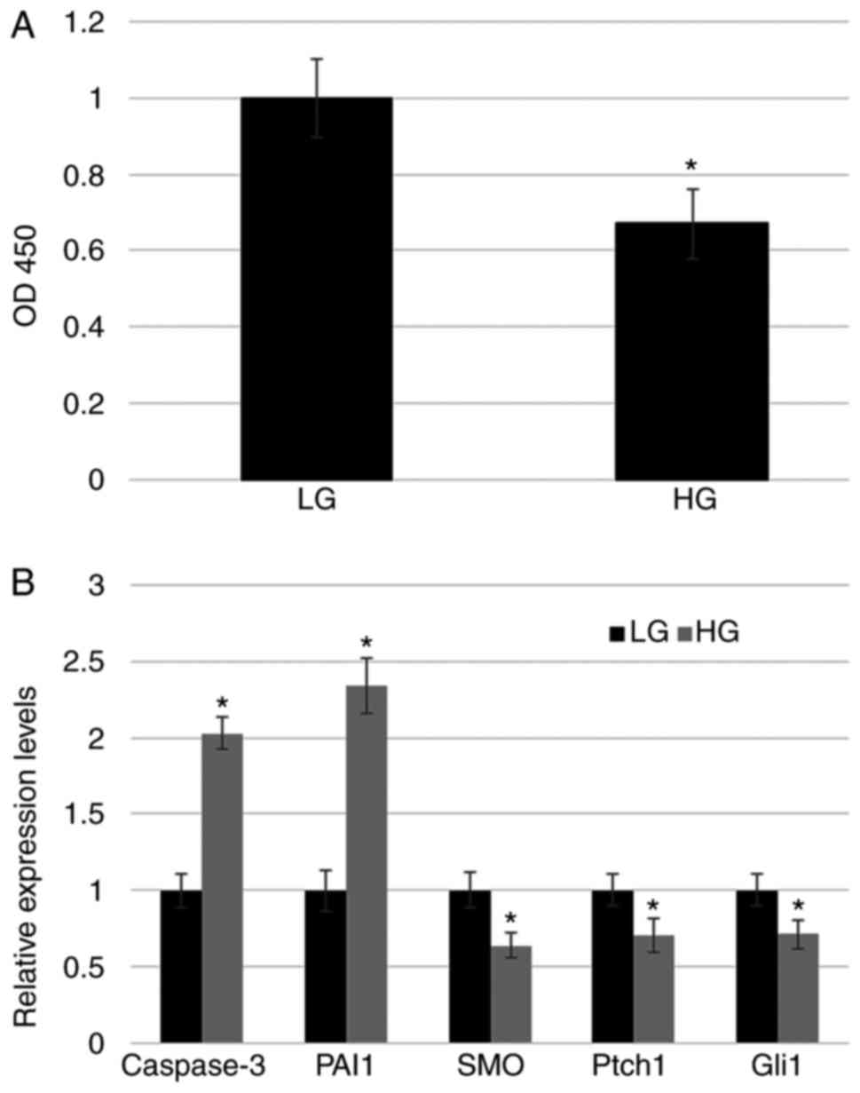 | Figure 1.Fibroblast proliferation rate and
marker gene expression upon HG stress. (A) Cell proliferation was
analyzed using a cell counting kit-8 assay. (B) Expression of
apoptosis marker (Caspase-3), inflammatory response marker
(PAI1), and hedgehog signaling-related markers (SMO,
Ptch1 and Gli1) was monitored in LG- and HG-treated
fibroblasts with reverse transcription-quantitative polymerase
chain reaction. *P<0.05 vs. LG. LG, low glucose concentration
(5.5 mM); HG, high glucose concentration (35 mM); PAI1, plasminogen
activator inhibitor-1; SMO, Smoothened; Ptch1, Patched1; Gli1, zinc
finger protein Gli1; OD, optical density. |
Shh treatment ameliorates the effect
of HG on fibroblasts
As HG suppressed hedgehog signaling and cell
proliferation, hedgehog signaling was activated via treatment with
Shh in the culture medium, and cell proliferation and hedgehog gene
expression were analyzed. Cell proliferation was activated by Shh
treatment in LG medium and significantly inhibited by HG stress.
Furthermore, addition of Shh with HG ameliorated the effect of HG
on cell proliferation, and cells exhibited similar proliferation as
cells grown under LG (Fig. 2A). The
effects of Shh on gene expression were subsequently analyzed. Shh
treatment induced Gli1 expression in LG growth medium,
whereas HG significantly suppressed Gli1 expression.
Treatment with Shh and HG eliminated the effect of HG on
Gli1 expression, resulting in similar gene expression as
that observed in cells grown under LG (Fig. 2B).
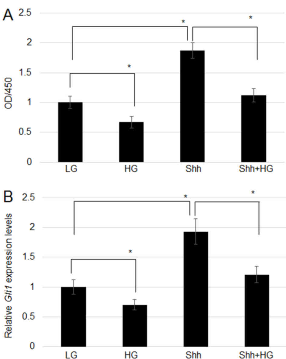 | Figure 2.Effect of Shh on cell proliferation
and gene expression. (A) Fibroblasts were treated with Shh (an
inducer of hedgehog signaling), HG and HG+Shh, and cell
proliferation was examined after 72 h. (B) Gli1 expression
level was tested following Shh, HG, and HG+Shh treatment.
Significant differences between groups are indicated with
*P<0.05, HG and Shh vs. LG; Shh+HG vs. Shh. Shh, Sonic hedgehog;
HG, high glucose concentration (35 mM); Gli1, zinc finger protein
Gli1; OD, optical density; LG, low glucose concentration (5.5
mM). |
JNK is associated with Shh-dependent
cell proliferation and hedgehog signaling-related gene
regulation
JNK modulates HG-mediated signaling in fibroblasts
(10). It is known that HG inhibits
JNK activity via suppression of p-JNK levels, without affecting
total JNK (t-JNK) levels (10). Shh
treatment induced a marked increase in the level of p-JNK (p-JNK)
in both LG- and HG-treated cells (Fig.
3A). However, Shh, HG, and HG+Shh treatments did not markedly
affect t-JNK levels (Fig. 3A).
Additionally, JNK function during Shh-induced cell proliferation
was examined. Compared with the LG group, treatment with sp600125,
a JNK-specific inhibitor, significantly inhibited cell
proliferation, whereas Shh treatment significantly promoted cell
proliferation. Furthermore, sp600125 treatment significantly
inhibited Shh-induced cell proliferation (Fig. 3B). As JNK has a role downstream of
Shh during cell proliferation, the role of JNK in hedgehog
signaling-related gene expression was further explored. Effects of
sp600126 treatment in LG-grown cells on SMO, Ptch1, and
Gli1 transcript levels were analyzed, indicating that
inhibition of JNK via sp600125 significantly suppressed SMO,
Ptch1, and Gli1 expression (Fig. 3C).
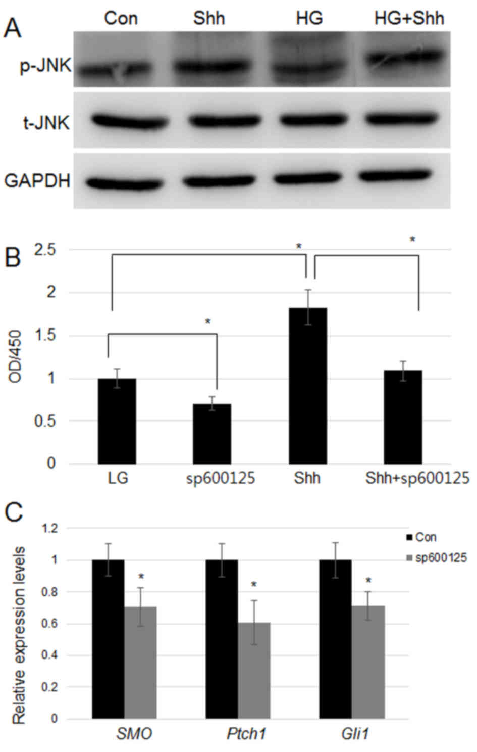 | Figure 3.Effect of Shh on JNK activity, and of
JNK inhibition on cell proliferation and hedgehog signaling-related
gene expression. (A) Fibroblasts were treated with Shh, HG and
HG+Shh, and p-JNK and total t-JNK levels were measured using
western blotting. GAPDH was utilized as an internal control. (B)
Fibroblasts were treated with Shh, sp600125, and HG+sp600125, and
cell proliferation was monitored. Significant differences are
marked with *P<0.05, sp60012 and Shh vs. LG; Shh+sp60012 vs.
Shh. (C) SMO, Ptch1, and Gli1 expression was analyzed
following treatment with sp600125. *P<0.05 vs. con. Shh, Sonic
hedgehog, JNK, c-Jun N-terminal kinase; HG, high glucose
concentration (35 mM); con, low glucose as the control group; p,
phosphorylated; t, total; SMO, Smoothened; Ptch1, Patched1; Gli1,
zinc finger protein Gli1; OD, optical density. |
Overexpression of Gli1 partially
eliminates HG and JNK inhibitor action
Gli1 expression was suppressed in fibroblasts
upon HG and sp100125 treatment (Figs.
1 and 3). Therefore, the
function of Gli1 was analyzed by overexpressing Gli1
in human fibroblasts. Following sequencing, the Lipofectamine 2000
system was used to transfect 2 µg pcDNA3-Gli1 or pcDNA3
empty plasmid into human foreskin primary fibroblasts. RT-qPCR
results demonstrated that Gli1 expression was significantly
increased in the Gli1-overexpressing cells than in cells
transfected with empty vector (Fig.
4A). Compared with empty vector transformation,
Gli1-overexpressing cells also exhibited a significant
increase in cell proliferation (Fig.
4B). Furthermore, RT-qPCR analysis of
Gli1-overexpressing cells treated with HG and sp600125
indicated that HG and sp600126 treatment did not significantly
affect Gli1 expression levels (Fig. 4A). In addition,
Gli1-overexpressing cells treated with sp600126 retained
their increased proliferation rate compared with control cells
(Fig. 4B), indicating that
Gli1 regulates fibroblast proliferation downstream of HG and
JNK signaling.
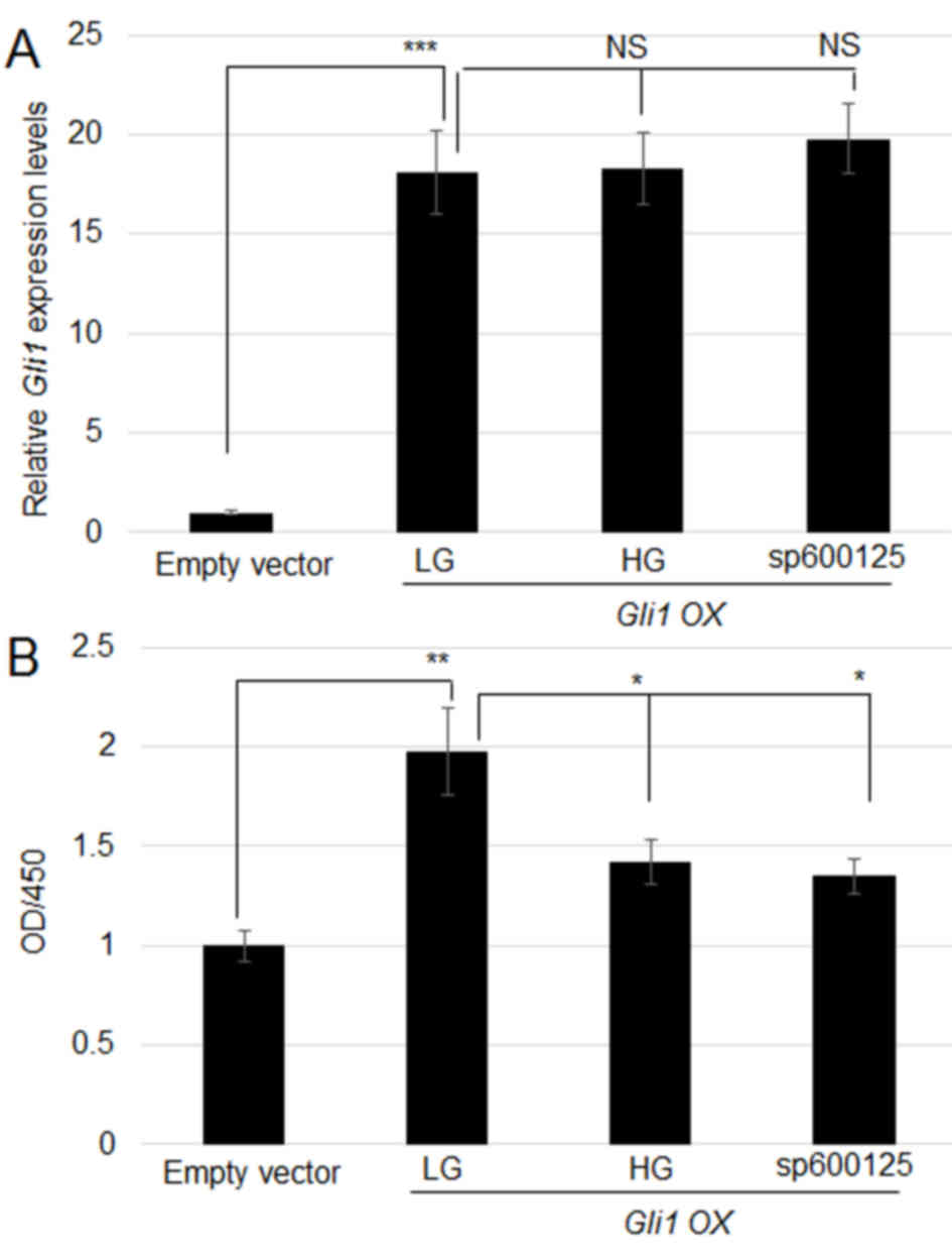 | Figure 4.Effect of Gli1 overexpression
on HG and sp600125-mediated inhibition of cell proliferation. (A)
Gli1 was overexpressed in human fibroblasts, and its
expression was analyzed with reverse transcription-quantitative
polymerase chain reaction. Gli1-overexpressing cells treated
with HG and sp600125, or without any treatment were compared.
pcDNA3 empty vector-transfected cells were used as a control.
***P<0.001, LG vs. Empty vector; NS, No significance. (B) Cell
proliferation was monitored in the pcDNA3 empty vector-transfected
cells, Gli1-overexpressing cells, and sp600125- or
HG-treated Gli1-overexpressing cells. Significant
differences are indicated with **P<0.01, LG vs. Empty vector;
*P<0.05, HG and sp600125 vs. LG. Gli1, zinc finger protein Gli1;
HG, high glucose concentration (35 mM); LG, low glucose
concentration (5.5 mM); OD, optical density; OX,
overexpression. |
Activation of hedgehog signaling
reduces HG-induced ROS generation
Previous reports have demonstrated that HG-induced
ROS production via inhibition of JNK and ROS levels is closely
associated with wound repair (10,22). To
further analyze the association between hedgehog signaling and
HG-induced ROS production, ROS levels were detected in fibroblasts
using DCFH-DA, which is a ROS indicator. LG-grown cells were used
as a control and were not observed to notably accumulate ROS.
However, HG-treated cells showed significantly increased ROS
accumulation, compared with LG. To analyze the effects of hedgehog
signaling on HG-induced ROS generation, Gli1-overexpressing
cells were treated with HG, which resulted in significantly lower
ROS levels (Fig. 5A and 5B). These
findings suggested that Gli1 overexpression reduced
HG-induced ROS production.
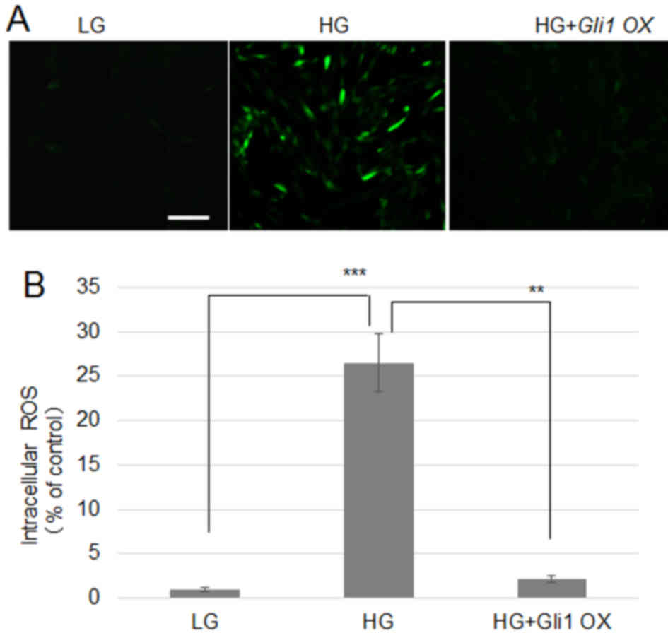 | Figure 5.Shh treatment or Gli1
overexpression reduces cellular ROS levels in fibroblasts. (A)
Cells were incubated for 1 h in LG, HG, or HG+Shh. Cellular ROS
levels were visualized using 2′,7′-dichlorofluorescein diacetate.
Scale bar=100 µm. (B) Fluorescence levels from 10 independent cells
in each treatment group were quantified (n=10). Significant
differences are shown with ***P<0.001, HG vs. LG; **P<0.01,
HG+Gli1 OX vs. HG. Shh, Sonic hedgehog; Gli1, zinc finger protein
Gli1; ROS, reactive oxygen species; LG, low glucose concentration
(5.5 mM); HG, high glucose concentration (35 mM); OX,
overexpression. |
Discussion
Skin ulcers induced by DM are difficult to repair
and afflict patients worldwide. Concentration of HG in in
vitro fibroblast experiments is typically higher than the
glucose levels in physiological levels of glucose in diabetic
patients (>11.1 mM); 25–50 mM HG has been used for DM study in
previous reports (10,23,24). In
the present study, 35 mM HG was used to perform the experiments.
Skin wound repair involves multiple steps including cell
proliferation, and requires the coordination of multiple cell
layers. Fibroblasts in the skin have an important role during wound
repair. Abnormal activation of Ras-related C3 botulinum toxin
substrate 1 and suppression of JNK were previously observed to be
associated with wound repair in HG-stressed skin fibroblasts
(10,25). In addition, transcriptome analysis
has revealed that several biological pathways are altered in
HG-damaged fibroblasts, including Wnt, hedgehog, and nuclear
factor-κB signaling (11). However,
to the best of our knowledge, the regulatory basis of hedgehog
signaling during HG damage in fibroblasts has not been
reported.
In the present study, it was observed that HG
inhibited cell proliferation and suppressed the expression of key
hedgehog signaling-related genes including SMO, Ptch1, and
Gli1. Treatment with Shh, an activator of hedgehog
signaling, was used to analyze the role of hedgehog in HG-damaged
fibroblasts. Shh treatment activated cell proliferation in LG and
HG growth conditions, and Gli1 expression was also induced
by Shh in both LG- and HG-grown cells. Notably, cell proliferation
ability and Gli1 expression were similar in LG and HG+Shh
conditions, suggesting that hedgehog may be a target of HG-mediated
signaling. To further examine the association of hedgehog signaling
in the HG-induced damage of fibroblasts, JNK activity was analyzed
in HG- and Shh-stimulated cells. The present results demonstrated
that JNK phosphorylation was activated by Shh and suppressed by HG.
Treatment with Shh and HG induced an increase in JNK
phosphorylation. In addition, sp600125, a JNK specific inhibitor,
was used, and cell proliferation was analyzed, which indicated that
sp600125 inhibited JNK activity, consequently inhibiting cell
proliferation, whereas Shh treatment partially rescued these
effects. These results indicate that JNK positively regulates cell
proliferation downstream of HG and Shh signaling. As a relatively
lower concentration of sp600125 may not completely suppress JNK
activity, Shh treatment may be able to partially rescue the
inhibited cell proliferation. As Shh stimulation activated JNK
activity, the effect of altered JNK function on hedgehog signaling
was further explored. sp600125 was added to fibroblasts in LG
medium and SMO, Ptch1, and Gli1 expression was
evaluated. The data indicate that JNK positively regulates the
expression of key hedgehog signaling-related genes and suggest that
hedgehog signaling is activated via Shh, which binds the receptor
to activate downstream signaling, and that JNK is downstream of
hedgehog activated by Shh stimuli. However, JNK also regulates
expression of SMO, Ptch1, and Gli1. These three genes
were induced by Shh stimulation and suppressed by HG stress,
implying that JNK may be a key downstream factor through which HG
and hedgehog signaling regulate expression of SMO, Ptch1,
and Gli1. Furthermore, Gli1 overexpression eliminated
HG- and sp600125-mediated effects on cell proliferation, further
suggesting that Gli1 function is downstream of HG and JNK
signaling.
Previously, it was demonstrated that HG-represses
JNK signaling to increase ROS production in human fibroblasts. A
previous study (10) determined that
as Shh treatment activates JNK activity, Gli1 overexpression
reduced impact of treatment with a JNK inhibitor. Therefore, the
ROS production in fibroblasts and in skin from diabetic rats was
analyzed. HG or diabetes highly induced ROS generation in
fibroblasts and rat skin, and the activation of hedgehog signaling
via Shh treatment and Gli1 overexpression in diabetic rat
skin and HG-stimulated fibroblasts, respectively, reduced ROS
production. These data suggested that activation of hedgehog
signaling reduces the effect of HG, possibly via inhibition of ROS
overproduction (10). Further
analysis is required to elucidate the associations between ROS
generation and hedgehog signaling in human fibroblasts. Although
the linkage between Gil1-JNK-ROS was identified in the
present study; however the regulation of this process was not
determined. Further study may be required to assess how HG stress
inhibits hedgehog signaling.
The findings of the present study are unique, to the
best of our knowledge, and provide important insights that may help
elucidate the regulatory basis of skin ulcers induced by diabetes.
Elucidating the mechanism of hedgehog signaling activation to
protect fibroblasts from HG damage may provide a molecular target
for clinical treatment.
Acknowledgements
Not applicable.
Funding
The present study was supported by National Natural
Science Foundation of China (grant no. 81602132), and grants from
the Department of Science and Technology of Henan Province (grant
nos. 172102310584 and 182102310242), the Education Department of
Henan Province (grant nos. 201610472017 and 201610472040). The
present study was also supported by Institute of Precision Medicine
Taihang Young Scholar Foundation of Xinxiang Medical University,
Doctoral Scientific Research Foundation of Xinxiang Medical
University (grant nos. XYBSKYZZ201513 and 201512).
Availability of data and materials
All data generated or analyzed during this study are
included in this paublished paper.
Authors' contributions
NS and HW coordinated the present study and wrote
the manuscript; TG, JQ, JY, YQ, QC, YZ, YC, QH and XM performed and
analyzed the experiments; and TS and ZF analyzed and reviewed the
data. All authors contributed to the interpretation of data,
manuscript revision and critical discussion.
Ethics approval and consent to
participate
The present study was approved by the Ethics
Committee of Xinxiang Medical University (Xinxiang, China), and
written informed consent was obtained from all patients.
Consent for publication
Written informed consent was obtained from all
patients.
Competing interests
The authors declare that they have no competing
interests.
References
|
1
|
Shi Y and Hu FB: The global implications
of diabetes and cancer. Lancet. 383:1947–1948. 2014. View Article : Google Scholar : PubMed/NCBI
|
|
2
|
Yach D, Stuckler D and Brownell KD:
Epidemiologic and economic consequences of the global epidemics of
obesity and diabetes. Nature Med. 12:62–66. 2006. View Article : Google Scholar : PubMed/NCBI
|
|
3
|
Braiman-Wiksman L, Solomonik I, Spira R
and Tennenbaum T: Novel insights into wound healing sequence of
events. Toxicol Pathol. 35:767–779. 2007. View Article : Google Scholar : PubMed/NCBI
|
|
4
|
Brem H and Tomic-Canic M: Cellular and
molecular basis of wound healing in diabetes. J Clin Invest.
117:1219–1222. 2007. View
Article : Google Scholar : PubMed/NCBI
|
|
5
|
Martin P: Wound healing-aiming for perfect
skin regeneration. Science. 276:75–81. 1997. View Article : Google Scholar : PubMed/NCBI
|
|
6
|
Goldin A, Beckman JA, Schmidt AM and
Creager MA: Advanced glycation end products: Sparking the
development of diabetic vascular injury. Circulation. 114:597–605.
2006. View Article : Google Scholar : PubMed/NCBI
|
|
7
|
Obayashi K, Akamatsu H, Okano Y, Matsunaga
K and Masaki H: Exogenous nitric oxide enhances the synthesis of
type I collagen and heat shock protein 47 by normal human dermal
fibroblasts. J Dermatol Sci. 41:121–126. 2006. View Article : Google Scholar : PubMed/NCBI
|
|
8
|
Loots MA, Lamme EN, Mekkes JR, Bos JD and
Middelkoop E: Cultured fibroblasts from chronic diabetic wounds on
the lower extremity (non-insulin-dependent diabetes mellitus) show
disturbed proliferation. Arch Dermatol Res. 291:93–99. 1999.
View Article : Google Scholar : PubMed/NCBI
|
|
9
|
Rowe DW, Starman BJ, Fujimoto WY and
Williams RH: Abnormalities in proliferation and protein synthesis
in skin fibroblast cultures from patients with diabetes mellitus.
Diabetes. 26:284–290. 1977. View Article : Google Scholar : PubMed/NCBI
|
|
10
|
Xuan YH, Huang BB, Tian HS, Chi LS, Duan
YM, Wang X, Zhu ZX, Cai WH, Zhu YT, Wei TM, et al: High-glucose
inhibits human fibroblast cell migration in wound healing via
repression of bFGF-regulating JNK phosphorylation. PLoS One.
9:e1081822014. View Article : Google Scholar : PubMed/NCBI
|
|
11
|
Pang L, Wang Y, Zheng M, Wang Q, Lin H,
Zhang L and Wu L: Transcriptomic study of high glucose effects on
human skin fibroblast cells. Mol Med Rep. 13:2627–2634. 2016.
View Article : Google Scholar : PubMed/NCBI
|
|
12
|
Bushman W: Hedgehog Signaling in
Development and CancerIn Prostate Cancer. Springer; pp. 107–118.
2007, View Article : Google Scholar
|
|
13
|
Taipale J and Beachy PA: The Hedgehog and
Wnt signalling pathways in cancer. Nature. 411:349–354. 2001.
View Article : Google Scholar : PubMed/NCBI
|
|
14
|
Jenkins D: Hedgehog signaling in
development and disease. Encyclopedia Cell Biol. 3:76–85. 2016.
View Article : Google Scholar
|
|
15
|
Beauchamp EM, Ringer L, Bulut G, Sajwan
KP, Hall MD, Lee YC, Peaceman D, Ozdemirli M, Rodriguez O,
Macdonald TJ, et al: Arsenic trioxide inhibits human cancer cell
growth and tumor development in mice by blocking Hedgehog/GLI
pathway. J Clin Invest. 121:148–160. 2011. View Article : Google Scholar : PubMed/NCBI
|
|
16
|
Wang K, Pan L, Che X, Cui D and Li C:
Sonic Hedgehog/GLI1 signaling pathway inhibition restricts cell
migration and invasion in human gliomas. Neurol Res. 32:975–980.
2010. View Article : Google Scholar : PubMed/NCBI
|
|
17
|
Huangfu D and Anderson KV: Signaling from
Smo to Ci/Gli: Conservation and divergence of Hedgehog pathways
from Drosophila to vertebrates. Development. 133:3–14. 2006.
View Article : Google Scholar : PubMed/NCBI
|
|
18
|
Rohatgi R, Milenkovic L and Scott MP:
Patched1 regulates hedgehog signaling at the primary cilium.
Science. 317:372–376. 2007. View Article : Google Scholar : PubMed/NCBI
|
|
19
|
Takebe N, Hunsberger S and Yang SX:
Expression of Gli1 in the hedgehog signaling pathway and breast
cancer recurrence. Chin J Cancer Res. 24:257–258. 2012. View Article : Google Scholar : PubMed/NCBI
|
|
20
|
Livak KJ and Schmittgen TD: Analysis of
relative gene expression data using real-time quantitative PCR and
the 2(-Delta Delta C(T)) method. Methods. 25:402–408. 2001.
View Article : Google Scholar : PubMed/NCBI
|
|
21
|
Walker SJ, Worst TJ and Vrana KE:
Semiquantitative real-time PCR for analysis of mRNA levels. Methods
Mol Med. 79:211–227. 2003.PubMed/NCBI
|
|
22
|
Tao L, Li X, Zhang L, Tian J, Li X, Sun X,
Li X, Jiang L, Zhang X and Chen J: Protective effect of
tetrahydroxystilbene glucoside on 6-OHDA-induced apoptosis in PC12
cells through the ROS-NO pathway. PLoS One. 6:e260552011.
View Article : Google Scholar : PubMed/NCBI
|
|
23
|
Zhu ZX, Cai WH, Wang T, Ye HB, Zhu YT, Chi
LS, Duan YM, Sun CC, Xuan YH and Jin LT: bFGF-regulating MAKPs are
involved in high-glucose-mediated ROS production and delay of
vascular endothelial cell migration. PLoS One. 10:e01444952015.
View Article : Google Scholar : PubMed/NCBI
|
|
24
|
Lamers ML, Almeida ME, Vicente-Manzanares
M, Horwitz AF and Santos MF: High glucose-mediated oxidative stress
impairs cell migration. PLoS One. 6:e228652011. View Article : Google Scholar : PubMed/NCBI
|
|
25
|
Yu P, Wang Z, Sun X, Chen X, Zeng S, Chen
L and Li S: Hydrogen-rich medium protects human skin fibroblasts
from high glucose or mannitol induced oxidative damage. Biochem
Biophys Res Commun. 409:350–355. 2011. View Article : Google Scholar : PubMed/NCBI
|


