Introduction
Ischemic stroke accounts for 75% of all stroke
patients; it is a long-term disability and a leading cause of death
worldwide (1). Thrombolytic and
neuroprotective therapy are the major therapeutic strategies for
ischemic stroke (2).
Tissue-plasminogen activator (t-PA) is the only FDA-approved
therapy for acute ischemic stroke, and must be used within a 3 h
time window (3). Unfortunately, only
1–2% patients are able to receive thrombolytic therapy within this
window. Around 60% stroke patients who receive intravenous tPA
suffer fatality or become severely disabled (4). The poor efficiency of this treatment
may be associated with the additional injury to the ischemic
penumbra caused by reperfusion itself (I/R injury) (5). I/R injury is believed to aggravate
cerebral injury through a series of inflammatory cascades,
including the infiltration and accumulation of neutrophils and
macrophages, the expression of certain cytokines, and the increased
production of nitric oxide (6).
Traditional Chinese medicines are believed to be
effective in treating patients with cerebral ischemia, and to have
few clinical side-effects. Panax ginseng is a widely used
medicinal herb, and its pharmacological effects have been
previously demonstrated in various types of cancer, diabetes and
cardiovascular diseases (7,8). It is also commonly used for promoting
immune function and central nervous system (CNS) function, and for
its antioxidant activities (7).
Ginsenosides are the major bioactive components of Panax
ginseng, and are a group of saponins with a dammarane
triterpenoid structure (8). Among
these ginsenosides, Rb1 has been demonstrated to have protective
effects on global cerebral I/R injury as well as acute myocardial
ischemia in rats (9). However, the
mechanism of the neuroprotective effect of Rb1 on focal cerebral
I/R injury remains to be characterized. In the present study, we
evaluated the influence of Rb1 on focal cerebral I/R injury
usingthrough a MCAO-reperfusion model, and investigated the
potential mechanisms underlying its protective effects.
Materials and methods
Experimental animals
A total of 50 Wistar rats (male; body weight,
270–330 g, age: 10 weeks-12 weeks) were obtained from the Animal
Center of Shandong University (Jinan, China). All rats were
maintained at 25±1°C, with 12 h light/12 h dark cycle of housing,
food and water available. All animal experiment protocols were
approved by the Institutional Animal Care Committee of Shandong
University (Jinan, China), and were performed in strict consistence
with its guidelines.
Ischemia-reperfusion model
After 1 week of accommodation, the rats were
subjected to middle cerebral artery occlusion (MCAO) surgery as
previously described (10,11). Briefly, following anesthetization
with 10% chloral hydrate (350 mg/kg; administered
intraperitoneally) the left common carotid artery (CCA) was
revealed and clipped using an artery clamp. During surgery, body
temperature was maintained at 36.5–37.0°C using a heating pad on
the surgical table. The incision region was disinfected with
povidone-iodine solution. The external carotid artery (ECA) was
separated and ligatured. A nylon suture with a blunted tip (0.35 mm
diameter) was drawn into the ECA and then into internal carotid
artery (ICA). The middle cerebral artery was occluded by the suture
18 mm distal from the carotid bifurcation. Ischemia reperfusion
injury was executed by removing the suture after 2 h of occlusion.
Following closure of the incision, the rats were returned to cages
with food and water available after the incision was closed.
Grouping and drug administration
Rb1 was dissolved in saline and intravenously
injected following initiation of ischemia. The animals were
randomized distributed into 5 groups according to the random number
table. Firstly, the rats were numbered by body weight. Second, we
chose the any row in the random number table and copy 50 random
numbers. Sort random numbers from small to large. Specify the first
10 numbers as the first group from the sorted numbers, followed by
analogy: i) sham control, the ECA was surgically prepared for
insertion of the filament as described above, but the filament was
not inserted and saline was received intravenously; ii) MCAO group,
subjected to MCAO and saline was received intravenously and iii)
Rb1 group, subjected to MCAO and 50, 100 or 200 mg/kg of Rb1 was
received intravenously. We conducted a pre-experiment to
investigate the effect of Rb1 100 mg/kg on the focal cerebral
ischemic reperfusion rats. The results manifested that Rb1 100
mg/kg remarkably decreased the ischemic injury. Therefore, we chose
Rb1 50, 100 and 200 mg/kg as the dose of Rb1.
Evaluation of neurological
deficits
Neurological examination was performed blindly 24 h
after reperfusion, according to Zea Longa's method (10). The scores of the neurological tests
were categorized according to 5 grades: 0, no neurological deficit;
1, unable to extend right forepaw fully upon lifting of the whole
body by the tail; 2, circling to the right; 3, falling to the
right, and 4, unable to walk spontaneously and reduced levels of
consciousness.
Triphenyltetrazolium chloride (TTC)
staining and infarct volume assessments
Coronal brain sections (2-mm thickness) were
incubated with 2% TTC at 37°C for 30 min with gentle agitation,
then fixed with 10% formalin in PBS. Pale unstained sections were
considered to be indicative of infarct regions, whereas red-stained
sections were indicative of normal tissue. The slices were
photographed from each side, and the infarct regions and were
detected both hemispheres using a morphological image-analysis
system (Jie Da software, China). Infarct volume was calculated as a
percentage of the contralateral hemisphere volume using an
‘indirect method’ (area of intact contralateral hemisphere-area of
intact regions of the ipsilateral hemisphere) to compensate for
edema formation in the ipsilateral hemisphere. The volume of
infarction was obtained according to the following formula, and
expressed as percentage of infarction in the ipsilateral hemisphere
(11):
V=∑i=1n-1(Ai+Ai+1)2×h
V=∑i=1n-1(Ai+Ai+1)2×h
V, volume of fraction; Ai, infarct area of each
slice, and h, slice thickness.
Histological examination and TUNEL
staining
After being anesthetized with 10% chloral hydrate
(350 mg/kg; injected intraperitoneally) the rats were sacrificed by
cardiac perfusion, the brains were immediately removed and the
bregma-3~3.8 mm areas were immobilized in 4% neutral buffered
formalin and embedded in paraffin. No peritonitis was observed in
the rats during the entire experimental protocol. The areas were
mounted onto slides, deparaffinized with xylene, rehydrated using a
graded alcohol series, stained with hematoxylin and eosin and
analyzed under a light microscope at magnification, ×100. The
brains were sliced into 10-µm thick coronal sections at the level
of the bregma. TUNEL staining was performed using an in situ
apoptosis detection kit (Nanjing KeyGen Biotech Co., Ltd., Nanjing,
China), according to the manufacturer's protocol. TUNEL staining
was detected under a fluorescence microscope (Olympus IX71; Olympus
Corporation, Tokyo, Japan). A total of 3 sections from each animal
were analyzed by 2 investigators, blinded to the origin of the
sections. For each section, TUNEL-positive cells were counted in 5
non-overlapping high-power fields at magnification, ×200.
Western blotting analysis
Protein samples were prepared as previously
described (12) and the protein
concentration was determined using the Bradford method. The protein
samples were heated at 95°C for 5 min, loaded at 30 µg per lane,
separated using 10% SDS-PAGE, and electrotransferred onto
polyvinylidene difluoride membranes. The membranes were incubated
with primary antibodies for cleaved caspase-3 (cat. no., 9664; Cell
Signaling Technology, Inc., Danvers, MA, USA), cleaved caspase-9
(cat. no., 9507; Cell Signaling Technology, Inc.), with β-actin
functioning as a loading control (cat. no., ab6276; Abcam,
Cambridge, UK), overnight at 4°C. Following washing with
Tris-buffered saline with Tween (TBS-T), the membranes were
incubated with a horseradish peroxidase-conjugated secondary
antibody for 1 h at room temperature, then washed again with TBS-T.
The antibodies were then visualized by enhanced chemiluminescence
and the density of the protein bands was analyzed using an
AlphaEaseFC system (ProteinSimple, San Jose, CA, USA).
ELISA
Cortex samples were homogenized in 1 ml
homogenization buffer and centrifuged at 14,000 × g for 10 min at
4°C. ELISA kits were used to verify the levels of high-mobility
group box 1 (HMGB1) and NF-κB p65, TNF-α, iNOS, NO and IL-6,
according to the manufacturer's instructions (Nanjing Jiancheng
Bioengineering Institute, Nanjing, China).
Statistical analysis
All data are expressed as the mean ± standard
deviation and analyzed using one-way analysis of variance followed
by the Least Significant Difference test. All the statistics
analyses were performed using SPSS software (v.18; SPSS, Inc.,
Chicago, IL, USA). P<0.05 was considered to indicate a
statistically significant difference.
Results
Rb1 attenuates neurological deficits
in MCAO animals
Neurological scores were determined 24 h after I/R
injury. No neurological deficitobserved in sham animals, whereas
MCAO animals suffered from I/R injury, displayed all the
characteristics of neuron damage and had relatively high
neurological deficit scores (2.07±0.24; Table I). The results also show that Rb1
treatment significantly improved the neurological deficits of MCAO
mice, and the deficit score in animals treated with 50, 100 and 200
mg/kg Rb1 were decreased to 1.71±0.43, 1.25±0.72 and 1.05±0.36,
respectively.
 | Table I.Effects of Rb1 on neurological
deficit scores in rats 24 h after reperfusion. |
Table I.
Effects of Rb1 on neurological
deficit scores in rats 24 h after reperfusion.
| Groups | Rat no. (n) | Neurological
scores |
|---|
| Sham | 8 |
0.00±0.00b |
| MCAO | 8 | 2.07±0.24 |
| Rb1 50 mg/kg | 8 | 1.71±0.43 |
| Rb1 100 mg/kg | 8 |
1.25±0.72a |
| Rb1 200 mg/kg | 8 |
1.05±0.36b |
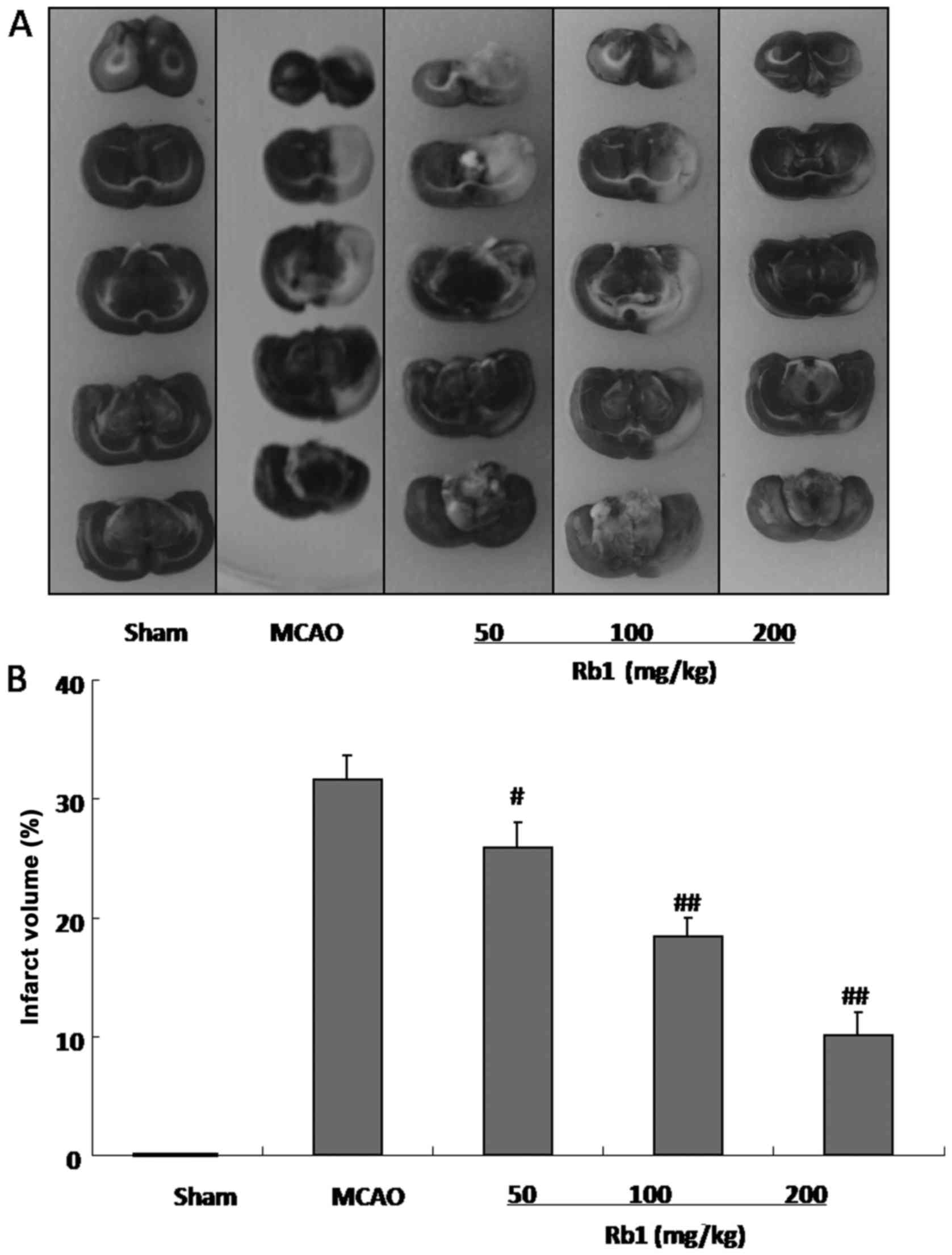
Rb1 reduces cerebral infarct volume in
the MCAO rat model
Infarct area of brain tissues from the animals
measured 24 h after I/R injury by TTC staining are presented in
Fig. 1. No infarct was observed in
sham animals, whereas in the MCAO group, the infarct area reached
31.56% the whole brain. However, as shown in Fig. 1B, Rb1-treatment decreased infarct
volumes in MCAO rats in a dose-dependent manner: 50, 100 and 200
mg/kg Rb1 treatment reduced the infarct volume to 25.89% (P<0.05
vs. MCAO animals), 18.35% (P<0.01 vs. MCAO animals) and 10.13%
(P<0.01 vs. MCAO animals), respectively.
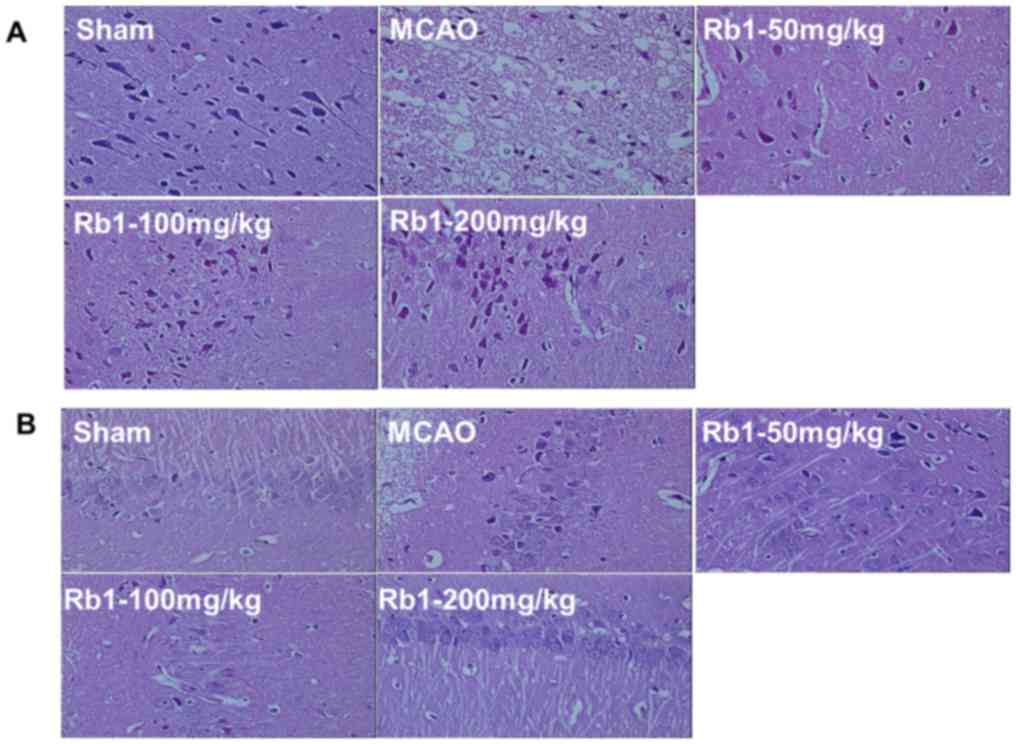
Rb1 treatment improves brain
histopathological abnormalities and neuron apoptosis
Hematoxylin and eosin staining was applied to
examine the histopathological abnormalities following focal
cerebral I/R (Fig. 2). No
histopathological damage was detected in the corext or pyramidal
neurons in the hippocampus CA1 region of sham animals (Fig. 2A and B). In the MCAO rat model, the
majority of the neurons in the infarct core were atrophied and/or
reduced in size, exhibiting a eosinophilic cytoplasm and
triangulated pycnotic nucleus compared with the intact and
well-arranged neurons with eumorphism in the sham group. However,
the number of pyramidal neurons in the MCAO model was significantly
decreased compared with the sham group, and large necrotic neurons
surrounding the infarct core and in the peri-infarct zone were
noted, exhibiting pycnotic shape and condensed nuclear material.
The necrotic tissue was notablyremarkably diminished following Rb1
treatment. This suggests a reduction in nerve injury, characterized
by the decreased number of cells with obvious historical change,
such as liquefaction necrosis, pycnosis, nucleoli abolition and
nuclear fragmentation.
Adjusted expression of HMGB1 and
inflammatory factors by Rb1
The level of HMGB1 in the peri-infarct zones of
ischemic cortex samples from each group was measured. A
significantly increased level of HMGB1 was identified in brain
tissue subjected to focal cerebral ischemia reperfusion.
Furthermore, an increased level of HMGB1 was observed in MCAO rats
compared with the sham group (P<0.01; Fig. 3A), which was notably decreased by
Rb1-treatment.
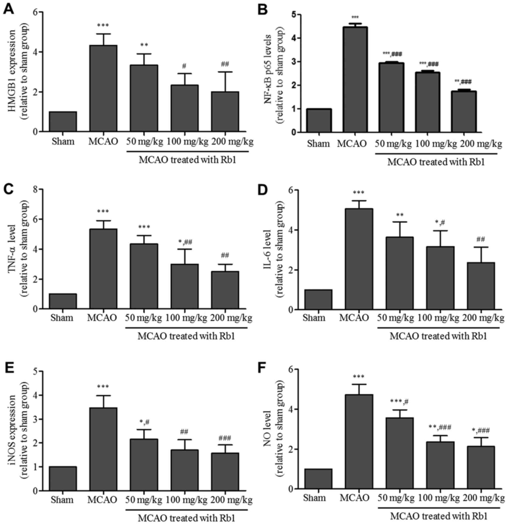 | Figure 3.Analysis of the levels of HMGB1 and
inflammatory factors. (A) HMGB1 levels, measured by ELISA. (B)
NF-κB expression levels. Levels of pro-inflammation factors, (C)
TNF-α, (D) IL-6, (E) iNOS and (F) NO measured by ELISA. *P<0.05,
**P<0.01, ***P<0.001 vs. sham group; #P<0.05,
##P<0.01, ###P<0.001 vs. MCAO group.
HMGB1, high-mobility group box 1; MCAO, middle cerebral artery
occlusion. |
High levels of TNF-α, iNOS, NO and IL-6 were also
identified in MCAO rats (P<0.05, sham vs. MACO; Fig. 3), which was significantly attenuated
in Rb1-treated animals.
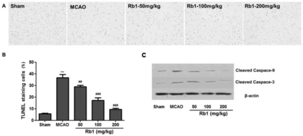
Decrease of neuronal cell apoptosis by
Rb1 treatment
Neuronal cell apoptosis was measured by TUNEL
staining in the rats of each group (Fig.
4). The number of TUNEL-positive neurons in the cortex region
was significantly increased in MCAO group after I/R injury compared
with the sham group (P<0.01). Following Rb1 treatment, the
number of TUNEL-positive neurons in the cortex region was
significantly reduced compared with the sham group (P<0.05;
Fig. 4). A higher proportion of
apoptotic neurons was detected in MCAO group, compared with the
sham group (P<0.01). Rb1 treatment at 50, 100 or 200 mg/kg
reduced the TUNEL-positive staining by 30.2, 19.6 and 9.2%,
respectively (P<0.01 vs. MCAO).
The expression levels of cleaved caspase-3 and
caspase-9 was also investigated. High expression of
apoptosis-related proteins was exhibited by brain tissues from the
MCAO group compared with the sham group (P<0.05; Fig. 4D). The elevated levels of cleaved
caspase-3 and caspase-9 were significantly attenuated with Rb1
treatment (Fig. 4C and D).
Discussion
Natural products can be used to modulate
cytokine-activity for treatment of diseases (5). In the present study, Rb1, one of the
major bioactive components of Panax ginseng, improved the
neurological function and decreased the infarct volume in brain
tissues of a MCAO rat model. HMGB1 levels in the brain tissue were
significantly decreased in MCAO rats after Rb1 treatment, and the
levels HMGB1-associated inflammatory factors, including TNF-α,
IL-6, iNOS and NO, were also reduced following Rb1 treatment. These
data suggested that Rb1 may have a neuroprotective effect against
I/R injury during stroke therapy.
Numerous studies have demonstrated the beneficial
effects of Rb1 in the treatment of ischemic stroke (9,13–15),
however, further research is required to understand the mechanisms
of Rb1 function. An inflammatory reaction in the brain causes
ischemic stroke, which occurs in ~80% stroke patients, and causes
the release of free radicals, resulting in oxidative damage of
brain tissues (13,14). High mobility group box1 (HMGB1) is a
highly conserved non-histone DNA-binding nuclear protein, and a
well-known damage-associated molecular pattern molecule which can
promote inflammatory injury. It is well established that
HMGB1-mediated inflammation may be a cause of cerebral I/R-induced
brain damage (16). Previous
research has demonstrated that inhibition of HMGB1 is associated
with suppression of infarct formation (17,18).
Other studies have implied that NF-κB may be a key regulator of
inflammation during and subsequent to brain damage (19,20). It
has been suggested that HMGB1 could rapidly bind to TLR-2 and
inhibit its expression, subsequently blocking NF-кB activation
induced by HMGB1 (21). In the
present study, significantly increased HMGB1 levels accompanied by
increased NF-кB levels in the MCAO model were demonstrated, which
is consistent with these previous reports. It was also demonstrated
that Rb1 administration markedly reduced the elevated HMGB1 and
NF-κB levels in the MCAO model. Therefore, we hypothesized that Rb1
treatment may lead to the downregulation of HMGB1 signaling and
that downstream molecules activate NF-κB p65, consequently
attenuating the I/R injury. Previous reports have demonstrated that
HMGB1 could upregulate the levels of TNF-α and IL-6, which can also
promote HMGB1 release via positive-feedback (22,23).
Indicators of inflammation were also identified in the present
study in the cerebral I/R, including TNF-α, IL-6 and iNOS, whose
levels were significantly decreased in the cortex tissue of MCAO
rats.
Furthermore, HMGB1 is a crucial proinflammatory
factor in ischemic stroke and the signal is transduced via its
putative receptors, such as toll-like receptors (TLRs), receptor
for advanced glycation end products (RAGE) and matrix
metalloproteinase (MMP) enzymes during ischemic stroke. The present
study suggests that Rb1 administration could markedly reduce the
elevated levels of HMGB1 and NF-κB in MCAO rats. However, the
effects of Rb1 on HMGB1-associated receptors remain to be
elucidated. HMGB1 may be a novel subject of brain-immune
communication and post-stroke immunomodulation research. In acute
ischemic stroke patients, the peripheral percentage of some subsets
of T-lymphocytes was associated with the level of neurological
deficit, and a predictive role of the peripheral percentage of
CD28-null cells in stroke diagnosis and TOAST subtyping was
suggested (24–25). Therefore, HMGB1 is a promising
therapeutic in promoting neurovascular repair and remodeling
following stroke.
I/R injury-induced oxidative stress and inflammation
also triggers multiple-cell apoptotic pathways responsible for cell
death by necrosis or apoptosis (26–28).
Previous research has demonstrated that HMGB1 could promote the
apoptosis of myocytes (22,29). In both MCAO rat models and stroke
patients, persistent NF-κB p65 activation has been indicated to
contribute to infarction and cell death induced by the I/R injury
(6,30). In the present study, Rb1 treatment
decreased neuronal cell apoptosis in MCAO rats in a dose-dependent
manner. Bcl-2 and Bax, belonging to the Bcl-2 family, are key
physiological and pathological regulators of cell apoptosis, and
act via the activation of caspase-triggered signaling cascades
(31,32). The present study demonstrated that
Rb1 markedly downregulated the levels of caspase-3 and caspase-9,
indicating that Rb1 suppressed cerebral I/R-induced cell apoptosis
in the brain tissue of MCAO rats by mediating caspase-3-associated
proteins.
In summary, the present study demonstrated that Rb1
has a protective effect on cerebral neurons in I/R injury. The
mechanisms underlying these actions are not well established,
however, our results suggest that the inhibition of inflammatory
HMGB1 signaling may serve an important role in the process.
Furthermore, Rb1 may be a promising neuroprotective candidate, and
requires further laboratory and clinical investigation.
Acknowledgements
The authors would like to thank the Central Research
Laboratory, The Second Hospital of Shandong University for
technical assistance and the generous support.
Funding
The present study was supported by the Natural
Science Foundation of China (grant no. 81402962).
Availability of data and materials
All of the materials used in the present study are
commercially available and all data included in the present study
were obtained by the co-authors.
Authors' contributions
AL, GH, ZC and LZ designed the study. HL, XX and WJ
performed the experiments. WZ, LS analyzed the data. HL and XX
wrote the manuscript.
Ethics approval and consent to
participate
All animal experiment protocols were approved by the
Institutional Animal Care Committee of Shandong University (Jinan,
China), and were performed in strict consistence with its
guidelines.
Patient consent for publication
Not applicable.
Competing interests
The authors declare that they have no competing
interests.
References
|
1
|
Zevallos J, Santiago F, González J,
Rodríguez A, Pericchi L, Rodríguez-Mercado R and Nobo U: Burden of
stroke in puerto rico. Int J Stroke. 10:117–119. 2015. View Article : Google Scholar : PubMed/NCBI
|
|
2
|
Shi GD, OuYang YP, Shi JG, Liu Y, Yuan W
and Jia LS: PTEN deletion prevents ischemic brain injury by
activating the mTOR signaling pathway. Biochem Biophys Res Commun.
404:941–945. 2011. View Article : Google Scholar : PubMed/NCBI
|
|
3
|
Cronin CA: Intravenous tissue plasminogen
activator for stroke: a review of the ECASS III results in relation
to prior clinical trials. J Emerg Med. 38:99–105. 2010. View Article : Google Scholar : PubMed/NCBI
|
|
4
|
Kirmani JF, Alkawi A, Panezai S and Gizzi
M: Advances in thrombolytics for treatment of acute ischemic
stroke. Neurology. 79:S119–S125. 2012. View Article : Google Scholar : PubMed/NCBI
|
|
5
|
Sun K, Fan J and Han J: Ameliorating
effects of traditional Chinese medicine preparation, Chinese
materia medica and active compounds on ischemia/reperfusion-induced
cerebral microcirculatory disturbances and neuron damage. Acta
Pharm Sin B. 5:8–24. 2015. View Article : Google Scholar : PubMed/NCBI
|
|
6
|
Xue X, Qu XJ, Yang Y, Sheng XH, Cheng F,
Jiang EN, Wang JH, Bu W and Liu ZP: Baicalin attenuates focal
cerebral ischemic reperfusion injury through inhibition of nuclear
factor κB p65 activation. Biochem Biophys Res Commun. 403:398–404.
2010. View Article : Google Scholar : PubMed/NCBI
|
|
7
|
Jung NP and Jin SH: Studies on the
physiological and biochemical effect of Korean ginseng. Korean J
Ginseng Sci. 20:431–471. 1996.
|
|
8
|
Li C, Zhu Y, Guo X, Sun C, Luo H, Song J,
Li Y, Wang L, Qian J and Chen S: Transcriptome analysis reveals
ginsenosides biosynthetic genes, microRNAs and simple sequence
repeats in Panax ginseng C. A. Meyer. BMC Genomics. 14:2452013.
View Article : Google Scholar : PubMed/NCBI
|
|
9
|
Zhang J, Han X, Li X, Luo Y, Zhao H, Yang
M, Ni B and Liao Z: Core-shell hybrid liposomal vesicles loaded
with panax notoginsenoside: Preparation, characterization and
protective effects on global cerebral ischemia/reperfusion injury
and acute myocardial ischemia in rats. Int J Nanomedicine.
7:4299–4310. 2012. View Article : Google Scholar : PubMed/NCBI
|
|
10
|
Longa EZ, Weinstein PR, Carlson S and
Cummins R: Reversible middle cerebral artery occlusion without
craniectomy in rats. Stroke. 20:84–91. 1989. View Article : Google Scholar : PubMed/NCBI
|
|
11
|
Wei X, Liu H, Sun X, Fu F, Zhang X, Wang
J, An J and Ding H: Hydroxysafflor yellow A protects rat brains
against ischemia-reperfusion injury by antioxidant action. Neurosci
Lett. 386:58–62. 2005. View Article : Google Scholar : PubMed/NCBI
|
|
12
|
Okuno S, Saito A, Hayashi T and Chan PH:
The c-Jun N-terminal protein kinase signaling pathway mediates Bax
activation and subsequent neuronal apoptosis through interaction
with Bim after transient focal cerebral ischemia. J Neurosci.
24:7879–7887. 2004. View Article : Google Scholar : PubMed/NCBI
|
|
13
|
Park EK, Choo MK, Oh JK, Ryu JH and Kim
DH: Ginsenoside Rh2 reduces ischemic brain injury in rats. Biol
Pharm Bull. 27:433–436. 2004. View Article : Google Scholar : PubMed/NCBI
|
|
14
|
Huang XP, Qiu YY, Wang B, Ding H, Tang YH,
Zeng R and Deng CQ: Effects of Astragaloside IV combined with the
active components of Panax notoginseng on oxidative stress injury
and nuclear factor-erythroid 2-related factor 2/heme oxygenase-1
signaling pathway after cerebral ischemia-reperfusion in mice.
Pharmacogn Mag. 10:402–409. 2014. View Article : Google Scholar : PubMed/NCBI
|
|
15
|
Ye R, Kong X, Yang Q, Zhang Y, Han J, Li
P, Xiong L and Zhao G: Ginsenoside rd in experimental stroke:
superior neuroprotective efficacy with a wide therapeutic window.
Neurotherapeutics. 8:515–525. 2011. View Article : Google Scholar : PubMed/NCBI
|
|
16
|
Zheng C, Liu C, Wang W, Tang G, Dong L,
Zhou J and Zhong Z: Ethanol extracts from Portulaca oleracea L.
attenuated ischemia/reperfusion induced rat neural injury through
inhibition of HMGB1 induced inflammation. Am J Transl Res.
8:5016–5024. 2016.PubMed/NCBI
|
|
17
|
Jin YC, Kim SW, Cheng F, Shin JH, Park JK,
Lee S, Lee JE, Han PL, Lee M, Kim KK, et al: The effect of
biodegradable gelatin microspheres on the neuroprotective effects
of high mobility group box 1 A box in the postischemic brain.
Biomaterials. 32:899–908. 2011. View Article : Google Scholar : PubMed/NCBI
|
|
18
|
Kim SW, Jin Y, Shin JH, Kim ID, Lee HK,
Park S, Han PL and Lee JK: Glycyrrhizic acid affords robust
neuroprotection in the postischemic brain via anti-inflammatory
effect by inhibiting HMGB1 phosphorylation and secretion. Neurobiol
Dis. 46:147–156. 2012. View Article : Google Scholar : PubMed/NCBI
|
|
19
|
Kim JW, Jin YC, Kim YM, Rhie S, Kim HJ,
Seo HG, Lee JH, Ha YL and Chang KC: Daidzein administration in vivo
reduces myocardial injury in a rat ischemia/reperfusion model by
inhibiting NF-kappaB activation. Life Sci. 84:227–234. 2009.
View Article : Google Scholar : PubMed/NCBI
|
|
20
|
Yang L, Tao LY and Chen XP: Roles of
NF-kappaB in central nervous system damage and repair, Neurosci.
Bull. 23:307–313. 2007.
|
|
21
|
Park JS, Svetkauskaite D, He Q, Kim JY,
Strassheim D, Ishizaka A and Abraham E: Involvement of toll-like
receptors 2 and 4 in cellular activation by high mobility group box
1 protein. J Biol Chem. 279:7370–7377. 2004. View Article : Google Scholar : PubMed/NCBI
|
|
22
|
Li X, Hu X, Wang J, Xu W, Yi C, Ma R and
Jiang H: Short-term hesperidin pretreatment attenuates rat
myocardial ischemia/reperfusion injury by inhibiting high mobility
group box 1 protein expression via the PI3K/Akt pathway. Cell
Physiol Biochem. 39:1850–1862. 2016. View Article : Google Scholar : PubMed/NCBI
|
|
23
|
Andersson U, Wang H, Palmblad K, Aveberger
AC, Bloom O, Erlandsson-Harris H, Janson A, Kokkola R, Zhang M,
Yang H and Tracey KJ: High mobility group 1 protein (HMG-1)
stimulates proinflammatory cytokine synthesis in human monocytes. J
Exp Med. 192:565–570. 2000. View Article : Google Scholar : PubMed/NCBI
|
|
24
|
ATuttolomondo A, Pecoraro R, Casuccio A,
Di Raimondo D, Buttà C, Clemente G, Della Corte V, Guggino G, Arnao
V, Maida C, et al: Peripheral frequency of CD4+ CD28-cells in acute
ischemic stroke: Relationship with stroke subtype and severity
markers. Medicine (Baltimore). 94:e812015.
|
|
25
|
Tuttolomondo A, Pedone C, Pinto A, Di
Raimondo D, Fernandez P, Di Sciacca R and Licata G: Gruppo Italiano
di Farmacoepidemiologia dell'Anziano (GIFA) researchers. Predictors
of outcome in acute ischemic cerebrovascular syndromes: The GIFA
study. Int J Cardiol. 125:391–396. 2008. View Article : Google Scholar : PubMed/NCBI
|
|
26
|
Hu GQ, Du X, Li YJ, Gao XQ, Chen BQ and Yu
L: Inhibition of cerebral ischemia/reperfusion injury-induced
apoptosis: Nicotiflorin and JAK2/STAT3 pathway. Neural Regen Res.
12:96–102. 2017. View Article : Google Scholar : PubMed/NCBI
|
|
27
|
Li P, Nijhawan D, Budihardjo I,
Srinivasula SM, Ahmad M, Alnemri ES and Wang X: Cytochrome c and
dATP-dependent formation of Apaf-1/caspase-9 complex initiates an
apoptotic protease cascade. Cell. 91:479–489. 1997. View Article : Google Scholar : PubMed/NCBI
|
|
28
|
Polster BM and Fiskum G: Mitochondrial
mechanisms of neural cell apoptosis. J Neurochem. 90:1281–1289.
2004. View Article : Google Scholar : PubMed/NCBI
|
|
29
|
Zhang HL, Gu ZL, Savitz SI, Han F,
Fukunaga K and Qin ZH: Neuroprotective effects of prostaglandin
A(1) in rat models of permanent focal cerebral ischemia are
associated with nuclear factor-kappaB inhibition and peroxisome
proliferator-activated receptor-gamma up-regulation. J Neurosci
Res. 86:1132–1141. 2008. View Article : Google Scholar : PubMed/NCBI
|
|
30
|
Hu X, Cui B, Zhou X, Xu C, Lu Z and Jiang
H: Ethyl pyruvate reduces myocardial ischemia and reperfusion
injury by inhibiting high mobility group box 1 protein in rats. Mol
Biol Rep. 39:227–231. 2012. View Article : Google Scholar : PubMed/NCBI
|
|
31
|
Nurmi A, Lindsberg PJ, Koistinaho M, Zhang
W, Juettler E, Karjalainen-Lindsberg ML, Weih F, Frank N,
Schwaninger M and Koistinaho J: Nuclear factor-kappaB contributes
to infarction after permanent focal ischemia. Stroke. 35:987–991.
2004. View Article : Google Scholar : PubMed/NCBI
|
|
32
|
Zhou JQ, Qiu T, Zhang L, Chen ZB, Wang ZS,
Ma XX and Li D: Allopurinol preconditioning attenuates renal
ischemia/reperfusion injury by inhibiting HMGB1 expression in a rat
model. Acta CBras. 31:176–182. 2016.
|