Introduction
By the end of the 21st century, the main demographic
trends have been characterized with declining birth rates,
stabilization in population size and elevation of the elderly
population (1). The aging of the
society is increasingly serious, and much attention has been paid
for the prevention of common diseases and improvement of the
quality of life of the elderly (2).
It is anticipated that affected populations and corresponding
health-care costs will be increased with population aging. Primary
osteoporosis, associated osteoporotic compression fractures and a
series of relevant complications are common in the elderly (in
postmenopausal women predominantly, but also found in men)
(3,4). Osteoporotic vertebral compression
fracture is a fragility fracture characterized by decreased in
distal forearm, hip, femur and other parts (5,6), most
common in the spine (7). In the
early years, frequent applicable approaches include bed rest, stent
assisted living and daily activities and the use of
anti-osteoporosis drugs. These measures are the most commonly used
for the treatment of this condition (7,8).
However, such conservative treatment has failed to improve
patients' severe pain and has always been accompanied by sleep
disorders, mental anxiety and other symptoms, seriously affecting
the quality of life of the patients (9,10).
Furthermore, long time physical dysfunction may not only lead to
severe bone loss and increase the progression of osteoporosis, but
also greatly improve risks of bedsore, related heart, lung
complications, and elevated mortality rates in elderly patients
during treatment.
The current treatment of osteoporotic vertebral
compression fracture is designed to relieve pain, restore the
activity of the elderly, and reduce the incidence of complications
(11,12). Vertebroplasty and kyphoplasty have
been generally accepted and implemented (13,14).
Previous evidence has supported that vertebroplasty is less
time-consuming and more cost-effective compared with kyphoplasty
(15). Kyphoplasty is considered to
be able to restore the vertebral height and reduce the risk of bone
cement leakage (16). However,
horizontal comparison of both methods has been widely debated.
Initial vertebroplasty was performed with the use of a hollow tube
and the push rod to inject bone cement into the vertebral body,
which is simple and effective (17,18).
However, this metod is associated with a higher rate of bone cement
leakage and recurrent fracture which is able to accurately control
cement injection volume and injection pressure (16,19).
Many scholars have tried to improve the bone cement injection
through other auxiliary devices, and new-type hydraulic delivery
vertebroplasty is one of the valid methods. Through the hydraulic
conveying bone cement injection device, precise control of bone
cement injection dosage and injection pressure restriction can be
achieved successfully, as well as the decreased risk of bone cement
leakage (20,21). Yet there were few relevant studies on
the clinical application of those methods in the treatment of
osteoporotic vertebral compression fractures.
Under the establishment of single segmental
osteoporotic compression fractures, a comparative analysis was
performed regarding the curative effect, physiological structure
recovery, and common complication rates such as bone cement leakage
of conventional pusher-type vertebroplasty, balloon kyphoplasty and
new-type hydraulic delivery vertebroplasty in the treatment of
single segmental osteoporotic vertebral compression fractures, so
as to evaluate corresponding safety and practicality in clinical
application for better guidance for the treatment of such kind of
disease.
Materials and methods
Ethical statement
All procedures performed in studies involving human
participants were in accordance with the ethical standards and with
the 1964 Helsinki declaration and its later amendments or
comparable ethical standards. Before enrollment, written informed
consents were provided from each participant before the performance
of the experiment, which involved the informing of the risk of
treatment procedures. The study was approved by the Ethics
Committee of the Third Affiliated Hospital of Guangzhou Medical
University (Guangzhou, China).
General information
The study was performed based on the incorporation
of osteoporotic vertebral compression fracture patients who had
operative indications in the Third Affiliated Hospital of Guangzhou
Medical University from May 2012 to September 2013. Following
screening and excluding, only patients with single segmental
osteoporotic vertebral compression fractures were included in the
study to avoid the mutual influence of multi-segment compression
fractures in the evaluation and statistical analysis for the
curative effect.
Inclusion criteria were: i) patients who were
positively diagnosed with single segmental osteoporotic vertebral
compression fractures in combination with clinical symptoms and
signs, X-ray film, and/or magnetic resonance imaging; ii) symptoms
appearing within 2 weeks and the magnetic resonance imaging results
were confirmed with fresh fracture; iii) vertebral compression
which did not exceed 75%; iv) spinal compression fractures without
causing symptoms of nerve compression; v) patients without severe
mental or neurological disorders, cardiac and pulmonary
dysfunction, or other disorders that may affect the outcome of
surgery or postoperative recovery; vi) patients who did not suffer
from femoral head necrosis, severe knee arthritis and other walking
activities that may affect the postoperative recovery of the
disease, and vii) operative indications covering the following
aspects: Vertebral compression exceeded 1/2 of the vertebral body;
angle of kyphosis over 30°; 1/3 intraspinal occupying; obvious
symptoms of vertebral instability; severe osteoporotic vertebral
fractures, and invalid conservative treatment.
Exclusion criteria were: i) imaging results showed
compression fracture in the fifth thoracic vertebral body and
above, but unclear during intraoperative X-ray imaging since the
lateral film was covered by the shoulder blade; ii) patients who
had poor blood pressure control during operation, or who were weak
and unable to keep pronation, as well as those patients who had
bone cement allergies or other situation that needed to suspend the
operation; iii) patients with symptoms such as femoral head
necrosis, severe knee arthritis and other walking activities, which
may affect the postoperative recovery of the disease; iv) patients
who had postoperative complications of bleeding, infection,
coagulation disorders and other disorders affecting the normal
course of treatment, and v) patients who were unable to cooperate
with the postoperative treatment or to adhere to 1 year-follow-up
due to economic or other medical reasons.
In strict accordance with the preset selection
criteria, the study was designed by prospective investigation.
There were a total of 188 cases of patients who were finally
incorporated in this cohort study, with a sum up of 259 segments.
Enrolled patients were classified into three groups by using
stratified-block randomized grouping method on the basis of sex
distribution, as well as affected/operated segments distributions
and positions. Patients were blinded to the study and were unaware
of the treatment assignment: i) group A, conventional pusher-type
vertebroplasty was carried out in 63 cases of patients with 90
segments within this group; ii) group B, patients in this group
were managed by balloon kyphoplasty, and a total of 58 cases of 81
segment were involved in; iii) group C, patients in this subgroup
received a new-type hydraulic delivery vertebroplasty treatment,
and there were 67 patients with 95 segments. Before operation, the
vertebroplasty and kyphoplasty groups had similar baseline back
pain based on the SF-36 physical component summary and Oswestry
disability index (ODI) scores assessment. After operation, all
patients received appropriate postoperative care, rehabilitation
exercise and guidance for the treatment of osteoporosis.
Follow-up information
In this study, the follow-up period lasted 12 months
after operation. At the end of the study, a total of 15 patients
were excluded from this study due to the exclusion criteria of the
cohort study; 4 of which showed blood pressure fluctuations during
operation and could not tolerate surgery and were further excluded
from this study; 4 patients with underlying disease progression
during the follow-up period were excluded from the study.
Additionally, 7 patients were lost during the follow-up period.
Finally, a total of 59 (59/63, 93.65%) patients were involved and
completed the study process in group A; 54 (54/58, 93.10%) patients
completed the study process in group B; 60 (60/67, 89.55%) patients
finished the study process in group C. The overall follow-up rate
was 92.02%.
Operative procedures
General information and procedures
within the three groups
The use of bone cement was the production of PMMA
MENDECSpine low viscosity bone cement in Italy (Tecres S.P.A.,
Sommacampagna, Italy). Surgical instruments were purchased from
Shanghai kailitai medical Polytron Technologies Inc. (Shanghai,
China). For three kinds of operation, patients were informed to
take the prone position in the surgical bed. Sterile surgical
towel, was used and foundation as well as sedative drugs were given
to relieve patients' anxiety. Surgery was conducted by unilateral
approach, and the side of the patient with conscious pain was
selected as the choice of surgical approach, following a 2%
lidocaine local anesthesia. Using a percutaneous procedure, the
needle was inserted in the direction of the pedicle of vertebral
arch (depending on different segments), and was adjusted under the
guidance of X-ray. The needle in the lateral spine development was
confirmed to be located in the pedicle of the vertebral arch, and
entered into the anterior-inferior 1/3 part along the pedicle of
vertebral arch. In order not to penetrate the inner wall of the
pedicle in the spinal normal development, the needle orientation
was kept at an approximately 10–15 abduction angle, and it was
ensured that the tip was as close to the midline of the vertebral
body as possible. Subsequently, following another confirmation of
the proper location of the puncture under fluoroscopic guidance,
the needle core was pulled out. Furthermore, after the finishing of
operational procedures in three groups, the operation time, number
of operations, as well as operation symptoms in the course of all
enrolled surgical patients were accurately recorded.
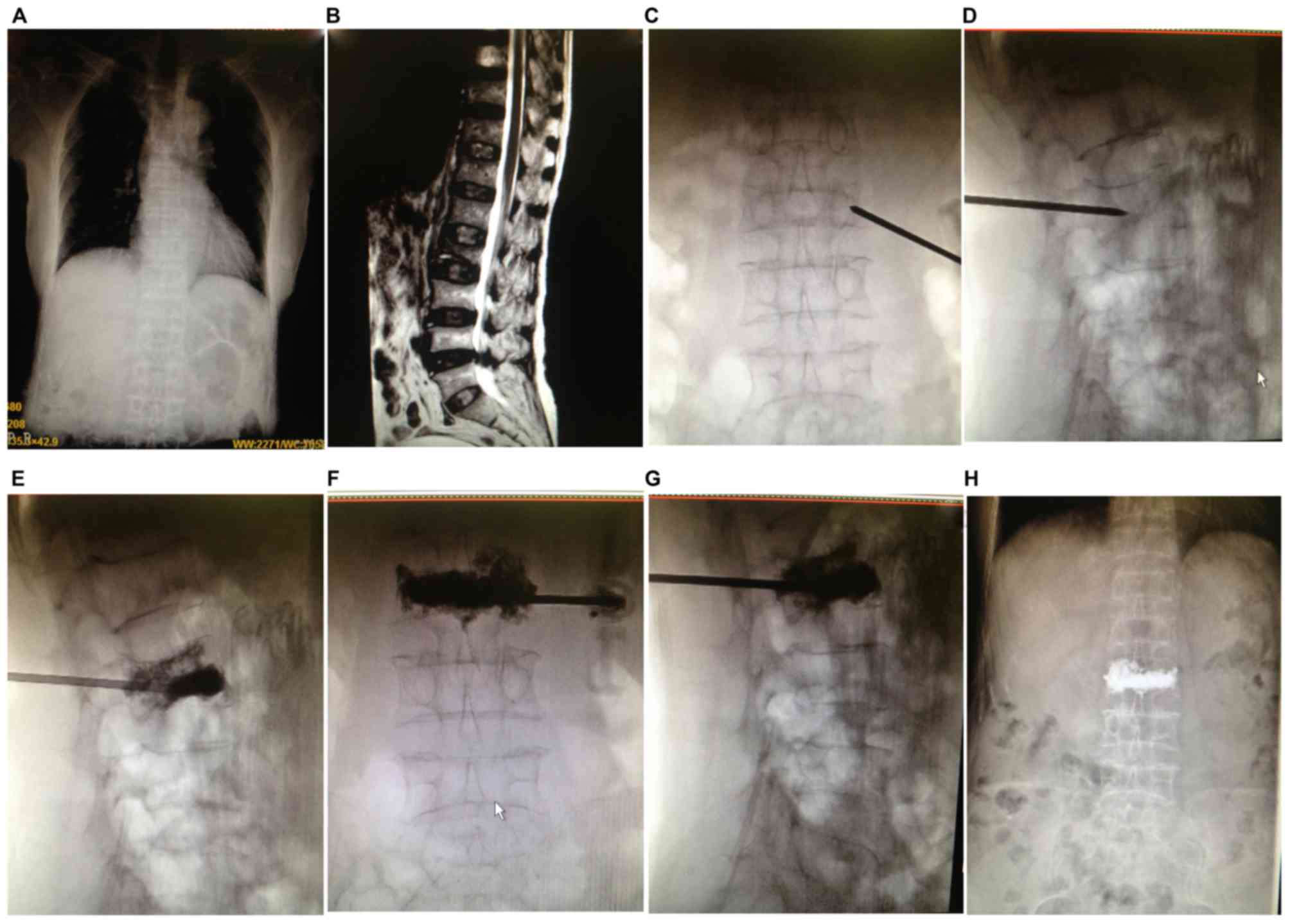
Conventional pusher-type
vertebroplasty
Typical images of patients and correlated surgical
procedures of conventional pusher-type vertebroplasty are shown in
Fig. 1. The equal proportion of bone
cement powder and liquid mixture were poured into the mixing bowl,
and cement was mixed into the early stage of the wire drawing under
rapid agitation. The hollow push pipes were filled with bone cement
(0.75 ml/pipe). Along the puncture needle, the pipes were inserted
and pushed into bone cement until bone cement was diffused
bilaterally and infiltration to posterior parts of the vertebral
bodies under the monitoring of the X-ray intermittent fluoroscopy
occurred. Of note, injection was stopped immediately if there was
an occurrence of bone cement leakage of the vertebral body before
satisfactory infiltration dispersion was reached. The above process
was controlled within 10 min after the bone cement mixture had been
completed. If the bone cement mixture had set within 15 min or full
hardening of the residual bone cement occurred, the puncture needle
was pulled out, followed by surgical dressing covering, and then
the operation was completed.
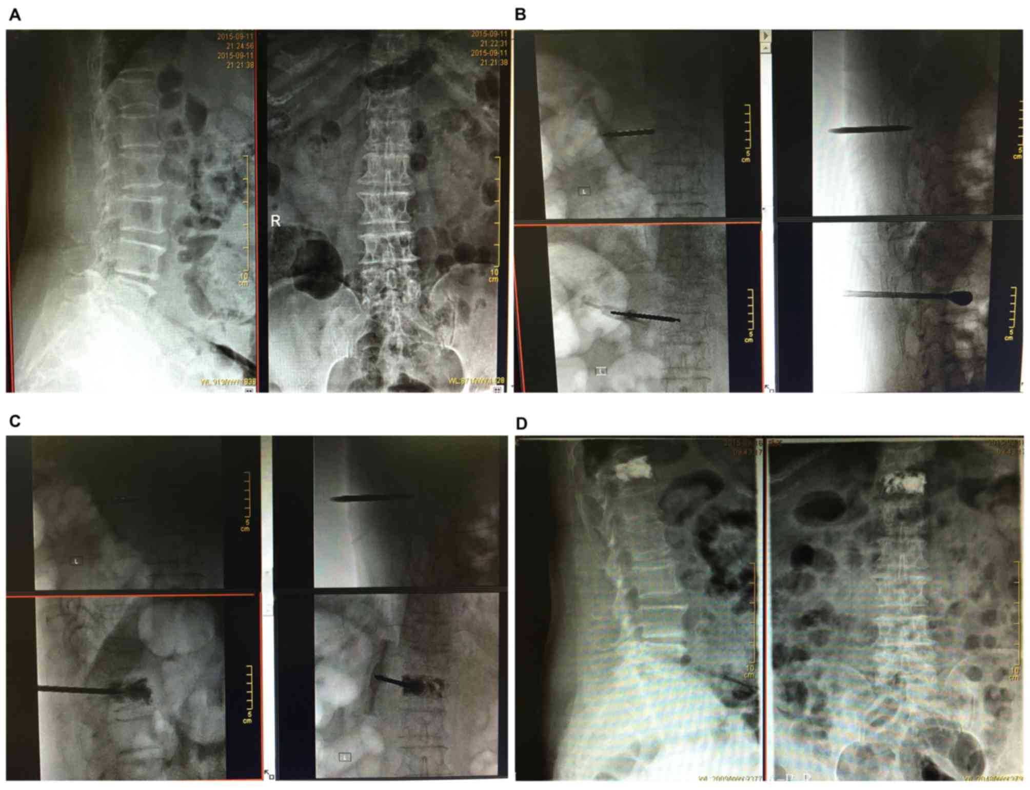
Balloon kyphoplasty
The size of dilation balloon was 15 or 20 mm
according to the size of the patient's injury. The puncture needle
was preset to be inserted into the vertebral body 1/3 part, and the
specific position was determined according to the size of the
vertebral injury and the size of dilation balloon. The guide wire
was placed from the outside of the needle tube, and the dilator was
inserted under the guidance of the guide wire to establish a
working channel. Fine bone drill was used to drill into the
vertebral body 1/3 part, and then the bone drill and vertebral body
bone fragments were removed. The dilation balloon was inserted via
the working channel, and it was confirmed that the balloon front
did not damage the vertebral body wall nor the trailing edge beyond
the outer tube of the puncture needle by the monitoring of the
X-ray intermittent fluoroscopy. After opening the tee valve,
injection of contrast agent was performed in 15 atmospheric
pressure to dilate the balloon, and the balloon was evacuated after
dilation was satisfactorily achieved after which the catheter was
removed. The equal proportion of bone cement powder and liquid
mixture were poured into the mixing bowl, and cement was mixed into
the early stage of the wire drawing. Subsequent procedures were the
same as those in group A (steps 1–5), followed by a surgical
dressing covering, indicating the operation in group B was
finished. Typical images related to the surgical procedures are
presented in Fig. 2.
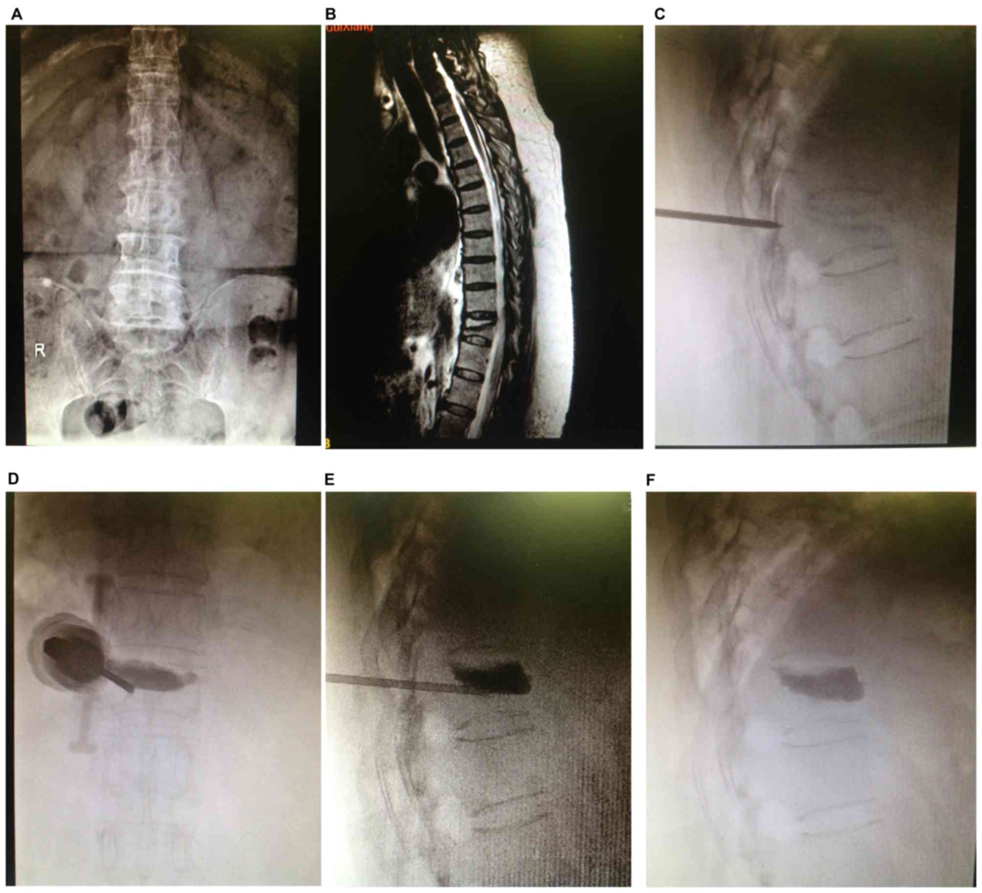
New-type hydraulic delivery
vertebroplasty
On the operating table, a 20-ml sterile saline
extracted with a syringe was injected into the hydraulic cylinder
to 0° of the water level calibration line, and then, the hydraulic
cylinder back cover was tightened. The equal proportion of bone
cement powder and liquid mixture were poured into the mixing bowl,
and cement was mixed into the early stage of the wire drawing.
Cylinder-connected injection tube was inverted and pressed into the
mixing drum, followed by clockwise rotation of the inverted barrel
collar, and stirring barrel of bone cement was transferred to the
injection tube. When the cylinder was arranged, the counter
rotating cylinder was rotated clockwise to take down the cylinder
and the mixing drum, and the quick joint of the hydraulic cylinder
was inserted into the back cover of the injection tube. The syringe
and needle tip were bolted down jointly. Clockwise rotation of the
hydraulic cylinder pushed the handle and bone cement in the
injection tube was slowly injected into the injured vertebra (0.3
ml/circle). The bone cement was injected into the posterior part of
the bone cement through the monitoring of X-ray, and the posterior
part of the vertebral body was diffused and infiltrated to the
posterior part of the bone cement. The above process was controlled
within 10 min after bone cement mixture was completed. Once the
bone cement mixture had set for 15 min or full hardening of the
residual bone cement occurred, the puncture needle was pulled out,
followed by surgical dressing covering, and then the operation was
completed. Details of the new-type hydraulic delivery
vertebroplasty typical images of patients and correlated surgical
procedures conducted step by step are shown in Fig. 3.
Evaluation of therapeutic effect
Before surgery, 1 day after the operation, one month
and six months after surgery, visual analogue scale (VAS) was used
to compare the degree of pain in patients, respectively (22). Corresponding scoring standards were
as follows: 0 point for painless, 10 points for intolerable pain.
Oswestry dysfunction indexes (ODI) (23) were used for functional recovery
assessment. Height of the center, vertebral body and percentage of
restoration in the vertebral body height were calculated
subsequently to measure the stability of the structure. The
incidence of bone cement leakage was recorded, and the risk of
surgical complications were further compared.
Statistical analysis
SPSS version 22.0 software (IBM Corp., Armonk, NY,
USA) was used. The age, sex ratio, operation time and bone cement
injection volume of different groups were verified by single factor
analysis of variance followed by post hoc test (Least Significant
Difference). The VAS score, ODI score and incidence of bone cement
leakage was verified by t-test before and after surgery. The
percentage of restoration in the vertebral body height was verified
by the Wilcoxon rank-sum test, and the difference was statistically
significant when P<0.05.
Results
General data comparison among the
three groups
There were 59 cases of patients with 85 segments
within group A. A total of 54 cases of 75 segment were involved in
group B, and 60 patients with 88 segments in group C. Corresponding
affected/operated segments of participants from each procedure are
provided in Table I, primarily
concentrated in the area of the thoracolumbar segments. Mean ages
of subjects from each group were 61.36±11.76, 59.73±10.71 and
64.38±12.00 years old, respectively. The sex ratios (male/female)
of each group were 14/49, 11/47 and 16/51, respectively. No
significant statistical difference was found regarding age and sex
among the three groups, suggesting a comparability in those groups
to some extent (all P>0.05). Furthermore, there was no
statistical significance in the operation time between group A and
group C, but there were statistical differences in that the
operation time in group B was obviously higher than that in groups
A and C (all P<0.05). In addition, considering the comparison of
VAS and ODI scoring results before and after surgery, as shown in
Table I, VAS and ODI scores were
significantly lower preoperatively than those postoperatively among
the three groups (all P<0.05). However, there was no statistical
difference in the comparison of different time-point before and
after operation within the three groups (all P>0.05).
 | Table I.Baseline characteristics and the
results of VAS and ODI scores before and after surgery among
groups. |
Table I.
Baseline characteristics and the
results of VAS and ODI scores before and after surgery among
groups.
| Variables | Group A (n=59) | Group B (n=54) | Group C (n=60) | F/χ2 | P-value |
|---|
| Age (years) | 61.36±11.76 | 59.73±10.71 | 64.38±12.00 | 2.409 | 0.093 |
| Sex
(male/female) | 12/47 | 11/43 | 13/47 |
| 0.980 |
| Affected/operated
segments |
|
|
| 0.261 | 0.992 |
| Upper-middle thoracic
vertebrae (T4-T9) | 26 | 21 | 26 |
|
|
| Thoracolumbar
(T10-L2) | 46 | 44 | 51 |
|
|
| Lower lumbar
vertebrae (L3-L5) | 18 | 16 | 18 |
|
|
| Operation time | 24.50±4.30 |
32.60±5.00a |
25.40±3.80b | 54.77 | <0.0001 |
| VAS score |
|
|
|
|
|
| Before
surgery | 8.10±2.40 | 8.10±2.50 | 8.20±2.20 | 0.035 | 0.966 |
| 1 day
after surgery | 2.60±0.30 |
2.30±0.40a |
2.20±0.40a |
| <0.0001 |
| 1 month
after surgery | 1.70±0.30 | 1.80±0.20 |
1.50±0.10a,b | 18.80 | <0.0001 |
| 6 months
after surgery | 1.50±0.20 | 1.40±0.30 |
1.40±0.20a | 3.497 | 0.033 |
| ODI score |
|
|
|
|
|
| Before
surgery | 78.24±13.07 | 77.52±12.27 | 78.12±12.83 | 0.051 | 0.950 |
| 1 day
after surgery | – | – | – |
|
|
| 1 month
after surgery | 34.55±8.77 |
32.14±10.10a | 35.27±11.35 | 1.465 | 0.234 |
| 6
months after surgery | 26.30±7.11 | 26.68±6.91 | 25.89±8.78 | 0.151 | 0.860 |
Total vertebral compression rates
comparison among the three groups
As shown in Table
II, the total vertebral compression rate of groups A and B was
43.31±8.93 and 43.17±6.66% respectively, compared with group C
(45.24±7.86%), and there was no significant difference among the
three groups (P>0.05). Furthermore, the bone cement injection
volumes in group A were significantly lower than those in groups B
and C, with statistical significance (both P<0.05). Yet there
was no significant difference between group B and group C
(P>0.05).
| Table II.The total vertebral compression and
vertebral height recovery rate comparison among the three
groups. |
Table II.
The total vertebral compression and
vertebral height recovery rate comparison among the three
groups.
| Variables | Group A (n=59) | Group B (n=54) | Group C (n=60) | P-value |
|---|
| Preoperative
vertebral compression rate (%) | 43.31±8.93 | 43.17±6.66 | 45.24±7.86 | 0.288 |
| Bone cement
injection volume (ml) | 3.41±1.30 |
4.60±1.00a |
4.40±0.90a | <0.0001 |
| Vertebral height
recovery rates (%) |
|
|
|
|
| 1 day after
surgery | 20.11±5.34 |
28.47±3.36a |
32.17±3.20a,b | <0.0001 |
| 1 year after
surgery | 16.14±3.33 |
26.24±2.12a |
29.82±5.13a,b | <0.0001 |
| Height restoration
loss rate (%) | 20.3 | 14.8 | 8.3 | <0.0001 |
One day after the operation, the vertebral height
recovery rates were 20.11±5.34, 28.47±3.36 and 32.17±3.20% in
groups A, B and C, respectively, showing statistical differences
(all P<0.05). After a year of follow-up, the vertebral height
recovery rate of the three groups were 16.14±3.33, 26.24±2.12 and
29.82±5.13%, respectively. The vertebral height recovery outcome in
group A was obviously poorer than that in groups B and C (both
P<0.05). Furthermore, a poorer outcome in group B was also found
when compared with group C (P<0.05). A year and one day after
operation, X-ray films showed the vertebral height restoration had
certain degree of loss, and compared with the recovery degree
results, the loss rate was 20.5, 14.0 and 7.5% in groups A, B and
C, respectively, while obviously lower in group C.
Bone cement dispersion comparison
among the three groups
X-ray was applied in the included patients to
confirm whether bone cement was over midline and reached the
opposite side more than 3/4 of the vertebral body or not, and bone
cement dispersion of the three different operative treatments was
classified into three levels, namely, satisfactory, unsatisfactory
and poor. There were 12 cases of poor dispersion and 20 cases of
unsatisfactory dispersion in group A. Furthermore, in group B, 9
cases of poor dispersion were found, and no cases of unsatisfactory
dispersion. In group C, there were 8 cases of poor dispersion and
no case of unsatisfactory dispersion. In comparison, the bone
cement dispersion in groups B and C were relatively better than
that in group A, with a statistical difference (both P<0.05). A
lower bone cement dispersion rate was also found in group C when
compared to that in group B.
Follow-up and postoperative
complications comparison
There were no complications such as vascular
embolization, spinal cord compression and other serious bone cement
leakage during the follow-up period of 1 year among the three
groups. There were a total of 17 segments showing bone cement
leakage event that had no symptoms or caused local mild symptoms: 7
segments were in group A (7/59, 11.86%), 6 segments in group B
(6/54, 11.11%) and 4 segments in group C (4/60, 6.67%). There were
8 cases of primary vertebral fractures, adjacent vertebral
fractures or other vertebral compression fractures in 1 year after
the operation with 3 cases in group A, 4 cases in group B, and 1
case in group C, while group C was significantly lower (Table III). In particular, 8 patients with
recurrent spinal fractures in one year after surgery were the same
as those with diffuse or poor dispersion. This may indicate that
there was a certain correlation between the dispersion of bone
cement and the high risk of re-fracture.
| Table III.Postoperative complications
classification and comparison among the three groups. |
Table III.
Postoperative complications
classification and comparison among the three groups.
| Complications | Types | Group A (n=59) n
(%) | Group B (n=54) n
(%) | Group C (n=60) n
(%) |
|---|
| Bone cement leakage
event | Peripheral
vertebral leakage | 1 (1.7) | 0 (0) | 0 (0) |
|
| Posterior vertebral
leakage | 3 (5.0) | 4 (7.4) | 2 (3.3) |
|
| Needle tract
leakage | 3 (5.0) | 2 (3.7) | 2 (3.3) |
| Recurrent spinal
fractures | Primary vertebral
fractures | 1 (1.7) | 0 (0) | 0 (0) |
|
| Adjacent vertebral
fractures | 0 (0) | 2 (3.7) | 0 (0) |
|
| Other vertebral
compression fractures | 2 (3.4) | 2 (3.7) | 1 (1.7) |
Discussion
In the study, the curative efficacy of conventional
pusher-type vertebroplasty, balloon kyphoplasty and new-type
hydraulic delivery vertebroplasty were compared in the treatment of
single segment osteoporotic vertebral compression fractures, to
evaluate the remission of symptoms, functional recovery,
reconstruction and prognosis assessment and thus to investigate the
safety and practicality correspondingly. All the incorporated
subjects were comparable among the three groups. Besides, enrolled
patients were confirmed with single segmental osteoporotic
vertebral compression fractures and underwent strict inclusion and
exclusion criteria to avoid possible selection bias and potential
interactions of mixed multiple segmental fracture affecting
therapeutic effects assessment and statistical analyses.
Additionally, thoracolumbar segments of the middle spinal are the
transition segment of the thoracic kyphosis to the lumbar lordosis,
upper undertaking of a relatively stable thoracic spine, lower
connecting of the lower lumbar vertebrae with a relatively strong
structure and supported by pelvis, which is the turning point in
the flexion position of spinal biomechanics, and fracture is
therefore frequent by external force.
In the comparison of pain relief related index VAS
score and functional recovery index ODI score, VAS and ODI scores
were significantly lower preoperatively than those postoperatively
among the three groups, yet no statistical difference was observed
comparing different time-points before and after operation within
the three groups. The aforementioned results suggested that all
approaches could significantly improve the postoperative VAS and
ODI scores, providing similar sustained improvement with regard to
pain and quality of life from baseline and remaining stable during
the follow-up. These improvements were statistically significant
and indicated certain clinical value regarding pain relief and
functional recovery. Yet, neither balloon kyphoplasty nor the
new-type hydraulic delivery vertebroplasty displayed their
superiority. However and most importantly, it is not sufficient to
simply evaluate the pain alleviation or short-term functional
recovery to comprehensively evaluate the therapeutic effect of
osteoporotic vertebral compression fractures. Vertebral body
structure stability plays an important role in the long-term
guarantee of the quality of life of patients after operation
(24). Further investigation on
postoperative recovery and complication rates supported the
significance of the new-type hydraulic delivery vertebroplasty in
the treatment of single segmental osteoporotic vertebral
compression fracture treatment.
Bone cement dispersion has been considered to be a
critical index in the evaluation of the treatment efficacy of
vertebroplasty and kyphoplasty (25–27).
Bone cement dispersion and distribution of the hydraulic delivery
vertebroplasty had been indicated to be a more ideal selection than
that of the conventional pusher-type vertebroplasty or balloon
kyphoplasty, which may also have a positive correlation with the
stability of the vertebral body and risk of complications
postoperatively. Furthermore, after a year of follow-up, results of
the height of the vertebral body also reflected the advantages of
the new-type hydraulic delivery vertebroplasty. Both the bone
cement injection volume and recovery rate of vertebral body height
postoperatively were compared, and the result of the hydraulic
delivery vertebroplasty group was better than that of the
conventional pusher-type vertebroplasty group or balloon
kyphoplasty group. Particularly noteworthy was that one year after
surgery, the recovery vertebral body height still appeared
significantly higher level in the patients of the hydraulic
delivery vertebroplasty group than that of the other two groups.
This may be likely to be a high-risk factor for the subsequent
fracture of the vertebral body or other vertebral bodies of the
latter two procedures, which may also be closely related to the
dispersion of the bone cement (28).
Furthermore, although more bone cement injection was
found in the hydraulic delivery vertebroplasty group than that in
the conventional pusher-type vertebroplasty group or balloon
kyphoplasty group. The leakage rate of the bone cement in the
former group did not increase and was even lower than the latter
two groups. Similar results were also found regarding complications
such as the recurrence of spine fracture, although the follow-up
time was relatively short, and the sample size was small. At least,
this part of the investigation proved the stability and security of
the new-type hydraulic delivery vertebroplasty in the management of
osteoporotic vertebral compression fractures. In addition, a
relatively higher bone cement leakage rate was found in the
conventional pusher-type vertebroplasty group than that in the
other two groups, and a possible reason may be that in the process
of injection of the bone cement, the loading of vertebral pressure
would inevitably result in the whole structure damage of the
original vertebral bone structure. If the bone cement dispersion
outcome was poor, the support effect would not be ideal, and
bearing unevenness would therefore cause loss of vertebral height
and increased refracture risk (28–30).
The major limitation in this study was the
relatively smaller sample size and relatively shorter follow-up
period, which may affect the statistical power of the results.
Furthermore, there is still no established standard for
vertebroplasty and kyphoplasty. However, health-care in the
perioperative period were kept in consistent. Of note, the
strengthening of this study lies in the detailed selection criteria
aimed to reduce selection bias, and the results of this trial
confirmed the effectiveness of the new-type hydraulic delivery
vertebroplasty at index levels was correlated with physical
examination, imaging findings and postoperative follow-up. It will
be better to verify the current study with further multicenter,
large sample size and long-term follow-up studies.
To sum up, three operation methods have equivalent
effects in the improvement of symptoms and functional recovery.
Significantly, new-type hydraulic delivery vertebroplasty has a
more concise operation and shorter operation time, displaying more
outstanding treatment outcomes in the spinal reconstruction and
reduction of complications by evaluating the diffusion of the bone
cement, vertebral height restoration rate and postoperative
complications than conventional pusher-type vertebroplasty and
balloon kyphoplasty.
Acknowledgements
Not applicable.
Funding
No funding was received.
Availability of data and materials
The datasets used and/or analyzed during the present
study are available from the corresponding author on reasonable
request.
Authors' contributions
PZ and JL designed the study. ZHZ and HTY collected
the patient data. WZ analyzed the patient data. PZ prepared the
manuscript. All authors read and approved the final manuscript.
Ethics approval and consent to
participate
The study was approved by the Ethics Committee of
the Third Affiliated Hospital of Guangzhou Medical University
(Guangzhou, China). Signed written informed consents were obtained
from the patients and/or guardians.
Patient consent for publication
Not applicable.
Competing interests
The authors declare that they have no competing
interests.
References
|
1
|
DESA, . World Population Prospects: The
2012 Revision. Population Division of the Department of Economic
and Social Affairs of the United Nations Secretariat. New York:
2013
|
|
2
|
Harper S: Economic and social implications
of aging societies. Science. 346:587–591. 2014. View Article : Google Scholar : PubMed/NCBI
|
|
3
|
Bliuc D, Alarkawi D, Nguyen TV, Eisman JA
and Center JR: Risk of subsequent fractures and mortality in
elderly women and men with fragility fractures with and without
osteoporotic bone density: The Dubbo Osteoporosis Epidemiology
Study. J Bone Miner Res. 30:637–646. 2015. View Article : Google Scholar : PubMed/NCBI
|
|
4
|
Fernández-Ruiz M, Guerra-Vales JM,
Trincado R, Medrano MJ, Benito-León J and Bermejo-Pareja F: Hip
fracture in three elderly populations of central Spain: Data from
the NEDICES study. Intern Emerg Med. 9:33–41. 2014. View Article : Google Scholar : PubMed/NCBI
|
|
5
|
Svensson HK, Olofsson EH, Karlsson J,
Hansson T and Olsson LE: A painful, never ending story: Older
women's experiences of living with an osteoporotic vertebral
compression fracture. Osteoporos Int. 27:1729–1736. 2016.
View Article : Google Scholar : PubMed/NCBI
|
|
6
|
Stauff MP and Carragee EJ: Vertebral
compression fracture rules. Spine J. 14:971–972. 2014. View Article : Google Scholar : PubMed/NCBI
|
|
7
|
Sinaki M: Yoga spinal flexion positions
and vertebral compression fracture in osteopenia or osteoporosis of
spine: Case series. Pain Pract. 13:68–75. 2013. View Article : Google Scholar : PubMed/NCBI
|
|
8
|
Ha KY, Park KS, Kim SI and Kim YH: Does
bisphosphonate-based anti-osteoporosis medication affect
osteoporotic spinal fracture healing? Osteoporos Int. 27:483–488.
2016. View Article : Google Scholar : PubMed/NCBI
|
|
9
|
Su FM, Chen YC, Cheng TT, Lin WC and Lui
CC: Is raloxifene associated with lower risk of mortality in
postmenopausal women with vertebral fractures after
vertebroplasty?: A hospital-based analysis. BMC Musculoskelet
Disord. 16:2092015. View Article : Google Scholar : PubMed/NCBI
|
|
10
|
Lazzari AA, Dussault PM, Thakore-James M,
Gagnon D, Baker E, Davis SA and Houranieh AM: Prevention of bone
loss and vertebral fractures in patients with chronic epilepsy -
antiepileptic drug and osteoporosis prevention trial. Epilepsia.
54:1997–2004. 2013. View Article : Google Scholar : PubMed/NCBI
|
|
11
|
Chen AT, Cohen DB and Skolasky RL: Impact
of non-operative treatment, vertebroplasty, and kyphoplasty on
survival and morbidity after vertebral compression fracture in the
medicare population. J Bone Joint Surg Am. 95:1729–1736. 2013.
View Article : Google Scholar : PubMed/NCBI
|
|
12
|
Chen HG, Zhang Z, Liang HP, Kong QZ, Chen
JH and Zhou Y: Clinical observation of effects and complications of
the mid-stage in treating osteoporotic vertebral compression
fracture with percutaneous kyphoplasty. Zhongguo Gu Shang.
23:743–745. 2010.(In Chinese). PubMed/NCBI
|
|
13
|
Ma XL, Xing D, Ma JX, Xu WG, Wang J and
Chen Y: Balloon kyphoplasty versus percutaneous vertebroplasty in
treating osteoporotic vertebral compression fracture: Grading the
evidence through a systematic review and meta-analysis. Eur Spine
J. 21:1844–1859. 2012. View Article : Google Scholar : PubMed/NCBI
|
|
14
|
Tseng YY, Su CH, Lui TN, Yeh YS and Yeh
SH: Prospective comparison of the therapeutic effect of
teriparatide with that of combined vertebroplasty with
antiresorptive agents for the treatment of new-onset adjacent
vertebral compression fracture after percutaneous vertebroplasty.
Osteoporos Int. 23:1613–1622. 2012. View Article : Google Scholar : PubMed/NCBI
|
|
15
|
Gu YF, Li YD, Wu CG, Sun ZK and He CJ:
Safety and efficacy of percutaneous vertebroplasty and
interventional tumor removal for metastatic spinal tumors and
malignant vertebral compression fractures. AJR Am J Roentgenol.
202:W298–W305. 2014. View Article : Google Scholar : PubMed/NCBI
|
|
16
|
Tomé-Bermejo F, Piñera AR, Duran-Álvarez
C, López-San Román B, Mahillo I and Alvarez L: Identification of
risk factors for the occurrence of cement leakage during
percutaneous vertebroplasty for painful osteoporotic or malignant
vertebral fracture. Spine. 39:E693–E700. 2014. View Article : Google Scholar : PubMed/NCBI
|
|
17
|
Lai PL, Tai CL, Chu IM, Fu TS, Chen LH and
Chen WJ: Hypothermic manipulation of bone cement can extend the
handling time during vertebroplasty. BMC Musculoskelet Disord.
13:1982012. View Article : Google Scholar : PubMed/NCBI
|
|
18
|
Boger A, Benneker LM, Krebs J, Boner V,
Heini PF and Gisep A: The effect of pulsed jet lavage in
vertebroplasty on injection forces of PMMA bone cement: An animal
study. Eur Spine J. 18:1957–1962. 2009. View Article : Google Scholar : PubMed/NCBI
|
|
19
|
Zhu SY, Zhong ZM, Wu Q and Chen JT: Risk
factors for bone cement leakage in percutaneous vertebroplasty: A
retrospective study of four hundred and eighty five patients. Int
Orthop. 40:1205–1210. 2016. View Article : Google Scholar : PubMed/NCBI
|
|
20
|
Wang CH, Ma JZ, Zhang CC and Nie L:
Comparison of high-viscosity cement vertebroplasty and balloon
kyphoplasty for the treatment of osteoporotic vertebral compression
fractures. Pain Physician. 18:E187–E194. 2015.PubMed/NCBI
|
|
21
|
Zhang L, Wang J, Feng X, Tao Y, Yang J,
Wang Y, Zhang S, Cai J and Huang J: A comparison of high viscosity
bone cement and low viscosity bone cement vertebroplasty for severe
osteoporotic vertebral compression fractures. Clin Neurol
Neurosurg. 129:10–16. 2015. View Article : Google Scholar : PubMed/NCBI
|
|
22
|
Langley GB and Sheppeard H: The visual
analogue scale: Its use in pain measurement. Rheumatol Int.
5:145–148. 1985. View Article : Google Scholar : PubMed/NCBI
|
|
23
|
Park SW, Shin YS, Kim HJ, Lee JH, Shin JS
and Ha IH: The dischargeable cut-off score of Oswestry Disability
Index (ODI) in the inpatient care for low back pain with
disability. Eur Spine J. 23:2090–2096. 2014. View Article : Google Scholar : PubMed/NCBI
|
|
24
|
Disch AC and Schmoelz W: Cement
augmentation in a thoracolumbar fracture model: Reduction and
stability after balloon kyphoplasty versus vertebral body stenting.
Spine. 39:E1147–E1153. 2014. View Article : Google Scholar : PubMed/NCBI
|
|
25
|
Chen C, Li D, Wang Z, Li T, Liu X and
Zhong J: Safety and efficacy studies of vertebroplasty,
kyphoplasty, and mesh-container-plasty for the treatment of
vertebral compression fractures: Preliminary Report. PLoS One.
11:e01514922016. View Article : Google Scholar : PubMed/NCBI
|
|
26
|
Hu X, Zhai X and Hirt T: A new concept for
more biocompliant bone cements for vertebroplasty and kyphoplasty.
Macromol Biosci. 9:195–202. 2009. View Article : Google Scholar : PubMed/NCBI
|
|
27
|
Hernández L, Parra J, Vázquez B, Bravo AL,
Collía F, Goñi I, Gurruchaga M and San Román J: Injectable acrylic
bone cements for vertebroplasty based on a radiopaque
hydroxyapatite. Bioactivity and biocompatibility. J Biomed Mater
Res B Appl Biomater. 88:103–114. 2009. View Article : Google Scholar : PubMed/NCBI
|
|
28
|
Shen Y, Ren H, Zhang Y, Zhi X, Ding W, Xu
J and Yang D: Correlative factor analysis of complications
resulting from cement leakage after percutaneous kyphoplasty in
treatment of osteoporotic vertebral body compression. Zhongguo Xiu
Fu Chong Jian Wai Ke Za Zhi. 24:27–31. 2010.(In Chinese).
PubMed/NCBI
|
|
29
|
Bolander JE, Choi S and Duddukuri SR:
Fracture of fiber-reinforced cement composites: Effects of fiber
dispersion. Int J Fract. 154:73–86. 2008. View Article : Google Scholar
|
|
30
|
Hulme PA, Boyd SK and Ferguson SJ:
Regional variation in vertebral bone morphology and its
contribution to vertebral fracture strength. Bone. 41:946–957.
2007. View Article : Google Scholar : PubMed/NCBI
|