Introduction
Chronic hepatitis B (CHB) is one of the most common
of chronic viral infectious diseases, which is mainly transmitted
by contacting the blood and body fluids of the infected people
(1). More than 350 million people
have been infected with CHB all over the world so far. CHB is the
leading cause of liver cirrhosis and hepatocellular carcinoma as
well as death of nearly 600,000 people every year, becoming one of
the worldwide primary health problems (2). Currently, the control rate of CHB is
<0.5% in some regions, and the disease mainly occurs among young
people. In some regions of Asia and Africa, mother-to-child
transmission is the most common route of transmission of CHB, with
a prevalence rate exceeding 10% (3).
CHB is characterized by recurrent attacks and prolonged courses,
and it can easily induce liver fibrosis, thus leading to liver
cirrhosis (4). The pathological
characteristic of liver fibrosis is excessive deposition of
extracellular matrix (ECM) in perisinusoidal space. Hepatitis B
virus (HBV) is the initiating agent of aggravated severity of CHB,
and apoptosis of liver cells induced by varying degrees of
inflammation plays an important role in the outcome of CHB
(5). Matrix metalloproteinase-9
(MMP-9) is a type of polypeptidase that can hydrolyze ECM (6), but the relationship of its level with
the severity of CHB still remains unclear at present. In this
study, the correlation of MMP-9 with inflammation was investigated
by means of analyzing the MMP-9 expression in different
inflammation grades and fibrosis stages of CHB, hoping to provide a
basis for controlling and reversing exacerbation of CHB degree.
Patients and methods
Clinical data
A total of 96 CHB patients admitted and treated in
Dongying People's Hospital (Dongying, China) from December 2016 to
November 2017 were selected as the observation group. The inclusion
criteria were as follows: i) patients who met the diagnostic
criteria of CHB (7), ii) patients
who did not receive antiviral therapy before admission to hospital,
and iii) patients who signed informed consent. The exclusion
criteria were as follows: i) patients with hepatitis A, C, D and E,
and ii) patients with malignant tumor as well as pregnant and
breast-feeding women. In addition, a total of 60 unrelated healthy
individuals in the physical examination center during the same time
period were selected as the control group. There were no
statistically significant differences in the general data of the
two groups of patients (P>0.05), and the data were comparable
(Table I).
 | Table I.Comparison of general data of the two
groups of patients. |
Table I.
Comparison of general data of the two
groups of patients.
| Items | Control group
(n=60) | Observation group
(n=60) | t/χ2 | P-value |
|---|
| Sex
(male/female) | 35/25 | 57/39 | 0.047 | 0.828 |
| Age (years) | 40–80 | 40–75 |
|
|
| Average age
(years) | 52.36±8.46 | 52.78±7.58 | 0.322 | 0.748 |
| BMI
(kg/m2) | 23.28±1.15 | 23.56±1.17 | 1.464 | 0.145 |
| Education level (n,
%) |
| Junior
high school and below | 16 (26.67) | 25 (26.04) | 0.126 | 0.939 |
| Senior
high school and special secondary school | 25 (41.67) | 38 (39.58) |
|
|
| College
and above | 19 (31.63) | 33 (34.38) |
|
|
This study was approved by the Ethics Committee of
Dongying People's Hospital. Signed informed consents were obtained
from the patients or guardians.
Methods
A total of 5 ml venous blood was collected from
every research object (fasted for 8 h from 7:00 a.m.), and was
centrifuged (Eppendorf Shanghai International Trade Co. Ltd.,
Shanghai, China) at 950 × g for 10 min and then stored in a
refrigerator at −20°C. Enzyme-linked immunosorbent assay (ELISA)
was used to detect the levels of MMP-9, tumor necrosis factor-α
(TNF-α) and interleukin-6 (IL-6) in the serum, relevant kits were
provided by Beijing Donggeboye Biological Technology Co., Ltd.
(Beijing, China) and the operations were performed in strict
accordance with the kit instructions. The sample (diluted at 1:2)
was added into the wells of microplate reader (Shenzhen SinoThinker
Technology Co., Ltd., Shenzhen, China), with 100 µl enzyme-labeled
solution in each well. Then the sample was incubated at 37°C for 90
min, and the microplate reader was washed 3 times. After that, 50
µl of color developing reagent solution Aand B was added and mixed
respectively, followed by incubation in the dark at room
temperature (20°C) for 15 min. Finally, the optical density (OD)
was measured at a wavelength of 450 nm with a microplate reader
within 15 min, and the levels of MMP-9, TNF-α and IL-6 were
calculated.
Evaluation criteria
Criteria of determining different degrees of CHB:
the liver tissue inflammation was divided into 5 grades and the
fibrosis was divided into 5 stages (8) (Table
II). A total of 5 ml venous blood was collected from every
research object in the two groups, and the serum MMP-9, TNF-α and
IL-6 levels were measured using ELISA.
| Table II.Criteria of determining degrees of
liver tissue inflammation and fibrosis. |
Table II.
Criteria of determining degrees of
liver tissue inflammation and fibrosis.
| Liver tissue
inflammation | Grade | Fibrosis degree | Stage |
|---|
| No inflammation | 0 | No fibrosis | 0 |
| Intralobar
degeneration and a few spotted and focal necrotic foci, and
inflammation in portal area | 1 | Expanded fibrosis in
portal area, localized fibrosis in lobule and perisinusoidal
space | 1 |
| Intralobar
degeneration and spotted and focal necrosis or eosinophilic bodies,
and mild piecemeal necrosis in portal area | 2 | Fibrosis around
portal area, formation of fibrous septa, and reservation of lobular
architecture | 2 |
| Intralobar
degeneration and confluent necrosis or bridging necrosis, and
moderate piecemeal necrosis in portal area | 3 | Formation of fibrous
septa associated with disorganized lobular architecture, without
liver cirrhosis | 3 |
| Extensive bridging
necrosis affecting several lobules, and severe piecemeal necrosis
in portal area | 4 | Liver cirrhosis at
early stage | 4 |
Statistical analysis
Statistical Product and Service Solutions (SPSS)
19.0 (IBM Corp., Armonk, NY, USA) software was applied to process
the data. The measurement data are presented as mean ± standard
deviation, and t-test was adopted. The enumeration data were
expressed by ratio, and χ2 test was performed. The
expression of MMP-9 in different degrees of lesion was examined via
rank sum test, and the P-value was calibrated using Bonferroni
method. Pearsons correlation coefficients were utilized to analyze
the correlations. P<0.05 was considered to indicate a
statistically significant difference.
Results
MMP-9, TNF-α and IL-6 expression in
the two groups of research objects
The levels of serum MMP-9, TNF-α and IL-6 in the
observation group were obviously higher than those in the control
group (P<0.05) (Table III).
| Table III.Comparison of MMP-9, TNF-α and IL-6
levels in the two groups of patients. |
Table III.
Comparison of MMP-9, TNF-α and IL-6
levels in the two groups of patients.
| Groups | n | MMP-9 (ng/ml) | TNF-α (ng/l) | IL-6 (ng/ml) |
|---|
| Observation | 96 | 256.95±15.13 | 79.97±6.47 | 56.76±6.38 |
| Control | 60 | 90.69±9.06 | 26.73±6.86 | 4.43±1.27 |
| t value |
| 76.884 | 48.853 | 77.932 |
| P-value |
| <0.001 | <0.001 | <0.001 |
MMP-9 expression in different
inflammation grades
The MMP-9 expression level was increased with the
aggravated inflammation activity, and the differences were
statistically significant among the 5 groups (P<0.05) (Table IV). It is indicated in the rank sum
test that there is statistical significance in the MMP-9 level
among different inflammation grades (P<0.05). Pairwise
comparisons using the Bonferroni method suggest that the
differences in MMP-9 level among the groups are statistically
significant (P<0.05).
| Table IV.Relations between MMP-9 level and
different inflammation grades. |
Table IV.
Relations between MMP-9 level and
different inflammation grades.
| Inflammation
grade | n | MMP-9 (ng/ml) | P-value |
|---|
| G0 | 16 | 98.95±9.13 | <0.001 |
| G1 | 30 | 143.69±9.56 |
|
| G2 | 26 | 218.95±13.13 |
|
| G3 | 18 | 283.69±16.06 |
|
| G4 | 6 | 318.69±20.06 |
|
MMP-9 expression in different fibrosis
stages
The MMP-9 expression level was elevated with the
increased fibrosis stage, and the differences among the 5 groups
were statistically significant (P<0.05) (Table V). It is indicated in the rank sum
test that there is statistical significance in the MMP-9 level in
different fibrosis stages (P<0.05). Pairwise comparison using
the Bonferroni method suggest that the differences in MMP-9 level
among the groups are statistically significant (P<0.05).
| Table V.Association of MMP-9 level with
different stages of fibrosis. |
Table V.
Association of MMP-9 level with
different stages of fibrosis.
| Fibrosis stage | n | MMP-9 (ng/ml) | P-value |
|---|
| S0 | 19 | 97.49±9.04 | <0.001 |
| S1 | 28 | 153.58±10.45 |
|
| S2 | 25 | 218.37±15.42 |
|
| S3 | 17 | 298.65±17.46 |
|
| S4 | 7 | 343.84±21.12 |
|
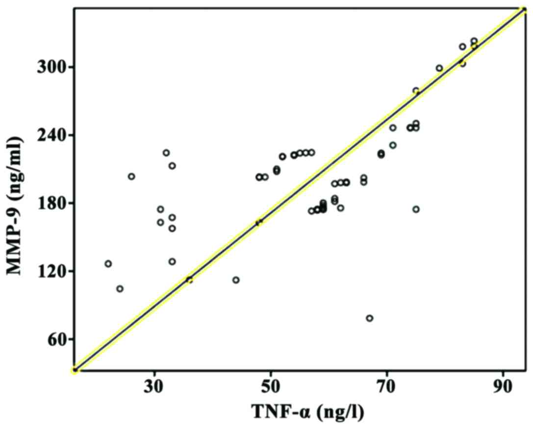
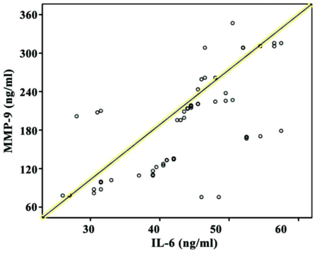
Analysis of correlation of MMP-9
expression level with inflammation
Pearsons correlation coefficient analysis showed
that the MMP-9 expression level was positively correlated with
TNF-α and IL-6 (P<0.05) (Figs. 1
and 2).
Discussion
The liver, a vital organ in the body that is
responsible for various kinds of biotransformation and storage,
plays an important role in the synthesis of coagulation factors,
storage and metabolism of fats and proteins as well as secretion
and detoxication of bile (9).
Multiple types of hepatitis occur easily after the liver is
infected by pathogens, dominated by CHB which has the highest
incidence rate among the infectious diseases of the digestive
system (10). According to the
report of the World Health Organization (WHO), there are
approximately 350 million CHB patients around the globe, including
nearly 28 million patients in China, where the incidence rate is
~2% (11). CHB can easily develop
into liver cirrhosis and result in liver cancer if it is not
treated in time (12). The
pathogenesis of CHB is very complex, which has not been elaborated
completely yet. It may be related to impacts of multiple factors,
such as hepatitis virus, apoptosis-related gene, autoimmunity and
cell molecules (13). The
pathological manifestations of CHB include massive apoptosis of
hepatocytes, severe liver dysfunction and abnormal biochemical
markers of liver function. Currently, there is no ideal therapeutic
method for CHB in clinic, and the major treatment strategies are
stimulation of the patient's immune responses and repression of HBV
replication (14).
MMP-9 is a kind of gelatinase substance secreted by
many cells, such as neutrophils, macrophages and capillary
endothelial cells, and it is a member of matrix metalloproteinases
(MMPs) (15). MMP-9 can participate
in the metabolism of collagen type IV in basement membrane, degrade
ECM and be widely involved in multiple types of tumor metastasis by
degrading ECM. In addition, it can induce basic fibroblast growth
factor and vascular endothelial growth factor and promote tumor
neovascularization (16). A study
has revealed that MMP-9 is closely related to CHB (17). TNF-α is the first inflammatory
mediator produced in the body, which plays a core role in the
occurrence and development of CHB and has crucial functions in the
induction of inflammatory responses, anti-infection and host
defense (18). IL-6 is a kind of
lymphocyte factor of acute phase reaction with diversified effects,
which accelerates the proliferation of myofibroblasts and leads to
liver fibrosis in CHB patients (19). The results of this study showed that
the levels of serum MMP-9, TNF-α and IL-6 in CHB patients were
significantly higher than those in normal population (P<0.05).
It is because the main pathological change of CHB is inflammation
in the liver. The peripheral blood mononuclear cell (PBMC) may
inhibit and attenuate the body's antiviral immune responses through
exogenous and endogenous apoptotic pathways, thus causing constant
replication of HBV. Moreover, it may activate inflammatory
responses, thus increasing the levels of TNF-α and IL-6 (20). The activities of MMPs in the body are
controlled strictly under normal conditions, and the MMP-9 remains
at a relatively low level. However, in CHB patients with
pathological inflammation, inflammatory factors may promote the
expression of MMP-9, thus increasing its level. It was indicated by
the results of this study that there were remarkable differences in
the MMP-9 expression in CHB patients with varying inflammation
grades (P<0.05), and that the MMP-9 expression level was
increased with the aggravated inflammation activity. The reason is
that MMP-9 is mainly secreted by inflammatory cells (macrophages
and T cells), and TNF-α, IL-6 and other inflammatory mediators
released at the early stage of inflammation can enhance the MMP-9
expression which is consistent with the severity of hepatic
inflammation.
An appropriate amount of MMP-9 can degrade ECM
deposited temporarily, and it can promote the reconstruction of
basement membrane, thus triggering liver fibrosis. However, the
liver fibrosis in CHB patients is a pathological repair process of
the liver parenchyma against chronic injuries. The results of this
study indicated that the differences in the MMP-9 expression in CHB
patients with different fibrosis stages were significant
(P<0.05). It is because only a small amount of MMP-9 is secreted
in the body at stage S0. At that moment, the production and
degradation of ECM is maintained in a state of homeostasis. With
the progression of CHB, however, the state of homeostasis is
broken, a large amount of ECM is accumulated in the liver, and
liver fibrosis is triggered, thus increasing the fibrosis stage. At
this time, massive MMP-9 is secreted in the body to decompose ECM,
and pathological repair is conducted by the body itself. MMP-9
level has a positive correlation with fibrosis stage, playing a
crucial role in the development of CHB.
In this study, Pearson correlation coefficient
analysis showed that the MMP-9 expression level was positively
correlated with TNF-α and IL-6 (P<0.05). This is because TNF-α
and IL-6 can influence the expression of MMP-9 and upregulate the
activity of MMP-9 in a concentration- and time-dependent manner,
while overexpressed MMP-9 can degrade ECM to destroy the basement
membrane and cause disorganized and loose tissue structures, thus
leading to further infiltration of TNF-α and IL-6. These factors
interact with each other and form a vicious cycle, further
resulting in pathological changes of the liver.
In conclusion, MMP-9 can be regarded as a
serological marker for judging the severity and progression of CHB,
which has very important clinical significance.
Acknowledgements
Not applicable.
Funding
No funding was received.
Availability of data and materials
All data generated or analyzed during this study are
included in this published article.
Authors' contributions
YL, HL and LX wrote the manuscript, performed ELISA,
as well as recorded and analyzed the data. All authors have read
and approved the final manuscript.
Ethics approval and consent to
participate
This study was approved by the Ethics Committee of
Dongying People's Hospital (Dongying, China). Signed informed
consents were obtained from the patients or guardians.
Patient consent for publication
Not applicable.
Competing interests
The authors declare that they have no competing
interests.
References
|
1
|
Zeisel MB, Lucifora J, Mason WS, Sureau C,
Beck J, Levrero M, Kann M, Knolle PA, Benkirane M, Durantel D, et
al: Towards an HBV cure: State-of-the-art and unresolved questions
- report of the ANRS workshop on HBV cure. Gut. 64:1314–1326. 2015.
View Article : Google Scholar : PubMed/NCBI
|
|
2
|
MacLachlan JH and Cowie BC: Hepatitis B
virus epidemiology. Cold Spring Harb Perspect Med. 5:a0214102015.
View Article : Google Scholar : PubMed/NCBI
|
|
3
|
Roberts H, Kruszon-Moran D, Ly KN, Hughes
E, Iqbal K, Jiles RB and Holmberg SD: Prevalence of chronic
hepatitis B virus (HBV) infection in U.S. households: National
Health and Nutrition Examination Survey (NHANES), 1988–2012.
Hepatology. 63:388–397. 2016. View Article : Google Scholar : PubMed/NCBI
|
|
4
|
Reddy KR, Bourlière M, Sulkowski M, Omata
M, Zeuzem S, Feld JJ, Lawitz E, Marcellin P, Welzel TM, Hyland R,
et al: Ledipasvir and sofosbuvir in patients with genotype 1
hepatitis C virus infection and compensated cirrhosis: An
integrated safety and efficacy analysis. Hepatology. 62:79–86.
2015. View Article : Google Scholar : PubMed/NCBI
|
|
5
|
Lamb C and Arbuthnot P: Activating the
innate immune response to counter chronic hepatitis B virus
infection. Expert Opin Biol Ther. 16:1517–1527. 2016. View Article : Google Scholar : PubMed/NCBI
|
|
6
|
Jakubowska K, Pryczynicz A, Iwanowicz P,
Niewiński A, Maciorkowska E, Hapanowicz J, Jagodzińska D, Kemona A
and Guzińska-Ustymowicz K: Expressions of matrix metalloproteinases
(MMP-2, MMP-7, and MMP-9) and their inhibitors (TIMP-1, TIMP-2) in
inflammatory bowel diseases. Gastroenterol Res Pract.
2016:24561792016. View Article : Google Scholar : PubMed/NCBI
|
|
7
|
Nguyen K, Pan C, Xia V, Hu J and Hu KQ:
Clinical course of chronic hepatitis B (CHB) presented with normal
ALT in Asian American patients. J Viral Hepat. 22:809–816. 2015.
View Article : Google Scholar : PubMed/NCBI
|
|
8
|
Bae CB, Kim SS, Ahn SJ, Cho HJ, Kim SR,
Park SY, Song GW, Kim DJ, Hwang SG, Yang JM, et al: Caspase-cleaved
fragments of cytokeratin-18 as a marker of inflammatory activity in
chronic hepatitis B virus infection. J Clin Virol. 58:641–646.
2013. View Article : Google Scholar : PubMed/NCBI
|
|
9
|
Himaja N and Shama SN: Herbal wealth for
hepatotoxicity: A review. ASIA J Pharm Sci. 8:3–9. 2015.
|
|
10
|
Price J: An update on hepatitis B, D, and
E viruses. Top Antivir Med. 21:157–163. 2014.PubMed/NCBI
|
|
11
|
Ofliver KA: Korean Association for the
Study of the Liver: KASL Clinical Practice Guidelines: Management
of chronic hepatitis B. Clin Mol Hepatol. 18:109–162. 2012.
View Article : Google Scholar : PubMed/NCBI
|
|
12
|
Karev VE: Fas, FasL, and bcl-2 expression
on hepatic intralobar lymphocytes in different variants of the
natural course of chronic HBV and HCV infection and in its
outcomes. Arkh Patol. 76:16–21. 2014.(In Russian). PubMed/NCBI
|
|
13
|
Peng CY, Hsieh TC, Hsieh TY, Tseng KC, Lin
CL, Su TH, Tseng TC, Lin HH, Wang CC and Kao JH: HBV-DNA level at 6
months of entecavir treatment predicts HBeAg loss in HBeAg-positive
chronic hepatitis B patients. J Formos Med Assoc. 114:308–313.
2015. View Article : Google Scholar : PubMed/NCBI
|
|
14
|
Tsamandas AC, Thomopoulos K, Zolota V,
Kourelis T, Karatzas T, Ravazoula P, Tepetes K, Petsas T, Karavias
D, Karatza C, et al: Potential role of bcl-2 and bax mRNA and
protein expression in chronic hepatitis type B and C: A
clinicopathologic study. Mod Pathol. 16:1273–1288. 2003. View Article : Google Scholar : PubMed/NCBI
|
|
15
|
Coffin CS, Osiowy C, Gao S, Nishikawa S,
van der Meer F and van Marle G: Hepatitis B virus (HBV) variants
fluctuate in paired plasma and peripheral blood mononuclear cells
among patient cohorts during different chronic hepatitis B (CHB)
disease phases. J Viral Hepat. 22:416–426. 2015. View Article : Google Scholar : PubMed/NCBI
|
|
16
|
Aoudjit F, Estève PO, Desrosiers M,
Potworowski EF and St-Pierre Y: Gelatinase B (MMP-9) production and
expression by stromal cells in the normal and adult thymus and
experimental thymic lymphoma. Int J Cancer. 71:71–78. 1997.
View Article : Google Scholar : PubMed/NCBI
|
|
17
|
Chen J, Xu W, Chen Y, Xie X, Zhang Y, Ma
C, Yang Q, Han Y, Zhu C, Xiong Y, et al: MMP-9 facilitates
hepatitis B virus replication through binding with IFNAR1 to
repress IFN/JAK/STAT signaling. J Virol. 91:e01824–16. 2017.
View Article : Google Scholar : PubMed/NCBI
|
|
18
|
Zhao ZH, Fan YC, Zhao Q, Dou CY, Ji XF,
Zhao J, Gao S, Li XY and Wang K: Promoter methylation status and
expression of PPAR-γ gene are associated with prognosis of
acute-on-chronic hepatitis B liver failure. Clin Epigenetics.
7:1152015. View Article : Google Scholar : PubMed/NCBI
|
|
19
|
De Simone V, Franzè E, Ronchetti G,
Colantoni A, Fantini MC, Di Fusco D, Sica GS, Sileri P, MacDonald
TT, Pallone F, et al: Th17-type cytokines, IL-6 and TNF-α
synergistically activate STAT3 and NF-κB to promote colorectal
cancer cell growth. Oncogene. 34:3493–3503. 2015. View Article : Google Scholar : PubMed/NCBI
|
|
20
|
Somal A, Aggarwal A and Upadhyay RC:
Effect of thermal stress on expression profile of apoptosis related
genes in peripheral blood mononuclear cells of transition Sahiwal
cow. Iran J Vet Res. 16:137–143. 2015.PubMed/NCBI
|