Introduction
Bladder cancer is the ninth most common malignant
tumor in the world (1). In 2015,
there were ~74,000 newly diagnosed cases and ~16,000 bladder
cancer-associated mortalities in the USA (2). In China, 80,500 cases of bladder cancer
were newly diagnosed and ~32,900 people succumbed to the disease in
2015 (3). Over 90% of cases are
bladder urothelial cancer (BUC) and the typical biological behavior
characteristics of BUC include recurrence and progression (4). Enabling the early detection of tumor
recurrence and metastasis is a major problem faced by researchers
and is of great significance for timely and appropriate clinical
treatment and judging tumor prognosis. Therefore, feasible in
vivo non-invasive visualization of tumors would be advantageous
for early diagnosis and monitoring of BUC.
Quantum dots (QDs) are a type of inorganic
nanoparticle with unique optical properties. Compared with
traditional organic fluorescent probes, QDs possess wide absorption
spectra, a narrow emission peak, high fluorescence intensity, long
fluorescent duration, stable long-term fluorescence and strong
resistance to photobleaching (5–8). Due to
their optical properties, QDs have a broad range of potential
applications in molecular and cellular labeling, imaging and
tracking in vitro and in vivo.
Prostate stem cell antigen (PSCA) is a type of
prostate tumor-associated antigen, which is homologous to the cell
surface antigen of the Thy-1/Ly-6 family (9). In normal prostate and bladder
epithelium, the expression of PSCA is low; however, PSCA is
overexpressed in prostate, kidney and bladder cancer (9–11).
In the present study, QD fluorescent probes with an
emission wavelength of 605 nm (QD605) conjugated to the PSCA
monoclonal antibody (QD605-PSCA) by covalent coupling were
prepared. QD-fluorescence labeling was used to observe the specific
imaging of the QD605-PSCA probes in the EJ human bladder urothelial
cancer cell line. The results were used to analyze the feasibility
of in vivo non-invasive targeted imaging of the probes in
bladder cancer.
Materials and methods
Materials and instruments
The human bladder urothelial carcinoma cell line EJ
was a gift from the Urology Department, Zhongnan Hospital of Wuhan
University (Wuhan, China), which is known to be cross-contaminated
with the T24 bladder carcinoma cell line (12,13).
Fetal bovine serum (FBS), RPMI-1640 and 0.25% trypsin were
purchased from Hyclone (GE Healthcare Life Sciences, Logan, UT,
USA). The mouse anti-human monoclonal antibody PSCA (cat. no.
ab56338) was purchased from Abcam (Cambridge, MA, USA). The QD605
Antibody Conjugation kit was purchased from Qingdao Jiayuan Group
Co., Ltd. (Wuhan, China). Succinimidyl-4-(N-maleimidomethyl)
cyclohexane-1-carboxylate (SMCC), dithiothreitol (DTT) and
2-(N-Morpholino) ethanesulfonic Acid (MES) were purchased from
Thermo Fisher Scientific, Inc. (Waltham, MA, USA). The fluorescence
microscope and inverted fluorescence microscope were purchased from
Olympus Corp. (Tokyo, Japan). The fluorescence spectrophotometer
was from PerkinElmer, Inc., (Waltham, MA, USA). The ultraviolet
spectrophotometer was from Shimadzu (Kyoto, Japan).
Cell culture
EJ cells were cultivated in RPMI-1640 medium
containing 10% FBS and 1% penicillin-streptomycin in an incubator
with humidified air under an atmosphere of 5% CO2 and a
temperature of 37°C. When the cells reached at least 80%
confluence, they were subcultured. EJ cells were maintained at 37°C
with in an atmosphere containing 5% CO2.
Preparation of QD605-PSCA probes
QD605-PSCA antibody probes were created using the
QD605 antibody conjugation kit according to the manufacturer's
protocol (all reagents and equipment mentioned subsequently were
included in this kit). The first step involved the activation and
purification of QDs. A total of 100 µl of amino QD605
(amine-functionalized CdSe/ZnS) at a concentration of 8 µM was
thoroughly mixed with 11 µl SMCC at a concentration of 10 mM in a 2
ml centrifuge tube. Following 1 h of shaking at room temperature,
the reaction liquid was transferred to a desalination column. The
colored elution was then collected with the MES elution buffer. The
second step involved the antibody reduction and purification. A
total of 300 µl of the PSCA monoclonal antibody (1 mg/ml) was
thoroughly mixed with 6.1 µl of DTT (1 M) in a 2 ml centrifuge
tube. Following 30 min of the reduction reaction, the colored
solution was collected by elution from the desalination column. The
next steps involved conjugation and purification. The elution
solutions collected in the first two steps were mixed for 1 h in
the conjugation reaction. Then, according to different molecular
sizes, molecular sieve column chromatography and ultrafiltration
were used to separate the unreacted antibody. Finally, the
QD605-PSCA probes were obtained.
Detection of the optical properties of
QD605-PSCA probes
The absorption and emission spectra of QD605 and
QD605-PSCA were measured with the ultraviolet spectrophotometer and
fluorescence spectrophotometer, respectively. The UV scanning
wavelength range was 400 to 700 nm with a 0.5 nm scanning interval.
The fluorescence excitation wavelength was 380 nm with a 1 nm scan
step. The reference solution was 10 mM PBS with a pH of 7.2.
Specific fluorescent labeling of EJ
fixed cells with QD605-PSCA probes
EJ cells in the logarithmic phase were trypsinized
and resuspended in RPMI 1640 medium. A 0.1 ml cell resuspension
(3×10⁵ cells) was inoculated onto a 6-well culture plate
and cells were cultivated in an incubator at 37°C with 5%
CO2. When the growth density was 50–70%, the cover glass
was removed, EJ cells were fixed by adding an ice-cold acetone
solution to cover each cover glass and the acetone underwent
natural drying. The slides were stored at −20°C.
The slides containing the EJ fixed cells were washed
lightly with TBS twice, incubated at 37°C with 0.1% Triton-X 100
for 10 min, washed twice with double distilled H2O,
washed twice with TBS and blocked with 2% bovine serum albumin
purchased form Wuhan Jiayuan Quantum Dot Technological Development
Co., Ltd. (Wuhan, China) for 30 min at 37°C. Each cover glass was
divided into two parts and the cells on each cover glass were
divided into three groups. 0.05 ml of the QD605-PSCA probe solution
that was diluted with TBS (1:50) was added the surface of the cover
glass in the experimental group. The labeling solution was replaced
with an equal volume of free QD605 or TBS in the control groups I
and II, respectively. Each group was set up in triplicate. The
cells were incubated at 4°C overnight. The labeling solution was
washed off and cells were lightly washed with TBS-T and TBS three
times to remove the unbound QD605-PSCA and QD605. A total of 0.05
ml DAPI solution was then diluted with TBS (1:300) and added to the
surface of the cover glass in all groups for 5 min at 37°C and
cells were lightly washed with TBS three times to remove unbound
DAPI. Finally, slides were placed on a glass slide with neutral
glycerin and a fluorescence microscope was used to observe the
labeling (magnification, ×400).
Fluorescent labeling of EJ living
cells with QD605-PSCA probes
Exponentially growing EJ cells (1×10⁵
cells/well) were inoculated onto a 12-well plate. These cells were
cultivated in an incubator at 37°C with 5% CO2. When the
growth density was ~70%, the fluorescent labeling experiment of
living cells was initiated. The key steps were as follows: The
original culture medium was discarded and cells were lightly washed
with PBS three times. Cells were divided into three groups. 0.1 ml
of the QD605-PSCA probe solution (10 nM) diluted with PBS (1:50)
was added to each well in the experimental group. The labeling
solution was replaced with an equal volume of free QD605 or PBS in
control groups I and II, respectively. Each group had three equal
wells. The cells were incubated in an incubator at 37°C with 5%
CO2 for 30 min. Then, the labeling solution was
discarded and the cells were lightly washed with PBS three times to
remove unbound QD605-PSCA or QD605. An inverted fluorescence
microscope was used to observe the labeling. After capturing images
of the cells, RPMI 1640 medium was added to the experimental group
for continued culturing and EJ living cells were observed with the
inverted fluorescence microscope at 6, 24 and 48 h (magnification,
×200).
Statistical analysis
All statistical analyses were performed using SPSS
17.0 (SPSS, Inc., Chicago, IL, USA). The statistical differences
between the groups were analyzed using the Rank sum test. P<0.05
was considered to indicate a statistically significant
difference.
Results
Preparation of QD605-PSCA probes
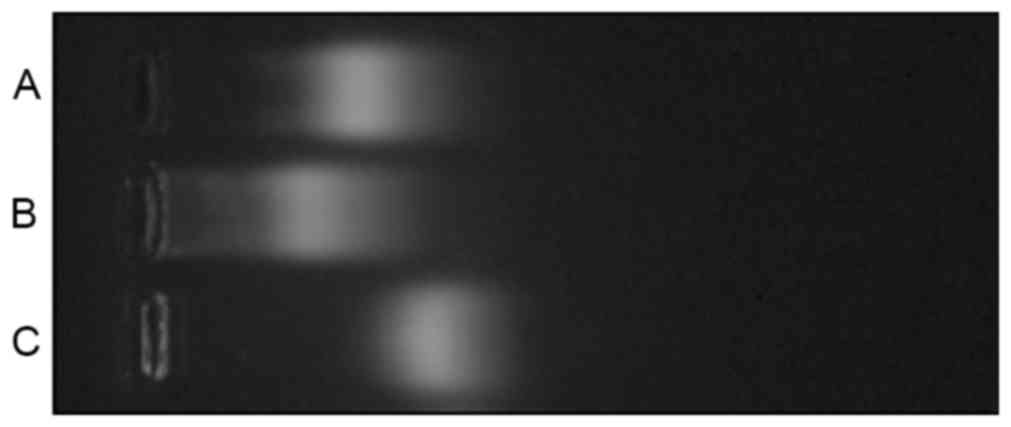
The coupling products were analyzed using Ready
Pouch™ Agerose Gels which was from Invitrogen (Carlsbad,
California, USA) The migration velocity is associated with the
surface charge, with more negative charges resulting in a faster
speed of moving to the anode (14).
In the activated reaction, the cationic amino group of QD605 was
bound to the carboxyl group on SMCC, which neutralized the positive
charges of QD605. In addition, SMCC was negatively charged in water
solutions due to the anionic sulfonic group. Therefore, the
migration speed of QD605-SMCC was faster than QD605. The thiol
group on reductive PSCA was connected with the maleimide group on
the intermediate of QD605-SMCC to produce the QD605-PSCA complex,
increasing the molecular weight and resulting in a slower migration
speed for QD605-PSCA compared with QD605-SMCC (Fig. 1).
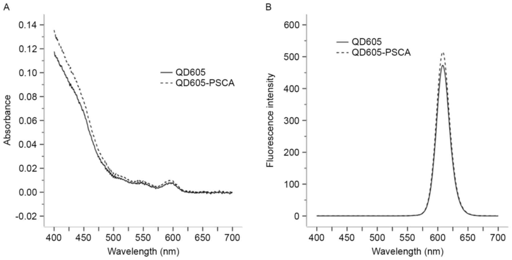
Optical properties of QD605-PSCA
probes
The absorption and emission spectra of the QD605 and
QD605-PSCA probes were determined using the ultraviolet
spectrophotometer and fluorescence spectrophotometer, respectively.
Following QD605 coupling with PSCA, the absorption spectrum of the
QD605-PSCA probes was wide and continuous. The absorbance spectrum
of QD605-PSCA was higher compared with that of QD605 (P<0.05;
Table I; Fig. 2A). The emission spectrum of
QD605-PSCA was narrow and symmetrical with an obvious fluorescence
emission peak at ~608 nm. No significant difference was observed
between the fluorescence intensities of QD605-PSCA and QD605
(Table I; Fig. 2).
 | Table I.Absorbance and fluorescence intensity
of QD605 and QD605-PSCA probes. |
Table I.
Absorbance and fluorescence intensity
of QD605 and QD605-PSCA probes.
| Group | Number | Absorbance (mean
rank) | Fluorescence
intensity (mean rank) |
|---|
| QD605 | 601 | 562.43 | 596.36 |
| QD605-PSCA | 601 | 640.57 | 606.64a |
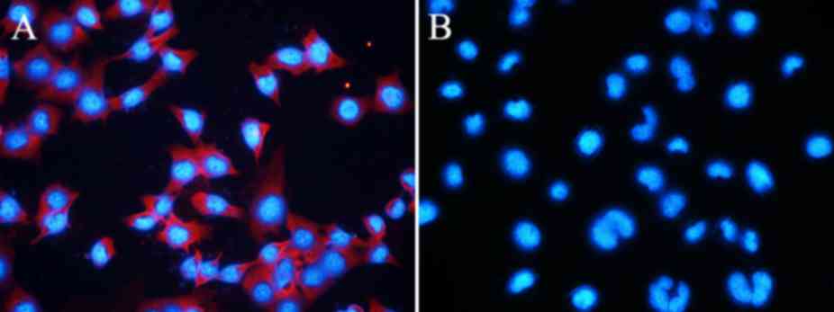
Specific fluorescent labeling of EJ
fixed cells with QD605- PSCA probes
The fluorescence of EJ cells labeled with QD605-PSCA
was examined using fluorescence microscopy. Orange-red fluorescence
of the QD605-PSCA probes was observed in the cell membrane and
cytoplasm, whereas the cell nucleus fluoresced blue following DAPI
staining (Fig. 3A). In the control
group, QD605 was not conjugated with the PSCA antibody. Due to
repeated washing, nonspecific adsorption of QDs and unbound QDs was
removed and no obvious orange-red fluorescence was observed
(Fig. 3B). Labeled slides were
stored at 4°C and fluorescence was observed every 24 h. The blue
fluorescence of the nuclei disappeared following 48 h, whereas
orange-red fluorescence of the QD605-PSCA probe signal was still
visible on the seventh day (data not shown).
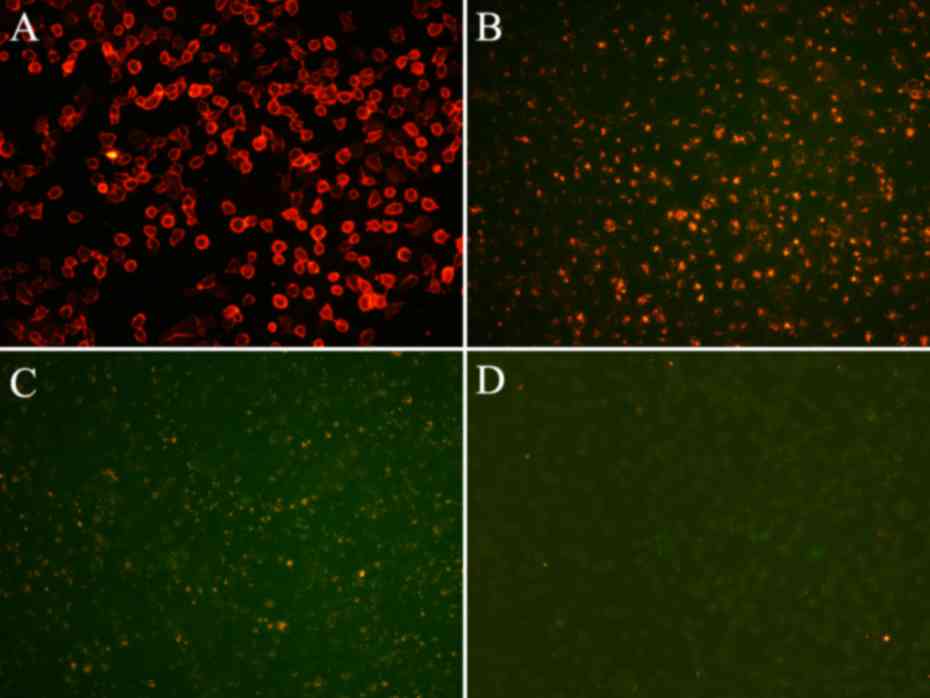
Fluorescent labeling of EJ living
cells with QD605-PSCA probes
The fluorescence of EJ living cells labeled with the
QD605-PSCA probes was observed using an inverted fluorescence
microscope (Fig. 4). The orange-red
fluorescence of the QD605-PSCA probes was observed in the cell
membrane (Fig. 4A). In the control
group, QD605 was not conjugated with PSCA and QD605 was not labeled
with EJ cells directly; as such, the fluorescence was consistent
with the background (Fig. 4D). Over
time, the QD605-PSCA probes were taken up into the cytoplasm. Due
to the influence of cell division and the culture medium, the
average fluorescence intensity was weakened gradually but remained
visible after 48 h (Fig. 4B and
C).
Discussion
In recent years, with the development of synthesis
technology and surface chemical modification, the toxicity,
biological compatibility and biological molecular coupling of QDs
have been effectively improved (15). QD-based fluorescent probes are widely
used in chemistry, biology, medicine and other fields due to their
favorable optical properties and biological compatibility (16,17).
Studies have revealed that QDs are the most ideal fluorescent
probes for surface receptor imaging in cellular labeling (18–20). In
imaging studies, QDs are able to produce biological effects through
modifying antibodies, peptides, proteins, nucleic acids or other
biological molecules (21–23).
QD coupling with biological molecules is typically
divided into non-covalent and covalent coupling. Non-covalent
coupling primarily includes electrostatic interactions and the
avidin-biotin complex method. Electrostatic interactions are a type
of early biological coupling method, where the positively charged
proteins are connected with the negatively charged QDs by
electrostatic attraction (24). This
method is quite simple, but vulnerable to the effects of pH and
ionic strength in biological environment (24). The avidin-biotin method is one of the
most widely applied biological coupling technologies, which
exhibits high selectivity and strong bonding (25). However, this method usually causes an
aggregated reaction (24–26). The combination of the two factors may
increase the molecular weight of the product with worse tissue
permeability, which would reduce the labeling efficiency (27).
The reaction between carboxyl and amino vs. maleic
imide and sulfur is commonly used for covalent coupling. The former
typically comprises the coupling of carboxyl on the QDs with the
amino on the molecules via an amide bond based on the EDC
[1-ethyl-3-(3-dimethylaminopropyl) carbodiimide]/NHS
(N-hydroxy-succinamide) (28). The
carboxyl and amino functional groups are rich in biological
molecules and can be easily introduced into the surface of the QDs
(28–30). However, during the binding reaction,
EDC and its intermediates will rapidly hydrolyze in the aqueous
medium and the total efficiency of the binding reaction is not high
(28). In addition, most peptides
and proteins containing a free carboxyl acid and/or amino acid are
not easy to use in the binding reaction due to unnecessary
cross-reaction between the constituents (28). The covalent bonding of maleic imide
and thiol depends on the formation of the thioether bond. The
binding reaction has high efficiency and strong specificity neutral
pH conditions (28). The
commercially available maleic imide reagent can be directly
combined to be exposed to a certain amino acid group in the QDs,
which enables these QDs to couple with biological molecules,
including thiol groups (28).
In the present study, the covalent binding of QD605
and the antibody was based on an SMCC coupling agent (31). SMCC is a type of cross-linking agent
with double functional groups that can activate the amino groups of
the QD605 to produce the QD605-SMCC polymer (31). In addition, under the action of DTT,
the antibody fragments were reduced to expose thiol groups. The
covalent bonding of antibodies onto QD605-SMCC occurred via the
thiol and maleic imide groups. Finally, QD605-PSCA was obtained.
The optical properties revealed that QD605-PSCA fluorescent probes
retained the original basic optical characteristics of QD605,
including a wide and continuous excitation spectrum, a narrow and
symmetrical emission spectrum, good fluorescence stability and long
fluorescence duration. The immune-fluorescence results revealed
that the QD605-PSCA probes retained the PSCA protein immune
activity and the PSCA was able to combine specifically with bladder
cancer cell surface antigens. In addition, in the living cell
labeling experiment, orange-red fluorescence was visible on the
cell membrane and bright fluorescence was still visible after
repeated laser irradiation. Over time, the fluorescence of QD605
appeared in the cytoplasm, which was considered to be indicative of
cellular proliferation and endocytosis. The results of the present
study demonstrate that QD605-PSCA probes are able to achieve
targeted labeling and their fluorescence intensity is high and
stable. The excellent optical properties and molecular targeting of
QD-antibody probes may be useful for in vivo non-invasive
targeted imaging and may contribute to early diagnosis, imaging
localization and targeted therapies for tumors.
Acknowledgements
The authors would like to thank D.W. Pang (College
of Chemistry and Molecular Sciences and State Key Laboratory of
Virology of Wuhan University) for his technical assistance in this
study. The present study was performed at the Key Laboratory of
Hubei Province for Digestive System Disease, Renmin Hospital of
Wuhan University (Hubei, China).
Funding
The present study was supported by the National
Natural Science Foundation of China (grant no. 81272826).
Availability of data and materials
The analyzed data sets generated during the present
study are available from the corresponding author on reasonable
request.
Authors' contributions
RY and TR were responsible for performing the
experiments, collecting the data and writing the manuscript. FC,
WY, YR, XZ and SL were responsible for the statistical analysis and
manuscript revision. FC designed the study.
Ethics approval and consent to
participate
Not applicable.
Patient consent for publication
Not applicable.
Competing interests
The authors declare that they have no competing
interests.
References
|
1
|
Torre LA, Bray F, Siegel RL, Ferlay J,
Lortet-Tieulent J and Jemal A: Global cancer statistic, 2012. CA
Cancer J Clin. 65:87–108. 2015. View Article : Google Scholar : PubMed/NCBI
|
|
2
|
Siegel RL, Miller KD and Jemal A: Cancer
statistics, 2015. CA Cancer J Clin. 65:5–29. 2015. View Article : Google Scholar : PubMed/NCBI
|
|
3
|
Chen W, Zheng R, Baade PD, Zhang S, Zeng
H, Bray F, Jemal A, Yu XQ and He J: Cancer statistics in china,
2015. CA Cancer J Clin. 66:115–132. 2016. View Article : Google Scholar : PubMed/NCBI
|
|
4
|
Sylvester RJ, van der Meijden AP,
Oosterlinck W, Witjes JA, Bouffioux C, Denis L, Newling DW and
Kurth K: Predicting recurrence and progression in individual
patients with stage Ta T1 bladder cancer using EORTC risk tables: A
combined analysis of 2596 patients from seven EORTC trials. Eur
Urol. 49:466–475. 2006. View Article : Google Scholar : PubMed/NCBI
|
|
5
|
Resch-Genger U, Grabolle M,
Cavaliere-Jaricot S, Nitschke R and Nann T: Quantum dots versus
organic dyes as fluorescent labels. Nat Methods. 5:763–775. 2008.
View Article : Google Scholar : PubMed/NCBI
|
|
6
|
Yu WW, Chang E, Drezek R and Colvin VL:
Water-soluble quantum dots for biomedical applications. Biochem
Biophys Res Commun. 348:781–786. 2006. View Article : Google Scholar : PubMed/NCBI
|
|
7
|
Smith AM, Ruan G, Rhyner MN and Nie S:
Engineering luminescent quantum dots for in vivo molecular and
cellular imaging. Ann Biomed Eng. 34:3–14. 2006. View Article : Google Scholar : PubMed/NCBI
|
|
8
|
Gonda K, Miyashita M, Higuchi H, Tada H,
Watanabe TM, Watanabe M, Ishida T and Ohuchi N: Predictive
diagnosis of the risk of breast cancer recurrence after surgery by
single-particle quantum dot imaging. Sci Rep. 5:143222015.
View Article : Google Scholar : PubMed/NCBI
|
|
9
|
Reiter RE, Gu Z, Watabe T, Thomas G,
Szigeti K, Davis E, Wahl M, Nisitani S, Yamashiro J, Le Beau MM, et
al: Prostate stem cell antigen: A cell surface marker overexpressed
in prostate cancer. Proc Natl Acad Sci USA. 95:1735–1740. 1998.
View Article : Google Scholar : PubMed/NCBI
|
|
10
|
Cunha AC, Weigle B, Kiessling A, Bachmann
M and Rieber EP: Tissue-specificity of prostate specific antigens:
Comparative analysis of transcript levels in prostate non-prostate
tissues. Cancer Lett. 236:229–238. 2006. View Article : Google Scholar : PubMed/NCBI
|
|
11
|
Amara N, Palapattu GS, Schrage M, Gu Z,
Thomas GV, Dorey F, Said J and Reiter RE: Prostate stem cell
antigen is overexpressed in human transitional cell carcinoma.
Cancer Res. 61:4660–4665. 2001.PubMed/NCBI
|
|
12
|
O'Toole CM, Povey S, Hepburn P and Franks
LM: Identity of some human bladder cancer cell lines. Nature.
301:429–430. 1983. View
Article : Google Scholar : PubMed/NCBI
|
|
13
|
Capes-Davis A, Theodosopoulos G, Atkin I,
Drexler HG, Kohara A, MacLeod RA, Masters JR, Nakamura Y, Reid YA,
Reddel RR and Freshney RI: Check your cultures! A list of
cross-contaminated or misidentified cell lines. Int J Cancer.
127:1–8. 2010. View Article : Google Scholar : PubMed/NCBI
|
|
14
|
Lee PY, Costumbrado J, Hsu CY and Kim YH:
Agarose gel electrophoresis for the separation of DNA fragment. J
Vis Exp. 20:e39232012.
|
|
15
|
Barak Y, Meir I, Shapiro A, Jang Y and
Lifshitz E: Fundamental properties in colloidal quantum dots. Adv
Mater. 19:e18014422018. View Article : Google Scholar
|
|
16
|
Jung S and Chen X: Quantum dot-dye
conjugates for biosensing, imaging, and therapy. Adv Healthc Mater.
7:e18002522018. View Article : Google Scholar : PubMed/NCBI
|
|
17
|
Matea CT, Mocan T, Tabaran F, Pop T,
Mosteanu O, Puia C, Iancu C and Mocan L: Quantum dots in imaging,
drug delivery and sensor applications. Int J Nanomedicine.
12:5421–5431. 2017. View Article : Google Scholar : PubMed/NCBI
|
|
18
|
Choi Y, Kim K, Hong S, Kim H, Kwon YJ and
Song R: Intracellular protein target detection by quantum dots
optimized for live cell imaging. Bioconjug Chem. 22:1576–1586.
2011. View Article : Google Scholar : PubMed/NCBI
|
|
19
|
Li H, Duan ZW, Xie P, Liu YR, Wang WC, Dou
SX and Wang PY: Effects of paclitaxel on EGFR endocytic trafficking
revealed using quantum dot tracking in single cells. PLoS One.
7:e454652012. View Article : Google Scholar : PubMed/NCBI
|
|
20
|
Allen PM, Liu W, Chauhan VP, Lee J, Ting
AY, Fukumura D, Jain RK and Bawendi MG: InAs(ZnCdS) quantum dots
optimized for biological imaging in the near-infrared. J Am Chem
Soc. 132:470–471. 2010. View Article : Google Scholar : PubMed/NCBI
|
|
21
|
Cai W, Shin DW, Chen K, Gheysens O, Cao Q,
Wang SX, Gambhir SS and Chen X: Peptide-labeled near-infrared
quantum dots for Imaging tumor vasculature in living subjects. Nano
Lett. 6:669–676. 2006. View Article : Google Scholar : PubMed/NCBI
|
|
22
|
Lu Z, Zhu Z, Zheng X, Qiao Y, Guo J and Li
CM: Biocompatible fluorescence-enhanced ZrO2-CdTe
quantum dot nanocomposite for in vitro cell imaging.
Nanotechnology. 22:1556042011. View Article : Google Scholar : PubMed/NCBI
|
|
23
|
Rosenthal SJ, Chang JC, Kovtun O, McBride
JR and Tomlinson ID: Biocompatible quantum dots for biological
applications. Chem Biol. 18:10–24. 2011. View Article : Google Scholar : PubMed/NCBI
|
|
24
|
Goldman ER, Balighian ED, Mattoussi H,
Kuno MK, Mauro JM, Tran PT and Anderson GP: Avidin: A natural
bridge for quantum dot-antibody conjugates. J Am Chem Soc.
124:6378–6382. 2002. View Article : Google Scholar : PubMed/NCBI
|
|
25
|
Boeneman K, Deschamps JR, Buckhout-White
S, Prasuhn DE, Blanco-Canosa JB, Dawson PE, Stewart MH, Susumu K,
Goldman ER, Ancona M and Medintz IL: Quantum dot DNA bioconjugates:
Attachment chemistry strongly influences the resulting composite
architecture. ACS Nano. 4:7253–7266. 2010. View Article : Google Scholar : PubMed/NCBI
|
|
26
|
Akerman ME, Chan WC, Laakkonen P, Bhatia
SN and Ruoslahti E: Nanocrystal targeting in vivo. Proc Natl Acad
Sci USA. 99:12617–12621. 2002. View Article : Google Scholar : PubMed/NCBI
|
|
27
|
Daniels TR, Bernabeu E, Rodríguez JA,
Patel S, Kozman M, Chiappetta DA, Holler E, Ljubimova JY, Helguera
G and Penichet ML: The transferrin receptor and the targeted
delivery of therapeutic agents against cancer. Biochim Biophys
Acta. 1820:291–317. 2012. View Article : Google Scholar : PubMed/NCBI
|
|
28
|
Hermanson GT: Bioconjugate techniques.
Academic Press; Amsterdam: 2013, View Article : Google Scholar
|
|
29
|
Susumu K, Oh E, Delehanty JB,
Blanco-Canosa JB, Johnson BJ, Jain V, Hervey WJ IV, Algar WR,
Boeneman K, Dawson PE and Medintz IL: Multifunctional compact
zwitterionic ligands for preparing robust biocompatible
semiconductor quantum dots and gold nanoparticles. J Am Chem Soc.
133:9480–9496. 2011. View Article : Google Scholar : PubMed/NCBI
|
|
30
|
Susumu K, Uyeda HT, Medintz IL, Pons T,
Delehanty JB and Mattoussi H: Enhancing the stability and
biological functionalities of quantum dots via compact
multifunctional ligands. J Am Chem Soc. 129:13987–13996. 2007.
View Article : Google Scholar : PubMed/NCBI
|
|
31
|
Li Z, Wang Y, Wang J, Tang Z, Pounds JG
and Lin Y: Rapid and sensitive detection of protein biomarker using
a portable fluorescence biosensor based on quantum dots and a
lateral flow test strip. Anal Chem. 82:7008–7014. 2010. View Article : Google Scholar : PubMed/NCBI
|