Introduction
Femtosecond laser is a kind of ultra-short pulse
laser with high instantaneous power, small focus size, strong
penetrability and high precision. It has been successfully applied
in refractive surgery and corneal transplantation, in recent years
(1,2). Since 2010, femtosecond laser has been
used in cataract surgery, which, assisted by a high-definition
ocular anterior segment imaging system, can complete the precise
anterior capsule circular incision, individualized surgical
incision, corneal release astigmatism correction and safe
pre-chopping, greatly improving the safety of cataract surgery and
the predictability of postoperative outcomes (3,4). The
success of this technique indicates that the cataract surgery is
quicker and safer, and the effect after intraocular lens (IOL)
implantation is more satisfactory.
A large number of studies have shown that some
patients have dry eye symptoms after receiving phacoemulsification
(phaco), such as dryness, foreign body sensation, burning sensation
and blurred vision (5–7). Microscopic ocular surface damage is a
pathogenic factor of dry eyes after cataract surgery, which can
cause ocular discomfort. This research aimed to study the dry eyes
of patients before and after femtosecond laser-assisted cataract
surgery (FLACS) and compare the effects of FLACS and conventional
cataract surgery on the ocular surface function of patients.
Patients and methods
Subjects of study
Patients with age-related cataract who underwent
phaco in Air Force Aviation Medicine Research Institute Affiliation
Hospital (Beijing, China) from January 2016 to December 2016 were
randomly divided into two groups. Patients in experimental group
(n=123, 150 eyes) received FLACS, while patients in phaco group
(n=110, 150 eyes) underwent conventional coaxial micro-incision
phaco and were implanted with foldable IOL. In the FLACS group,
there were 60 males (67 eyes) and 63 females (83 eyes) aged 54–78
years (65.74±11.80 years on average). In the phaco group, there
were 51 males (62 eyes) and 59 females (88 eyes) aged 56–81 years
(69.05±12.61 years on average). There were no statistically
significant differences in the sex ratio, average age and
preoperative routine eye examination results, between the two
groups (Table I). All patients
underwent preoperative routine eye examinations (including
uncorrected visual acuity, best corrected visual acuity,
intraocular pressure measurement, slit-lamp examination, ocular A
ultrasound, ocular B ultrasound and IOL power measurement) and
other relevant auxiliary examinations (including blood routine,
stool routine, urine routine, hepatic and renal function tests,
blood glucose test, eight-item detection before blood transfusion,
electrocardiogram and anterioposterior and lateral chest film) in
the Department of Ophthalmology.
 | Table I.Demographics and baseline
characteristics of patients undergoing cataract surgery. |
Table I.
Demographics and baseline
characteristics of patients undergoing cataract surgery.
| Parameter | FLACS group
(n=150) | Phaco group
(n=150) | P-value |
|---|
| Age (years) | 65.74±11.80 | 69.05±12.61 | 0.848 |
| Sex (n, %) |
| Male | 67 (44.7) | 62 (41.3) | 0.656 |
|
Female | 83 (55.3) | 88 (58.7) |
|
| Mean IOP (mmHg) | 12.50±4.69 | 13.86±3.07 | 0.809 |
| AL (mm) | 24.19±1.34 | 23.87±1.06 | 0.852 |
| ACD (mm) | 2.85±0.33 | 2.79±0.42 | 0.911 |
| Mean K (D) | 44.13±1.95 | 43.80±1.76 | 0.900 |
None of the patients had dry eyes
The diagnostic criteria for dry eyes were based on
the Japanese Diagnostic Criteria for Dry Eye (8), namely the following three criteria:
Schirmer's I test (SIt) ≤10 mm, breakup time (BUT) ≤5 sec; corneal
fluorescein staining (CFS) ≥1; symptoms, such as dryness, foreign
body sensation and burning sensation.
All patients suffered from age-related cataract
without active inflammation, such as conjunctivitis and keratitis,
within 3 months before operation. No eye-drop irritation symptoms
occurred locally. Patients did not wear contact lenses, and had no
history of ophthalmic surgery, eye trauma, eyelid varus-valgus,
incomplete closure, dacryocystitis, uveitis, glaucoma, fundus
diseases or other eye diseases; they were not complicated by other
systemic metabolic and immune diseases affecting tear secretion;
they did not use eye drops or oral glucocorticoid hormones
affecting the tear film function locally within 6 months. Those
patients with bilateral cataract received surgery at an interval
>1 month, so as to avoid dry eye symptoms in both eyes and cross
influence of signs. All patients were followed up for >3 months
after operation, and all objects of study were informed of the
study and signed the informed consent. This study was approved by
the Ethics Committee of Air Force Aviation Medicine Research
Institute Affiliation Hospital.
Surgical methods
FLACS: under surface anesthesia (Benoxil; Santen
Pharmaceutical Co., Ltd., Beijing, China), the patients lay on the
operating bed, and the femtosecond laser was used to make the main
corneal incision (2.3 mm) and side incision (1.0 mm), perform
capsulorhexis (4.6 mm/4.8 mm) and chop nucleus (energy parameter,
10 µJ) (LenSx; Alcon Laboratories, Inc., Fort Worth, TX, USA). The
corneal incision was bluntly separated using an iris restorer into
the anterior chamber, the anterior chamber was filled with
viscoelastic agent, and the anterior capsule was removed and
separated using water. After the energy was adjusted to 100% and
the suction to 450 mmHg (1 mmHg = 0.133 kPa), phaco was performed
(INFINITI phaco instrument; Alcon Laboratories, Inc.), residual
cortices were sucked in I/A mode, and IOL was implanted into a
capsular bag and adjusted to the normal position. Then viscoelastic
agent was injected, and the puncture site was hydrated, forming the
anterior chamber. Tobramycin and Dexamethasone eye ointments
(Yangtze River Pharmaceutical Group Co., Ltd., Taizhou, China) were
used and the operative eyes were bandaged.
Phaco: under surface anesthesia (Benoxil; Santen
Pharmaceutical Co., Ltd.), a transparent corneal incision was made
at 2.2 mm above the temple or nose, and an auxiliary corneal
incision was made at 2.00 cm on the lateral side. After injection
of viscoelastic agent into the anterior chamber, continuous
circular capsulorhexis, hydrodissection and in situ chopping
method in the capsular bag, phaco was performed for lens nucleus
(ultrasound energy of 2–30%, 8% on average). After removal of
cortices, the capsular bag was implanted with a foldable IOL, and
Tobradex eye ointment (Yangtze River Pharmaceutical Group Co.,
Ltd.) was used in the conjunctival sac after operation.
All operations were performed by one experienced
cataract specialist. The same equipment and instruments were used
during operation, and Tobramycin, Dexamethasone and Pranoprofen eye
drops (Yangtze River Pharmaceutical Group Co., Ltd.) were used
after operation for 4 weeks (4 times/day in the 1st week, and then
decreased progressively by 1 day every week).
Examination methods
At 1 day before operation, and at 1 day, 1 week, 1
month and 3 months after operation, the ocular surface disease
index (OSDI), BUT and tear meniscus height (TMH) were evaluated,
and the SIt and CFS were performed; the observation data were
recorded.
OSDI was used for dry eye symptoms according to a
previous study (9). The
questionnaire included 12 major questions about eye discomfort
(questions 1–3), visual function (questions 4–9) and environmental
triggers (questions 10–12). The score for each question was based
primarily on the duration of symptoms in patients: 4 points, all
the time; 3 points, most of the time; 2 points, half of the time; 1
point, a short time (occasionally); 0 point, never. Patients did
not need to answer all 12 questions. The total score of OSDI was
0–100. Total score of OSDI = total scores for all questions ×25/the
number of questions answered.
In SIt, standard 5 mm × 35 mm Whatman filter paper
(YZB/0360-2004; Tianjin Jingming New Technological Development Co.,
Ltd., Tianjin, China) was used; patients sat with their backs to
the light in a dark room; one end of the filter paper was folded
and placed at 1/3 junction of lower-eyelid conjunctival sac, and
the other end naturally drooped; patients closed their eyes gently,
and the filter paper was removed after 5 min. The length of tears
soaking the filter paper was measured with a ruler in
millimeters.
In CFS, the cornea was observed under the blue light
of slit lamp. The cornea was divided into four quadrants (superior
temporal, inferior temporal, superior nasal and inferior nasal) to
observe the presence of fluorescein staining in each quadrant. No
staining, 0 point; dotted and small pieces of staining, 1 and 2
points; block staining, 3 points; a total of 0–12 points.
In non-invasive tear-film assessment, a newly
developed corneal topographer (Oculus Optikgeräte GmbH, Wetzlar,
Germany) was used to measure the TMH, and non-invasive tear film
BUT according to previous studies (10–12),
including the first and the average BUT. The procedures were
conducted as per the protocol in previous literature.
All examinations were performed by the same
experienced physician in the same quiet examination room.
Statistical analysis
Statistical Product and Service Solutions (SPSS)
13.0 software (SPSS, Inc., Chicago, IL, USA) was used for
statistical processing. W test showed that the test indexes in this
study were normally distributed, continuous variables were
presented as mean ± standard deviation (SD), and Levene's test
showed the homogeneity of variance. One-way repeated measurement
data analysis of variance was used for the comparison of test index
between FLACS and phaco group at different time-points before and
after operation, and least significant difference (LSD) t-test was
used for the pairwise comparison. Independent-samples t-test was
used for the comparison of the sample mean between two groups at
the same time-point, and Chi-square test was used for the
categorical data. P<0.05 was considered to indicate a
statistically significant difference.
Results
OSDI
Compared with that before operation, OSDI in both
groups at 1 day and 1 week after operation was significantly
increased, and the differences were statistically significant
(P<0.05). The main symptom in patients was foreign body
sensation. There was a statistically significant difference in OSDI
in the FLACS group at 1 month after operation compared with that
before operation (P=0.011), but there was no statistically
significant difference in the phaco group compared with that before
operation (P=0.094). There were no statistically significant
differences in OSDI in both groups at 3 months after operation
compared with that before operation (P>0.05). Compared with the
phaco group, OSDI in the FLACS group at 1 day and 1 week after
operation was increased, and the differences were statistically
significant (P<0.05); the foreign body sensation and discomfort
were more severe than those in the phaco group; there were no
statistically significant differences between the two groups at 1
month and 3 months after operation (P>0.05) (Table II).
| Table II.OSDI before and after cataract surgery
in the operated eyes of the study patients. |
Table II.
OSDI before and after cataract surgery
in the operated eyes of the study patients.
| Time | FLACS group | Phaco group | P-value |
|---|
| Baseline | 0.45±0.23 | 0.47±0.39 | 0.965 |
| Postoperative 1
day | 5.34±0.49 | 4.04±0.33 | 0.028 |
| Postoperative 1
week | 4.99±0.53 | 3.47±0.55 | 0.048 |
| Postoperative 1
month | 2.23±0.66 | 1.83±0.71 | 0.680 |
| Postoperative 3
months | 0.59±0.30 | 0.51±0.39 | 0.394 |
SIt value
Before operation, SIt values in the FLACS and the
phaco group were 10.92±4.13 and 9.37±3.98 mm, respectively. SIt
values in both groups were significantly decreased at 1 week after
operation (7.55±3.69 and 7.20±3.29 mm, respectively). SIt values
increased at 1–3 months after operation and returned to
preoperative levels at 3 months after operation (Table III). There was no statistically
significant difference in SIt between the two groups
(P>0.05).
| Table III.SIt values before and after cataract
surgery in the operated eyes of the study patients (mm). |
Table III.
SIt values before and after cataract
surgery in the operated eyes of the study patients (mm).
| Time | FLACS group | Phaco group | P-value |
|---|
| Baseline | 10.92±4.13 | 9.37±3.98 | 0.829 |
| Postoperative 1
day | 11.27±4.87 | 10.68±3.73 | 0.923 |
| Postoperative 1
week | 7.55±3.69 | 7.20±3.29 | 0.944 |
| Postoperative 1
month | 8.83±2.58 | 8.04±2.73 | 0.834 |
| Postoperative 3
months | 11.15±4.99 | 10.08±5.35 | 0.884 |
CFS score
CFS scores in both groups after operation were
significantly increased compared with those before operation, and
the differences were statistically significant (P<0.05). CFS
scores were the highest at 1 day after operation, gradually
decreased afterwards, and returned to preoperative levels at 3
months after operation. CFS scores in the FLACS group at 1 day, 1
week and 1 month after operation were higher than those in the
phaco group, and the differences were statistically significant
(P=0.008, 0.017 and 0.046, respectively) (Table IV).
| Table IV.CFS scores before and after cataract
surgery in the operated eyes of the study patients. |
Table IV.
CFS scores before and after cataract
surgery in the operated eyes of the study patients.
| Time | FLACS group | Phaco group | P-value |
|---|
| Baseline | 0.46±0.20 | 0.38±0.22 | 0.788 |
| Postoperative 1
day | 2.34±0.31 | 1.22±0.28 | 0.008 |
| Postoperative 1
week | 1.88±0.29 | 1.02±0.21 | 0.017 |
| Postoperative 1
month | 0.97±0.20 | 0.48±0.14 | 0.046 |
| Postoperative 3
months | 0.51±0.39 | 0.46±0.35 | 0.775 |
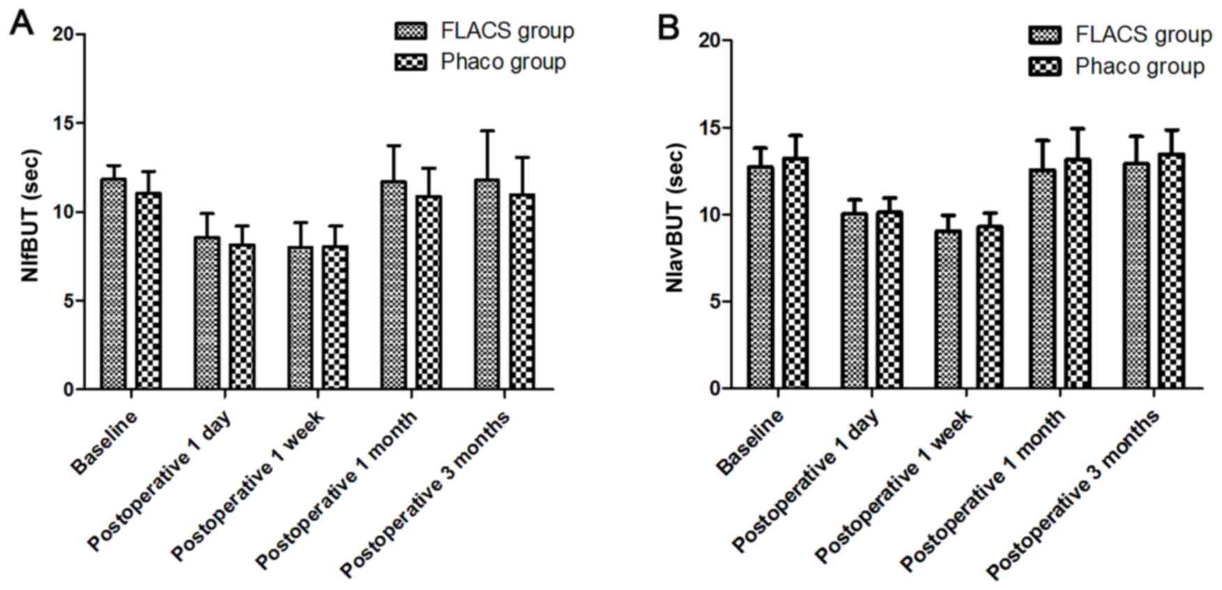
First and average BUT
In this study, the first and average BUT in both
groups at 1 week after operation were significantly shortened
compared with those before operation, and the differences were
statistically significant (P<0.05); indicating that the
stability of tear film is decreased after operation. BUT returned
to the preoperative level at 3 months after operation, and there
were no statistically significant differences at 1 and 3 months
after operation compared with those before operation (P>0.05).
First and average BUT had no statistically significant differences
between the two groups (Fig. 1;
Table V).
| Table V.NIfBUT and NIavBUT values before and
after cataract surgery in the operated eyes of the study patients
(sec). |
Table V.
NIfBUT and NIavBUT values before and
after cataract surgery in the operated eyes of the study patients
(sec).
| Parameters | FLACS group | Phaco group | P-value |
|---|
| NIfBUT |
|
Baseline | 11.83±0.79 | 11.04±1.24 | 0.592 |
|
Postoperative 1 day | 8.54±1.36 | 8.16±0.04 | 0.825 |
|
Postoperative 1 week | 8.01±1.38 | 8.07±1.13 | 0.973 |
|
Postoperative 1 month | 11.68±2.05 | 10.89±1.57 | 0.760 |
|
Postoperative 3 months | 11.80±2.77 | 10.97±2.11 | 0.812 |
| NIavBUT |
|
Baseline | 12.71±1.09 | 13.21±1.33 | 0.771 |
|
Postoperative 1 day | 10.01±0.81 | 10.12±0.82 | 0.924 |
|
Postoperative 1 week | 9.03±0.92 | 9.34±0.88 | 0.808 |
|
Postoperative 1 month | 12.56±1.68 | 13.15±1.77 | 0.809 |
|
Postoperative 3 months | 12.90±1.59 | 13.44±1.43 | 0.801 |
TMH
TMH in both groups was slightly increased at 1 day
after operation; it was decreased at 1 week after operation
compared with that before operation, and the difference was
statistically significant (P<0.05). At 1 and 3 months after
operation, TMH basically returned to the preoperative levels, and
the differences were not statistically significant (P>0.05).
There was no statistically significant difference in TMH between
the two groups (Table VI).
| Table VI.TMH values before and after cataract
surgery in the operated eyes of the study patients (mm). |
Table VI.
TMH values before and after cataract
surgery in the operated eyes of the study patients (mm).
| Time | FLACS group | Phaco group | P-value |
|---|
| Baseline | 0.37±0.09 | 0.35±0.08 | 0.868 |
| Postoperative 1
day | 0.41±0.13 | 0.44±0.11 | 0.860 |
| Postoperative 1
week | 0.22±0.07 | 0.20±0.06 | 0.828 |
| Postoperative 1
month | 0.32±0.05 | 0.30±0.06 | 0.798 |
| Postoperative 3
months | 0.36±0.07 | 0.37±0.06 | 0.925 |
Discussion
Tear film is a basis for maintaining the normal
structure and function of ocular surface epithelium, which can
moisten and protect the corneal and conjunctival epithelium. The
stability of normal ocular surface tear film depends on the normal
quality and quantity of tear film lipid layer, aqueous layer and
mucinous layer and normal tear kinetics. When a variety of reasons
lead to tear film instability and abnormal tear secretion, ocular
surface changes can be caused, leading to dry eyes. Studies have
found that all ophthalmic surgeries can cause tear film instability
and postoperative dry eye syndrome (12,13).
Whether tear film and ocular surface will be affected after FLACS,
as a kind of surgery with small trauma and fast recovery, is rarely
studied.
The results of this study showed that the first BUT,
average BUT and TMH in both groups were shortened significantly,
and OSDI and CFS scores of dry eyes were significantly increased at
1 day and 1 week after operation compared with those before
operation, which may be associated with the direct damage of phaco
combined with IOL implantation to ocular surface. Currently,
scholars world-wide consider that the reasons why phaco combined
with IOL implantation reduces the stability of tear film are the
following: frequent application of eye drops for preoperative local
surface anesthesia; mechanical damage to ocular surface epithelium
caused by preoperative irrigation and improper surgical skills;
peripheral nerve injury at the incision and decreased local corneal
sensation caused by transparent corneal incision (14); damage of preservatives in hormonal
eye drops and local eye drops used after surgery to the ocular
surface epithelial tissues (15,16);
postoperative exposure of ocular surface to hard light and repeated
washing with lavage fluid, and damage to ocular surface goblet cell
density and shortening of BUT (17);
local and surrounding tissue edema, and patients' emphasis on the
surgery and psychological factors.
It was found that patient usually have most of the
chief complaints at 1 day after operation when the bandage was
opened. The reason may be that the tear component in an eye-closed
state after bandage is different from that when the eyes are
opened. When eyes are opened in the daytime, the tear film is in a
dynamic balance, promoting the secretion of fresh tears and
removing tears. But the removal rate of tears becomes slow when
eyes are closed at night. Besides, tears contain a large amount of
immunoglobulin A (IgA), albumin concentration is also increased,
plasminogen activators convert C3 into C3c,
and a large number of neutrophils are aggregated, which is known as
subclinical inflammation, and such a change is the most obvious at
3–5 h after the eyes are closed (18,19). In
the present study, the patients' eyes were bandaged for a long
time, and the stress of operative wound and trauma made the
epithelial cells secrete a large number of inflammatory factors
accumulating in the tears, aggravating the inflammatory response.
Inflammation leads to ocular surface epithelial damage, decreased
stability of tear film, changes in tear component, and more main
complaints of discomfort.
Each index in both groups returned to preoperative
level at 90 days after operation, and the possible reasons are the
following: the local eye drops applied after operation have a
certain anti-inflammatory effect; when inflammation occurs in
patients receiving phaco combined with IOL implantation, abundant
lymphocyte infiltration can be found on ocular surface in patients
with dry eyes, and ocular surface epithelial cells will produce a
large number of pro-inflammatory cytokines, which will induce the
phospholipase A2 in corneal and conjunctival epithelium to catalyze
the phopholipid layer and convert it into arachidonic acid;
furthermore, cyclooxygenase (COX) will decompose arachidonic acid
to produce prostaglandins and thromboxane, and these products will
further induce the inflammatory response and aggravate dry eyes.
Non-steroidal anti-inflammatory drugs (NSAIDs) can inhibit the
occurrence of inflammation through inhibiting the decomposition of
arachidonic acid via COX, so they have a certain repair effect on
the ocular surface. Secondly, the operative incision is small and
it is located at the 11 o'clock direction of cornea; corneal nerves
controlling the corneal sensation are mainly at 3 and 9 o'clock
directions, and only a few nerve fibers are cut off during
operation. Some studies have also shown that nerve growth factors
are released onto the corneal epithelial regenerating axon at 25
days after operation, producing new nerve cells and healing the
cornea (20), which also explains
why dry eye symptoms are improved in early stage.
It was found in our study that CFS scores and OSDI
in the FLACS group at 1 day and 1 week after operation were
increased more significantly than those in the phaco group, but
there were no statistically significant differences at 3 months
after operation, indicating that the effect on ocular surface
function of patients in the FLACS group was larger than that in the
phaco group, and the dry eye symptoms after operation were also
more obvious. This may be related to the peri-conjunctival injury
caused by the suction ring used in femtosecond laser, which is
similar to the pathological changes in dry eyes caused by
laser-assisted in situ keratomileusis (LASIK) (21,22). In
addition, the irregular corneal surface damage due to surgical
incision made by Sinskey hook is also an important cause, which is
consistent with the corneal epithelial infiltration and
fluorescence staining around the surgical incision that we observed
in most patients after operation.
There were still many shortcomings in this study.
The sample size was too small. In order to study the effect of
FLACS on ocular surface function, more subjects of clinical study
should be enrolled for large-sample data analysis; there are many
influencing factors of ocular surface dysfunction after FLACS, such
as the preoperative and postoperative application of local eye
drops, intraoperative application of local surface anesthetics and
operation time; but these factors affecting the experimental
results were not taken into account in this study, so the
influences of these factors should be fully lowered to reduce the
error and deviation in subsequent studies; moreover, patients were
followed up for a very short time, so the postoperative follow-up
time should be prolonged in a future research to explore the
long-term impact of FLACS on ocular surface.
In conclusion, both FLACS and conventional phaco can
reduce the tear film stability of patients in a short term, thus
affecting the ocular surface function. The effects of FLACS on CFS
and dry eye symptoms are greater than those of conventional
phaco.
Acknowledgements
Not applicable.
Funding
No funding was received.
Availability of data and materials
All data generated or analyzed during this study are
included in this published article.
Authors' contributions
DS and HW designed the study and performed the
experiments; DS, XZ, WS and PC collected the data; WC and HW
analyzed the data; DS prepared the manuscript. All authors read and
approved the final manuscript.
Ethics approval and consent to
participate
This study was approved by the Ethics Committee of
Air Force Aviation Medicine Research Institute Affiliation Hospital
(Beijing, China). Patients or their guardians provided written
informed consent for publication.
Patient consent for publication
Not applicable.
Competing interests
The authors declare that they have no competing
interests.
References
|
1
|
Salomão MQ and Wilson SE: Femtosecond
laser in laser in situ keratomileusis. J Cataract Refract Surg.
36:1024–1032. 2010. View Article : Google Scholar : PubMed/NCBI
|
|
2
|
Moshirfar M, Hatch BB, Chang JC, Kurz CJ,
Eugarrios MF and Mifflin MD: Prospective, contralateral comparison
of 120-µm and 90-µm LASIK flaps using the IntraLase FS60
femtosecond laser. J Refract Surg. 27:251–259. 2011. View Article : Google Scholar : PubMed/NCBI
|
|
3
|
Sutton G, Bali SJ and Hodge C: Femtosecond
cataract surgery: Transitioning to laser cataract. Curr Opin
Ophthalmol. 24:3–8. 2013. View Article : Google Scholar : PubMed/NCBI
|
|
4
|
He L, Sheehy K and Culbertson W:
Femtosecond laser-assisted cataract surgery. Curr Opin Ophthalmol.
22:43–52. 2011.PubMed/NCBI
|
|
5
|
Sutu C, Fukuoka H and Afshari NA:
Mechanisms and management of dry eye in cataract surgery patients.
Curr Opin Ophthalmol. 27:24–30. 2016. View Article : Google Scholar : PubMed/NCBI
|
|
6
|
Kasetsuwan N, Satitpitakul V, Changul T
and Jariyakosol S: Incidence and pattern of dry eye after cataract
surgery. PLoS One. 8:e786572013. View Article : Google Scholar : PubMed/NCBI
|
|
7
|
Cho YK and Kim MS: Dry eye after cataract
surgery and associated intraoperative risk factors. Korean J
Ophthalmol. 23:65–73. 2009. View Article : Google Scholar : PubMed/NCBI
|
|
8
|
Homma M, Tojo T, Akizuki M and Yamagata H:
Criteria for Sjögren's syndrome in Japan. Scand J Rheumatol
(Suppl). 61:26–27. 1986.PubMed/NCBI
|
|
9
|
Ozcura F, Aydin S and Helvaci MR: Ocular
surface disease index for the diagnosis of dry eye syndrome. Ocul
Immunol Inflamm. 15:389–393. 2007. View Article : Google Scholar : PubMed/NCBI
|
|
10
|
Jiang Y, Ye H, Xu J and Lu Y: Noninvasive
keratograph assessment of tear film break-up time and location in
patients with age-related cataracts and dry eye syndrome. J Int Med
Res. 42:494–502. 2014. View Article : Google Scholar : PubMed/NCBI
|
|
11
|
Hong J, Sun X, Wei A, Cui X, Li Y, Qian T,
Wang W and Xu J: Assessment of tear film stability in dry eye with
a newly developed keratograph. Cornea. 32:716–721. 2013. View Article : Google Scholar : PubMed/NCBI
|
|
12
|
Yu EY, Leung A, Rao S and Lam DS: Effect
of laser in situ keratomileusis on tear stability. Ophthalmology.
107:2131–2135. 2000. View Article : Google Scholar : PubMed/NCBI
|
|
13
|
Albietz JM, Lenton LM and McLennan SG:
Effect of laser in situ keratomileusis for hyperopia on tear film
and ocular surface. J Refract Surg. 18:113–123. 2002.PubMed/NCBI
|
|
14
|
Müller LJ, Vrensen GF, Pels L, Cardozo BN
and Willekens B: Architecture of human corneal nerves. Invest
Ophthalmol Vis Sci. 38:985–994. 1997.PubMed/NCBI
|
|
15
|
Pisella PJ, Pouliquen P and Baudouin C:
Prevalence of ocular symptoms and signs with preserved and
preservative free glaucoma medication. Br J Ophthalmol. 86:418–423.
2002. View Article : Google Scholar : PubMed/NCBI
|
|
16
|
Herreras JM, Pastor JC, Calonge M and
Asensio VM: Ocular surface alteration after long-term treatment
with an antiglaucomatous drug. Ophthalmology. 99:1082–1088. 1992.
View Article : Google Scholar : PubMed/NCBI
|
|
17
|
Cetinkaya S, Mestan E, Acir NO, Cetinkaya
YF, Dadaci Z and Yener HI: The course of dry eye after
phacoemulsification surgery. BMC Ophthalmol. 15:682015. View Article : Google Scholar : PubMed/NCBI
|
|
18
|
Yokoi N, Takehisa Y and Kinoshita S:
Correlation of tear lipid layer interference patterns with the
diagnosis and severity of dry eye. Am J Ophthalmol. 122:818–824.
1996. View Article : Google Scholar : PubMed/NCBI
|
|
19
|
White KM, Benjamin WJ and Hill RM: Human
basic tear fluid osmolarity. II. Importance of processing strategy.
Acta Ophthalmol (Copenh). 71:530–538. 1993. View Article : Google Scholar : PubMed/NCBI
|
|
20
|
Shaheen BS, Bakir M and Jain S: Corneal
nerves in health and disease. Surv Ophthalmol. 59:263–285. 2014.
View Article : Google Scholar : PubMed/NCBI
|
|
21
|
Salomão MQ, Ambrósio R Jr and Wilson SE:
Dry eye associated with laser in situ keratomileusis: Mechanical
microkeratome versus femtosecond laser. J Cataract Refract Surg.
35:1756–1760. 2009. View Article : Google Scholar : PubMed/NCBI
|
|
22
|
Rodriguez-Prats JL, Hamdi IM, Rodriguez
AE, Galal A and Alio JL: Effect of suction ring application during
LASIK on goblet cell density. J Refract Surg. 23:559–562.
2007.PubMed/NCBI
|