Introduction
The cerebral blood supply comes from the internal
carotid artery system (anterior circulation) and vertebrobasilar
system (posterior circulation). Not only sex, age, diabetes,
hypertension, hyperlipidemia and other recognized factors affecting
morbidity but also posterior communicating artery (PCoA) is an
important collateral vessel linking anterior and posterior
circulation blood, which may be related to posterior circulation
ischemia (PCI) (1–5). Functional posterior communicating
artery (F-PCoA) may be divided into the following 3 types: F-PCoA
was consistent with anatomical posterior communicating artery
(A-PCoA), F-PCoA was inconsistent with A-PCoA, and mixed type,
which is different from F-PCoA and A-PCoA. For example, A-PCoA is
only a potential component, namely, the same blood pressure at both
ends, no flow in blood vessels, or too small vascular diameter, and
very low shunt flow volume, indicating the presence of A-PCoA but
absence of F-PCoA. The hemodynamic information of the F-PCoA, such
as blood flow direction, velocity and volume, can be quantitatively
analyzed via phase contrast magnetic resonance angiography (PC-MRA)
without using the contrast agent, which is of important value in
evaluating the collateral circulation function of PCoA (6). PC-MRA is a non-radioactive detection
method for cerebral blood flow, which can detect the acute ischemic
lesions and provide both morphological and hemodynamic information
simultaneously. The velocity-time curve of target vessels can be
obtained, and such hemodynamic parameters as the mean flow velocity
and mean flow volume can be calculated using the flow analysis
software, which is of important value in evaluating the collateral
compensatory F-PCoA (7).
In this study, the hemodynamic features of PCoA in
PCI patients were studied via PC-MRA to provide clinical
references. This study is helpful to identify subjects with
clinically suspected PCI but negative results in routine MR plain
scan, or subjects without clinical symptoms but with decreased
posterior circulation blood flow volume.
Patients and methods
Enrolment of patients
Patients receiving magnetic resonance imaging (MRI)
plain scan and PC-MRA in Shunde Hospital, Southern Medical
University (The First People's Hospital of Shunde Foshan) (Foshan,
China) from April 2015 to March 2017 were collected. Exclusion
criteria for the study were: i) those with abnormal collateral
circulation or vascular malformation in the brain, ii) those with
hemorrhagic cerebrovascular disease, or iii) with brain tumor,
brain trauma or a history of brain surgery. Finally, 51 subjects
were enrolled. The study was approved by the Ethics Committee of
Shunde Hospital, Southern Medical University (The First People's
Hospital of Shunde Foshan). Signed informed consents were obtained
from the patients or the guardians.
The functional PCoA (F-PCoA) was defined as the
complete arterial blood vessel with a diameter >1 mm and both
ends connecting to the cerebral anterior and posterior circulation
vessels, whose flow volume and velocity could be measured via
PC-MRA. The anatomical PCoA (A-PCoA) was defined as the artery
between the anterior circulation vessel (internal carotid artery,
middle cerebral artery or anterior cerebral artery) and the P1
segment of the ipsilateral posterior cerebral artery, including
dysplastic vessels.
Among those with F-PCoA, 27 subjects diagnosed with
PCI based on the ‘Chinese Guidelines for Diagnosis and Treatment of
Acute Ischemic Stroke in 2014’ (8)
as the golden standard were included into research group, including
16 males and 11 females at an average age of 65.56±13.47 years, and
their clinical data were reviewed in detail one by one. Eleven
non-PCI subjects clinically diagnosed according to the same golden
standard at the similar age and with similar blood pressure, blood
glucose and blood lipid to those in research group were included
into control group 1, including 4 males and 7 females at an average
age of 59.45±7.13 years. The remaining 13 non-PCI subjects without
F-PCoA were included as control group 2, including 5 males and 8
females at an average age of 64.92±7.35 years. Control groups 1 and
2 were combined as control group (n=24).
Data collection
Philips Intera Achieva 1.5T MRI, Fast CINE PC-MRA
sequence and pulse peripheral gating were used. On the
three-dimensional time-of-flight MRA maximum signal projection
reconstructed images, combined with two-dimensional tomographic
source images, the basal artery (BA), left and right vertebral
arteries, internal carotid artery and F-PCoA were located and
detected. Fast CINE PC-MRA sequence parameters are as follows: the
shortest TR and TE were selected automatically, FOV (mm): 100×100,
NSA=2, and layer thickness: 2 mm. The target vessel, perpendicular
to the long axis of the measuring segment of target vessel, was
placed in the central position of the layer, and the appropriate
heart phase value was adjusted according to the finger pulse. The
velocity encoding (Venc) was set as 100 cm/sec, and the total
sequence running time was approximately 3 min and 30 sec.
Mean flow volume, mean flow velocity, minimum flow
volume, maximum flow volume, minimum flow velocity and maximum flow
velocity of target vessels were recorded one by one, the peak
height of flow volume (maximum flow volume - minimum flow volume)
and flow velocity (maximum flow velocity - minimum flow velocity)
were calculated, and the blood flow direction was determined
according to the plus-minus sign of flow velocity. The relationship
between F-PCoA and A-PCoA was recorded.
Statistical analysis
Statistical Product and Service Solutions (SPSS)
13.0 software was used. In statistical inference, t-test was used
for measurement data in line with the normal distribution and
homogeneity of variance, and rank sum test was used otherwise.
Chi-square test was used for constituent ratio data. Two-sided
tests were used. P<0.05 was considered to indicate a
statistically significant difference.
Results
PC-MRAs of F-PCoA
Among subjects receiving PC-MRA, there were 38
subjects with a total of 52 F-PCoAs, and they were divided into the
following 3 types: i) F-PCoA was consistent with A-PCoA, accounting
for 84.21% (32/38), ii) F-PCoA was inconsistent with A-PCoA,
accounting for 13.16% (5/38), and iii) mixed type, accounting for
2.63% (1/38). The blood flow curve of F-PCoA displayed
sawtooth-like changes, and there could be either unidirectional
blood flow or bidirectional blood flow in one cardiac cycle. Among
the 16 subjects with bilateral F-PCoA, the bilateral blood flow
direction was the same in 10 cases (62.50%) (Table I).
 | Table I.PC-MRAs of F-PCoA. |
Table I.
PC-MRAs of F-PCoA.
| Factors | Left F-PCoA | Right F-PCoA | Total |
|---|
| Blood flow
direction |
|
Bidirectional blood flow
(n) | 7 | 7 | 14 |
| Backwards
blood flow (n) | 16 | 19 | 35 |
| Forwards
blood flow (n) | 3 | 0 | 3 |
| Mean flow
(ml/min) |
| (Min,
Max) | (0.24, 81.6) | (1.92, 72.0) | (0.24, 81.6) |
|
Median | 10.44 | 7.50 | 20.31 |
| Mean velocity
(cm/sec) |
| (Min,
Max) | (−9.67, 11.2) | (−5.98, 19.10) | (−9.67, 19.10) |
|
Median | 1.07 | 1.27 | 4.01 |
| Cross-sectional area
(cm2) |
| (Min,
Max) | (0.04, 0.14) | (0.02, 0.21) | (0.02, 0.21) |
|
Median | 0.07 | 0.07 | 0.08 |
| Diameter (cm) |
| (Min,
Max) | (0.11, 0.24) | (0.11, 0.31) | (0.11, 0.31) |
|
Median | 0.16 | 0.16 | 0.16 |
Parameters
There were no significant differences in sex
composition, age distribution, total flow volume of bilateral
internal carotid arteries, total flow volume of bilateral vertebral
arteries, and mean flow volume, mean flow velocity, minimum flow
volume, minimum flow velocity and peak height of flow velocity of
BA between control groups 1 and 2. However, the cross-sectional
area, diameter, maximum flow volume, peak height of flow volume and
maximum flow velocity of BA had significant differences (Table II).
| Table II.Statistical analysis of 24 cases
without PCI. |
Table II.
Statistical analysis of 24 cases
without PCI.
| Characteristics | F-PCoA (n=11) | Non-F-PCoA
(n=13) | Statistical
analysis |
|---|
| Sex
(male/female) | 4/7 | 5/8 |
χ2=0.011 | P=0.916 |
| Age (years) | 59.45±7.13 | 64.92±7.35 | t=1.840 | P=0.079 |
| BA |
|
Cross-sectional area
(mm2) | 12.95±2.98 | 19.11±6.85 | t=2.762 | P=0.011a |
| Diameter
(mm) | 2.86±0.66 | 3.49±0.42 | t=2.846 | P=0.009a |
| Mean flow
volume (ml/min) | 144.16±44.01 | 165.18±24.11 | t=1.483 | P=0.152 |
| Mean
velocity (cm/sec) | 19.30±6.72 | 15.25±4.67 | t=2.099 | P=0.147 |
| Minimum
flow volume (ml/min) | 81.98±40.73 | 87.97±18.73 | t=13.538 | P=0.661 |
| Maximum
flow volume (ml/min) | 214.58±56.98 | 271.80±47.10 | t=2.695 | P=0.013a |
| Peak
height of flow volume (ml/min) | 132.60±31.00 | 183.83±42.65 | t=3.308 | P=0.003a |
| Minimum
velocity (cm/sec) | 30.91±9.46 | 25.12±7.17 | t= −1.706 | P=0.102 |
| Maximum
velocity (cm/sec) | 71.80±15.76 | 59.62±11.49 | t= −2.186 | P=0.040a |
| Peak
height of velocity (cm/sec) | 40.89±9.36 | 34.51±7.33 | t= −1.875 | P=0.074 |
| Flow
volume of bilateral internal carotid arteries (ml/min) | 520.68±89.00 | 461.43±64.14 | t= −1.858 | P=0.077 |
| Flow
volume of bilateral vertebral arteries (ml/min) | 211.16±72.94 | 221.27±46.60 | t=0.345 | P=0.735 |
There were no significant differences in sex
composition, age distribution, total flow volume of bilateral
internal carotid arteries, total flow volume of bilateral vertebral
arteries and cross-sectional area of BA between research group and
control group. However, the mean flow volume, mean flow velocity,
minimum flow volume, maximum flow volume and minimum flow velocity
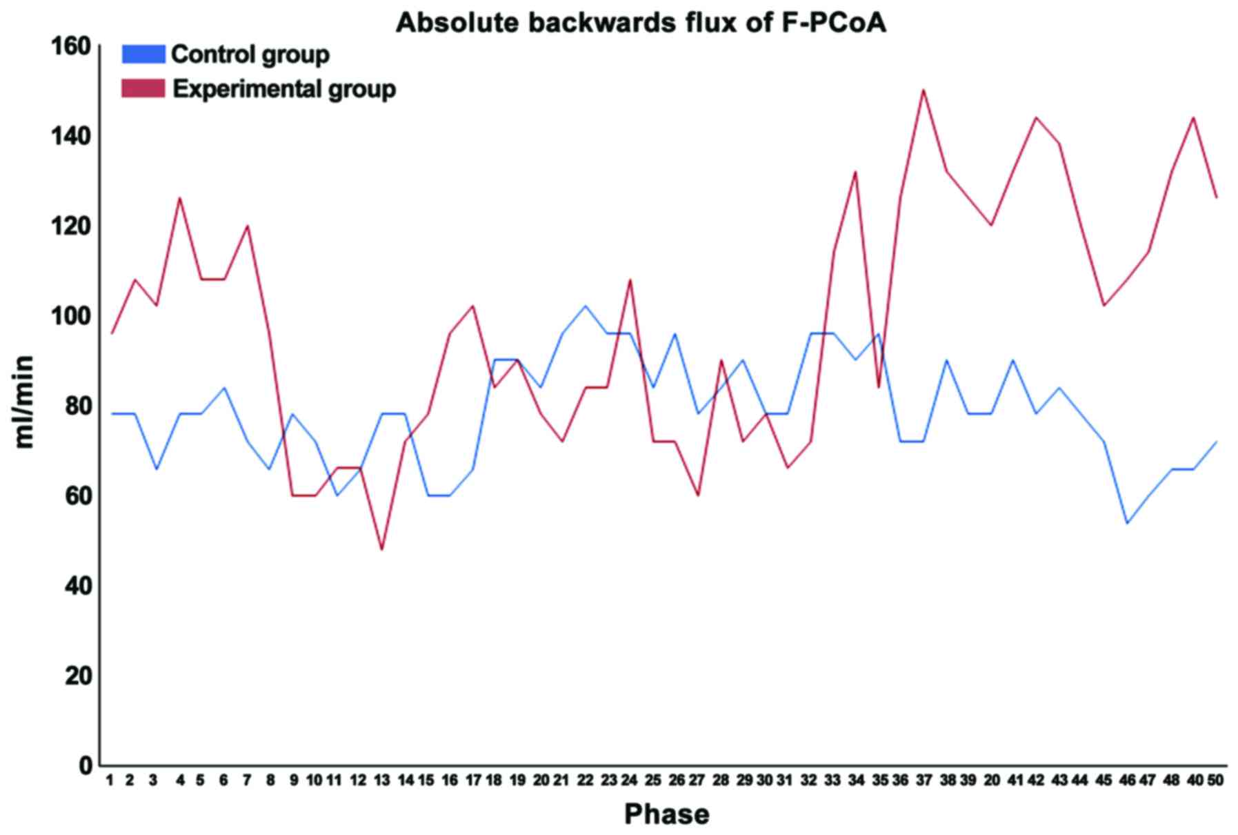
of BA, and forward posterior shunt flow volume of F-PCoA had
significant differences (Table
III).
| Table III.Statistical analysis of 51 cases. |
Table III.
Statistical analysis of 51 cases.
| Characteristics | Experimental group
(n=27) | Control group
(n=24) | Statistical
analysis |
|---|
| Sex
(male/female) | 16/11 | 9/15 |
χ2=2.407 | P=0.121 |
| Age (years) | 65.56±13.47 | 62.42±7.62 | t= −1.038 | P=0.305 |
| BA |
|
Cross-sectional area
(mm2) | 20.20±9.46 | 16.28±6.18 | t= −1.768 | P=0.084 |
| Mean flow
volume (ml/min) | 119.38±33.78 | 155.55±35.49 | t=3.726 | P=0.001a |
| Mean
velocity (cm/sec) | 11.69±5.33 | 17.10±5.94 | t=3.432 | P=0.001a |
| Minimum
flow volume (ml/min) | 54.33±33.21 | 85.23±30.23 | t=3.458 | P=0.001a |
| Maximum
flow volume (ml/min) | 208.31±69.67 | 245.58±58.46 | t=2.055 | P=0.045a |
| Peak
height of flow volume (ml/min) | 153.98±73.52 | 160.35±45.24 | t=1.178 | P=0.278 |
| Minimum
velocity (cm/sec) | 21.06±7.47 | 27.77±8.63 | t=2.976 |
P=0.005a |
| Maximum
velocity (cm/sec) | 59.61±23.62 | 65.20±14.67 | t=1.001 | P=0.322 |
| Peak
height of velocity (cm/sec) | 38.54±20.88 | 37.43±8.75 | t=0.556 | P=0.456 |
| F-PCoA |
|
Backward shunt flow
(ml/min) | 27.15±35.33 | 15.12±29.05 | t=6.216 |
P=0.013a |
| Flow
volume of bilateral internal carotid arteries (ml/min) | 451.24±124.41 | 487.19±79.92 | t=1.191 | P=0.239 |
| Flow
volume of bilateral vertebral arteries (ml/min) | 191.28±48.35 | 216.51±58.66 | t=1.392 | P=0.173 |
Cases
One case in the research group involved a
54-year-old male patient suffering from repeated dizziness for 1
year, which was aggravated for 10 days. MRI manifestations were
softening lesion in the left cerebellum, multiple stenosis in the
bilateral posterior cerebral arteries, and segmental stenosis in
the left vertebral artery. PC-MRA manifestations showed moderate
decline in the total blood flow volume in the posterior
circulation, slight decline in the mean flow velocity of BA, and
compensatory changes in the posterior circulation blood-supply area
in the bilateral PCoA.
One case in the control group was a 77-year-old
female patient suffering from repeated dizziness for 6 months,
which was aggravated for 2 days. No abnormalities were identified
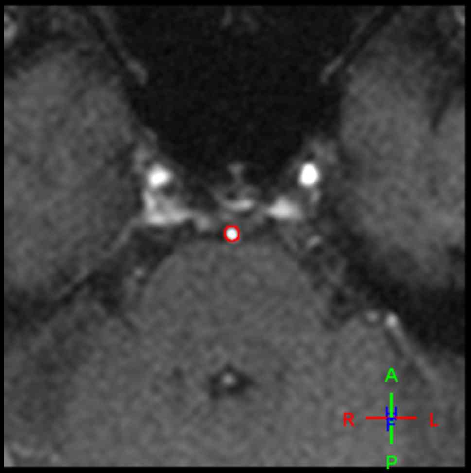
in MRI and PC-MRA (Figs. 1–5).
Discussion
Research methods of cerebral hemodynamics (9–13)
include positron mission tomography/computer tomography (PET/CT),
transcranial Doppler ultrasonography (TCD), CT/MRI perfusion and
PC-MRA. PET/CT is the golden standard for clinical detection of
cerebral blood flow volume, which can provide morphological and
metabolic parameters. However, the examination cost is high, only a
few large hospitals are qualified for the examination, and a
control study for healthy individuals does not conform to medical
ethics. The single target vessel can be detected via TCD, but PCoA
has a small diameter, tortuous walking and complex variation, so it
is difficult to locate it. Currently, PCoA has not been applied in
the clinical detection yet. The whole or local macroscopic cerebral
blood flow information can be obtained via CT or MRI perfusion, but
it is more difficult to study the responsible vessels in a targeted
manner. PC-MRA is a non-radioactive detection method for cerebral
blood flow, which can detect the acute ischemic lesions and provide
both morphological and hemodynamic information simultaneously. The
velocity-time curve of target vessels can be obtained, and such
hemodynamic parameters as the mean flow velocity and mean flow
volume can be calculated using flow analysis software, which is of
important value in evaluating the collateral compensatory function
of PCoA (7). In addition, digital
subtraction angiography (DSA) is characterized by high spatial
resolution, temporal resolution, as well as sensitivity and
specificity, which is the gold standard for the vascular
morphological examination. However, DSA cannot be used in the
examination for normal control subjects, because it can cause large
trauma, and the contrast agent can only be injected in sequence to
display the left and right PCoA one by one, with poor overall
view.
F-PCoA is an important collateral blood vessel
linking the anterior-posterior circulation blood flow. Under
different conditions, there may be forward posterior, backward
anterior or bidirectional shunt in F-PCoA, and the influence of
F-PCoA cannot be ignored. Table II
indicates that in the presence of F-PCoA, the diameter and
cross-sectional area of BA are small, and the maximum flow volume
and peak height of flow volume are reduced, but the maximum flow
velocity is increased, which can be regarded as a kind of adaptive
change that the internal carotid artery blood flow reaches the
blood-supply area of posterior circulation through forward
posterior shunt of F-PCoA to maintain the stability of the total
blood flow volume in the posterior circulation. There is no
necessary correlation between the smaller diameter of BA and the
occurrence of PCI. Whether there are shunt and compensation between
the anterior and posterior circulations is important clinical
diagnostic information. The following 4 conditions can indicate no
shunt between the anterior and posterior circulation based on the
PC-MRA results in this study: i) there is no PCoA, ii) PCoA is only
a potential, namely the same blood pressure at both ends, no flow
in blood vessels, or too small vascular diameter, and very low
shunt flow volume, indicating the presence of A-PCoA but absence of
F-PCoA, iii) the blood pressure at both ends of F-PCoA is similar,
and the bidirectional blood flow reaches the dynamic balance in one
cardiac cycle, and iv) the flow volume of bilateral F-PCoA is the
same but the flow direction is the opposite. The above results
suggest that the differences and relationship between F-PCoA and
A-PCoA should be paid attention to, and the theory in previous
studies (1,6,9) that
there is shunt in the presence of PCoA and PCoA is a protective
blood flow pathway for BA stenosis should be further improved.
Results revealed that in the presence of F-PCoA,
there were no significant differences in the total blood flow of
bilateral internal carotid arteries and vertebral arteries and the
diameter of BA between PCI and non-PCI patients, but the mean flow
volume, mean flow velocity, minimum flow volume, maximum flow
volume and minimum flow velocity of BA in PCI patients were all
decreased. At the same time, the forward posterior shunt flow
volume of F-PCoA was significantly increased (Table III). Results also showed that there
was a more obvious main peak of F-PCoA in the research group than
that in the control group, indicating that the forward and backward
shunt flow volume in the systolic phase is increased in one cardiac
cycle, while that in the diastolic phase is similar to that in
control group. The above results suggest that the blood flow in
F-PCoA can compensate for the decreased blood flow of BA to a
certain degree and play a protective role in PCI, and such effects
are more significant in the systolic phase.
If the blood flow of BA is reduced, forward
posterior shunt is detected in F-PCoA, and there are clinical
manifestations of PCI, but no positive infarct is detected via
conventional MRI, it may be in the decompensatory stage of
posterior circulation blood flow, and active intervention should be
made for risk factors of PCI. Kaspera et al (14) showed that in the bilateral internal
carotid arterial occlusion, the anterior circulation ischemia can
be partially compensated by posterior circulation blood flow
through backward anterior shunt of PCoA. According to further
research (1,6,15), such
a shunt may increase the risk of posterior circulation ischemia. In
this study, the shunt from backward to forward was detected in some
F-PCoAs, the blood flow of internal carotid artery was reduced,
there was no significant or slight increase in the blood flow of
BA, there were clinical manifestations of posterior circulation
ischemia, or posterior circulation infarct was detected via MRI,
thereby providing additional evidence for the above theory.
This study is useful in the identification of
subjects with clinically suspected PCI but negative results in
routine MR plain scan, or subjects without clinical symptoms but
with decreased posterior circulation blood flow volume, the former
of which is the incomplete compensation of blood flow and the
latter of which is the complete compensation of blood flow. In
either case, however, if subjects are not closely followed up and
risk factors are not controlled in a timely manner, PCI may
progress to irreversible cerebral infarction. This study also had
some limitations: i) the sample size was small, and the supporting
evidence for statistical analysis was insufficient. ii) There was a
lack of reliable and recognized gold standard for the PC-MRA
results, and it was only a single-center study; thus, results
remain to be further verified.
In conclusion, this study indicates that hemodynamic
characteristics of F-PCoA can be analyzed using the mature
technique of PC-MRA. The forward posterior shunt flow volume of
F-PCoA can provide references for the clinical auxiliary
diagnosis.
Acknowledgements
Not applicable.
Funding
This study was supported by the Medical Science and
Technology Breakthrough project of Foshan Science and Technology
Bureau (nos. 2015AB002553, 2014AB002553).
Availability of data and materials
The datasets used and/or analyzed during the present
study are available from the corresponding author on reasonable
request.
Authors' contributions
WZ drafted the manuscript. WZ, ML and JL contributed
to collecting and interpreting the data. FC, QH and SY were
responsible for statistical analysis. All authors read and approved
the final manuscript.
Ethics approval and consent to
participate
The study was approved by the Ethics Committee of
Shunde Hospital, Southern Medical University (The First People's
Hospital of Shunde Foshan) (Foshan, China). Signed informed
consents were obtained from the patients or the guardians.
Patient consent for publication
Not applicable.
Competing interests
The authors declare that they have no competing
interests.
References
|
1
|
Hu XY, Li ZX, Liu HQ, Zhang M, Wei ML,
Fang S, Chen W, Pan H, Huang JX, Zhu YM, et al: Relationship
between vertebral artery hypoplasia and posterior circulation
stroke in Chinese patients. Neuroradiology. 55:291–295. 2013.
View Article : Google Scholar : PubMed/NCBI
|
|
2
|
Iqbal S: A comprehensive study of the
anatomical variations of the circle of Willis in adult human
brains. J Clin Diagn Res. 7:2423–2427. 2013.PubMed/NCBI
|
|
3
|
Nouh A, Remke J and Ruland S: Ischemic
posterior circulation stroke: A review of anatomy, clinical
presentations, diagnosis, and current management. Front Neurol.
5:302014. View Article : Google Scholar : PubMed/NCBI
|
|
4
|
Zhou W, Xu YK, Lu MR, Liang YJ, Chen F, Hu
QG and Liu J: MRA Study on correlation of posterior communicating
artery variation and posterior circulation infarction. J Sun Yatsen
Univ (Med Sci). 37:277–282. 2016.(In Chinese).
|
|
5
|
Zhou W and Xu YK: MRA evaluation of
variation of posterior communicating artery and its correlation
with posterior circulation infarction. Int J Med Radiol.
39:373–377. 2016.(In Chinese).
|
|
6
|
Goerlitz J, Wenz H, Al-Zghloul M, Kerl HU,
Groden C and Förster A: Anatomical variations in the posterior
circle of Willis and vascular pathologies in isolated unilateral
thalamic infarction. J Neuroimaging. 25:983–988. 2015. View Article : Google Scholar : PubMed/NCBI
|
|
7
|
Orosz L, Hoksbergen AW, Molnár C, Siró P,
Cassot F, Marc-Vergnes JP and Fülesdi B: Clinical applicability of
a mathematical model in assessing the functional ability of the
communicating arteries of the circle of Willis. J Neurol Sci.
287:94–99. 2009. View Article : Google Scholar : PubMed/NCBI
|
|
8
|
Division of Cerebrovascular Disease and
Branch of Neurology: Chinese Medical Association: Chinese guidance
for diagnosis and treatment of acute ischemic stroke in 2014. Chin
J Neurol. 48:246–257. 2015.
|
|
9
|
Leung J, Behpour A, Sokol N, Mohanta A and
Kassner A: Assessment of intracranial blood flow velocities using a
computer controlled vasoactive stimulus: A comparison between phase
contrast magnetic resonance angiography and transcranial Doppler
ultrasonography. J Magn Reson Imaging. 38:733–738. 2013. View Article : Google Scholar : PubMed/NCBI
|
|
10
|
Kim HJ, Kim TW, Ryu SY, Yang PS, Kwon MJ,
Kim JC, Lee YS, Lee HJ, Yang JH, Kim JK, et al:
Acetazolamide-challenged perfusion magnetic resonance imaging for
assessment of cerebrovascular reserve capacity in patients with
symptomatic middle cerebral artery stenosis: Comparison with
technetium-99m-hexamethylpropyleneamine oxime single-photon
emission computed tomography. Clin Imaging. 35:413–420. 2011.
View Article : Google Scholar : PubMed/NCBI
|
|
11
|
Rijbroek A, Wisselink W, Vriens EM,
Barkhof F, Lammertsma AA and Rauwerda JA: Asymptomatic carotid
artery stenosis: Past, present and future. How to improve patient
selection? Eur Neurol. 56:139–154. 2006.PubMed/NCBI
|
|
12
|
Ursino M, Lodi CA and Russo G: Cerebral
hemodynamic response to CO(2) tests in patients with internal
carotid artery occlusion: Modeling study and in vivo validation. J
Vasc Res. 37:123–133. 2000. View Article : Google Scholar : PubMed/NCBI
|
|
13
|
Purkayastha S and Sorond F: Transcranial
Doppler ultrasound: Technique and application. Semin Neurol.
32:411–420. 2012. View Article : Google Scholar : PubMed/NCBI
|
|
14
|
Kaspera W, Ładziński P, Larysz P,
Majchrzak H, Hebda A, Kopera M, Tomalski W and Ślaska A:
Transcranial color-coded Doppler assessment of cerebral
arteriovenous malformation hemodynamics in patients treated
surgically or with staged embolization. Clin Neurol Neurosurg.
116:46–53. 2014. View Article : Google Scholar : PubMed/NCBI
|
|
15
|
Asif A, Leon C, Merrill D, Bhimani B,
Ellis R, Ladino M and Gadalean FN: Arterial steal syndrome: A
modest proposal for an old paradigm. Am J Kidney Dis. 48:88–97.
2006. View Article : Google Scholar : PubMed/NCBI
|