Introduction
Systemic lupus erythematosus (SLE) is a diffuse
connective tissue disease transmitted by autoimmunity as a medium
(1), characterized by long duration,
easy recurrence, and high mortality. It has not been clearly
defined yet and is still incurable (2). The disease occurs in young women of
childbearing age and the prevalence of females accounts for 90% of
the total number of affected patients. The peak age of the disease
is 15–40 years (3,4). SLE usually affects the whole body, and
there are multiple autoantibodies in the patient's serum as
clinical features. The course of SLE, the number of affected organs
and the prognosis of survival are negatively correlated (5). These autoantibodies, which are immune
complexes, bind to their corresponding antigens to form
antigen-antibody complexes which then are deposited in the kidneys,
blood vessels, subcutaneous tissue, or even the nervous system,
promoting multiple organ and tissue damage (6). Currently, steroids, immunosuppressants
and other drugs are used to treat SLE. However, in addition to
relieving the disease, many adverse reactions are also caused to
the body (7). Therefore, it is of
great significance to detect multiple autoantibodies in SLE
patients' sera and provide evidence for clinical rational drug
use.
The antinuclear antibody (ANA) is a general term for
autoantibodies against various nuclear components (8). ANA can be seen in all kinds of
rheumatic diseases (9). The
currently recognized autoantibody markers for the diagnosis of SLE
are anti-double-stranded DNA (ds-DNA) antibodies, because high
concentrations of anti-ds-DNA antibodies are almost exclusively
present in SLE patients, anti-ds-DNA antibodies are SLE-specific
(10). C3 and C4 are important
components in the complement system and play an important role in
complement activation, participating in immunity and maintaining
internal environment stability (11).
The purpose of this study was to detect ANA,
anti-ds-DNA antibody, and complement C3, C4 in the serum of 194
patients with SLE, 106 patients with non-SLE, and 120 healthy
volunteers, and to investigate the diagnostic value of these four
factors, either alone or in combination, for SLE.
Materials and methods
Sample collection
Retrospective methods were adopted. A total of 194
patients with SLE who were admitted to the Department of
Rheumatology at Yantaishan Hospital of Yantai (Yantai, China) from
January 2012 to December 2017 were selected as SLE group. The
average age was 40.82±13.46 years, including 23 males and 171
females. All cases were diagnosed according to the revised
standards of the American Rheumatism Association (12). A total of 106 patients with non-SLE
rheumatism were selected as disease control group, with an average
age of 51.27±11.42 years, including 18 males and 88 females. In the
same period, 120 healthy subjects were selected as healthy control
group with average age of 40.28±10.85 years, including 20 males and
100 females. Patients during pregnancy, lactating, suffering from
other immune disease, or having incomplete clinical information
were excluded. The heart, liver and kidney functions of all
subjects enrolled were normal. The study was approved by the Ethics
Committee of Yantaishan Hospital of Yantai. Signed informed
consents were obtained from the patients or the guardians.
Results showed that there was no significant
difference in age, sex, marriage and childbirth status between the
three groups (p>0.05). The average red blood cell volume,
distribution width of red blood cells, platelets, Hcy level, total
cholesterol, and urine protein were significantly different
(p<0.001). General information is shown in Table I.
 | Table I.General information. |
Table I.
General information.
| Factor | SLE group
(n=194) | Disease control group
(n=106) | Control group
(n=120) | F/χ2 | P-value |
|---|
| Age (years) |
| ≥40 | 72
(37.11) | 52 (49.06) | 46
(38.33) | 4.378 | 0.112 |
|
<40 | 122 (62.89) | 54 (50.94) | 74
(61.67) |
|
|
| Sex |
| Male | 23
(11.86) | 18 (16.98) | 20
(16.67) | 2.072 | 0.355 |
|
Female | 171 (88.14) | 88 (83.02) | 100 (83.33) |
|
|
| Marriage and
childbirth |
| Yes | 161 (82.99) | 82 (77.36) | 86
(71.67) | 5.680 | 0.058 |
| No | 33
(17.01) | 24 (22.64) | 34
(28.33) |
|
|
| Average volume of red
blood cells (fl) | 84.65±3.30 | 86.72±3.12 | 89.68±0.28 | 124.90 | <0.001 |
| Erythrocyte
distribution width (%) | 15.51±2.57 | 13.42±0.81 | 12.56±0.76 | 106.10 | <0.001 |
| Platelets
(109/l) | 85.42±0.73 | 164.21±18.94 | 198.74±32.21 | 1,360.00 | <0.001 |
| Hcy (µmol/l) | 14.31±2.13 | 11.25±2.48 | 9.15±2.41 | 194.9 | <0.001 |
| Total cholesterol
(mmol/l) | 5.75±2.45 | 5.03±1.05 | 4.12±0.85 | 30.31 | <0.001 |
| Urine protein (g/24
h) | 4.13±3.12 | 2.21±1.86 | 0.05±0.02 | 115.7 | <0.001 |
Reagents and equipment
ANA and anti-ds-DNA antibody kits were purchased
from Trinity Biotech, Inc. (Jamestown, NY, USA); Microplate reader
was purchased from Bio-Rad Laboratories, Inc. (Hercules, CA, USA);
complement C3 and C4 kits were purchased from Beckman Coulter, Inc.
(Brea, CA, USA); Hitachi automatic biochemical analyzer was
purchased from Hitachi, Ltd. (Tokyo, Japan).
Detection methods
All subjects gave fasting venous blood (4 ml) and
serum was centrifuged at 2,080 × g for 8 min at 4°C. ANA and
anti-ds-DNA antibody were determined by ELISA, and velocity
nephelometry was used to determine complement C3 and C4. All test
items were operated strictly in accordance with the manufacturer's
instructions.
Interpretation of the results
The OD value of ANA and anti-ds-DNA antibody
measured by a microplate reader was determined by the formula: OD
value of sample/(mean OD of standard solution × correction factor);
≥1.1 was positive, and ≤0.9 was negative (13). The number of positive cases of
complement C3 and C4 was determined based on the ROC curve.
Statistical analysis
SPSS 16.0 software was used for statistical analysis
(Shanghai Kabei Information Technology Co., Ltd., Shanghai, China).
Chi-square test was used for enumeration data and t-test was used
for measurement data. One-way ANOVA was used for comparison among
groups and SNK-q test was used as a post-hoc test for group
analysis. ROC curves were used to analyze the diagnostic value of
complement C3 and C4. P<0.05 was considered to indicate a
statistically significant difference.
Results
Positive rate of autoantibody
detection in SLE and disease control groups
The positive rates of ANA and anti-ds-DNA antibody
in SLE group were 91.75 and 67.01%, respectively. The positive
rates of ANA and anti-ds-DNA antibody in disease control group were
43.40 and 3.77%, respectively. The positive rates in the disease
control and SLE groups were higher than that in the control group,
and the differences were statistically significant (p<0.05). The
difference between the positive rates in the disease control and
SLE group was statistically significant (p<0.05). The
sensitivity and specificity of ANA in the diagnosis of SLE were
91.75% (178/194) and 79.65% (180/226), respectively. The
sensitivity and specificity of anti-ds-DNA antibody in the
diagnosis of SLE were 67.01% (130/194), 98.23% (222/226) (Tables II and III).
| Table II.Comparison of the positive rate of
autoantibody detection in SLE and disease control groups (%). |
Table II.
Comparison of the positive rate of
autoantibody detection in SLE and disease control groups (%).
| Group | No. of cases | ANA | Anti-ds-DNA
antibody |
|---|
| SLE group | 194 | 178
(91.75)a | 130
(67.01)a |
| Disease control
group | 106 | 46
(43.40)a,b | 4 (3.77)a,b |
| Control group | 120 | 0 (0) | 0 (0) |
| χ2 |
| 52.34 | 110.90 |
| P-value |
| <0.001 | <0.001 |
| Table III.Diagnostic sensitivity and specificity
of ANA and anti-ds-DNA antibody for SLE (%). |
Table III.
Diagnostic sensitivity and specificity
of ANA and anti-ds-DNA antibody for SLE (%).
| Antibody | No. of positive
cases | Sensitivity | Specificity |
|---|
| ANA | 178 | 91.75 | 79.65 |
| Anti-ds-DNA | 130 | 67.01 | 98.23 |
Complement test results
The expression levels of C3 and C4 in SLE and
disease control groups were lower than those in control group, and
the differences were statistically significant (p<0.001). The
expression levels of C3 and C4 in the disease control group were
significantly different from those in the SLE group (p<0.05)
(Table IV).
| Table IV.Complement test results (g/l). |
Table IV.
Complement test results (g/l).
| Group | No. of cases | C3 | C4 |
|---|
| SLE group | 194 |
0.58±0.24a |
0.19±0.08a |
| Disease control
group | 106 |
0.64±0.23a,b |
0.23±0.04a,b |
| Control group | 120 | 0.97±0.15 | 0.29±0.11 |
| F |
| 128.30 | 54.39 |
| P-value |
| <0.001 | <0.001 |
Comparison of the sensitivity and
specificity of complement C3 and C4 in SLE diagnosis
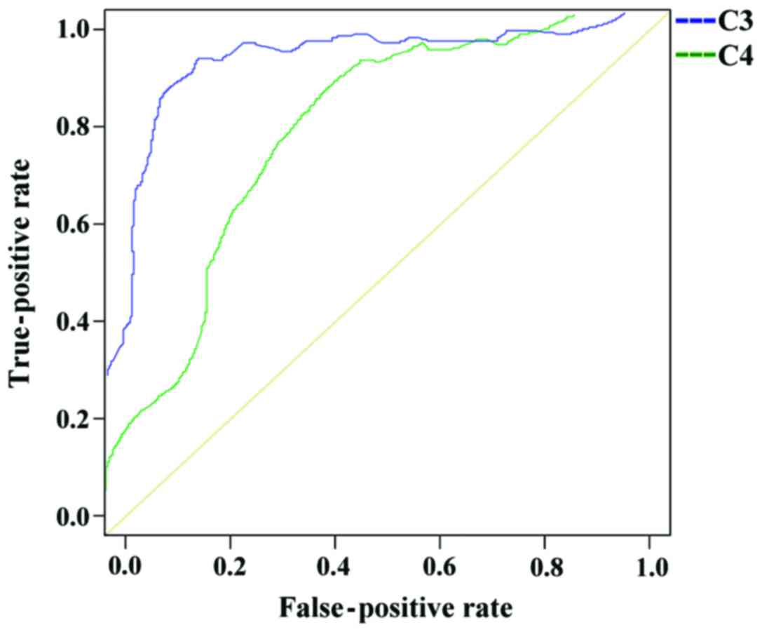
The ROC curve analysis of the diagnosis of SLE with
complement C3 showed that the AUC for the diagnosis of SLE with C3
was 0.903, the progressive 95% confidence interval was 0.869–0.937,
the cut-off value was 0.663, the SLE group positive case number was
169, the number of positive cases in the disease control group was
39, the sensitivity was 87.11% and the specificity was 82.74%.
The ROC curve analysis of the diagnosis of SLE with
complement C4 showed that the AUC was 0.763, the progressive 95%
confidence interval was 0.705–0.820, the cut-off value was 0.434,
the SLE group included 172 positive cases, the number of positive
cases in the disease control group was 51, the sensitivity was
88.66% and the specificity was 77.43%.
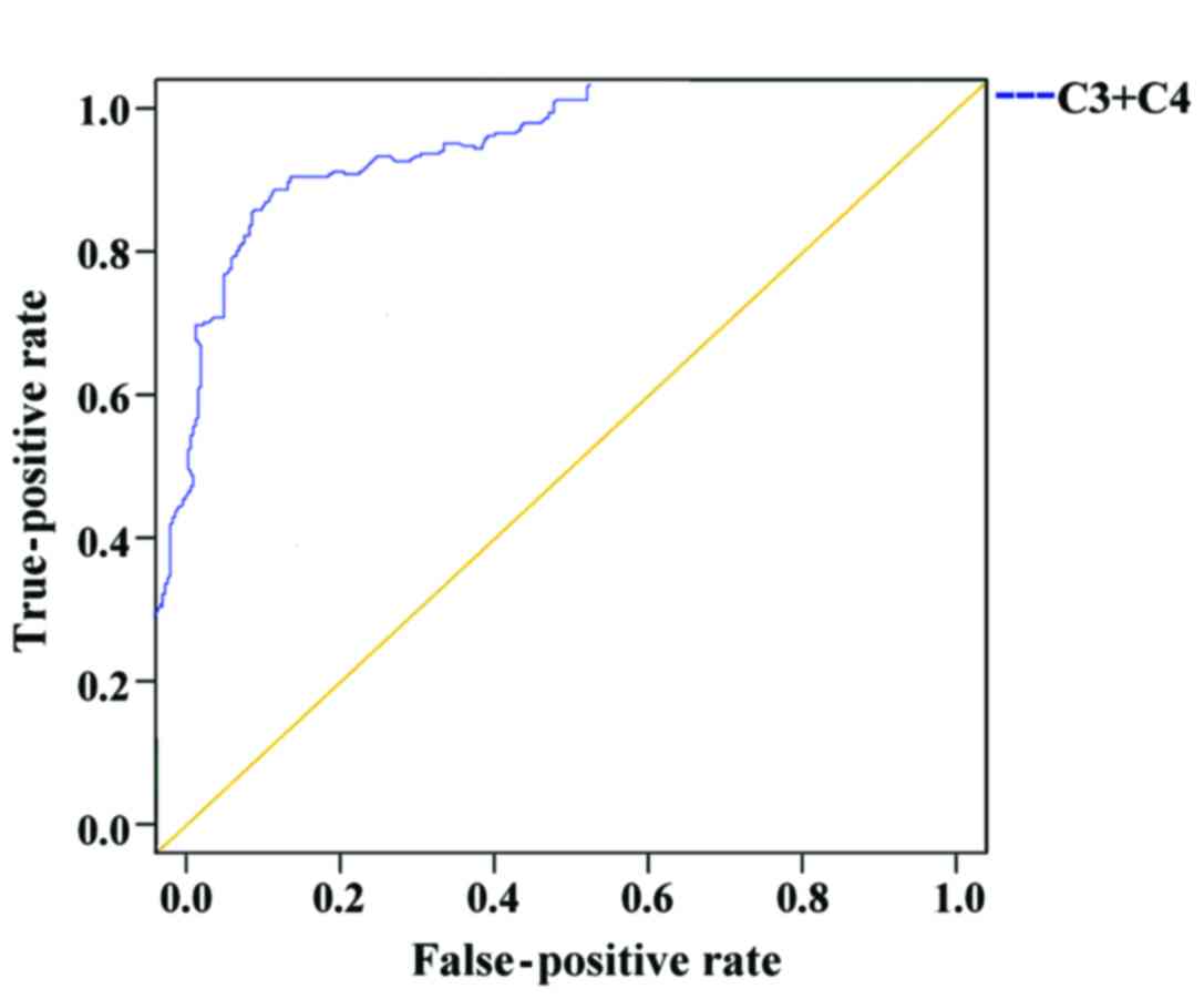
The ROC curve analysis of the diagnosis of SLE by
combination of C3 and C4 showed that the AUC of the combined
diagnosis of SLE was 0.931, the progressive 95% confidence interval
was 0.904–0.958, the cut-off value was 0.727, the SLE group
included 180 positive cases, the number of positive cases in the
disease control group was 47, the sensitivity was 92.78%, and the
specificity was 79.20% (Figs. 1 and
2, and Table V).
| Table V.Diagnostic sensitivity and
specificity of complement C3 and C4 for SLE (%). |
Table V.
Diagnostic sensitivity and
specificity of complement C3 and C4 for SLE (%).
| Complement | No. of positive
cases | Sensitivity | Specificity |
|---|
| C3 | 169 | 87.11 | 82.74 |
| C4 | 172 | 88.66 | 77.43 |
| C3+C4 | 180 | 92.78 | 79.20 |
Comparison of the sensitivity of the
combined application in SLE diagnosis
The sensitivity and specificity of ANA and ds-DNA in
the diagnosis of SLE were 95.36% (185/194) and 96.90% (219/226),
respectively; of C3 and C4 were 92.78% (180/194) and 79.20%
(179/226); and of all the four factors were 97.42% (189/194) and
80.97% (183/226), respectively (Table
VI).
| Table VI.Combined sensitivity in SLE diagnosis
(%). |
Table VI.
Combined sensitivity in SLE diagnosis
(%).
| Item | n | No. of positive
cases | Sensitivity | Specificity |
|---|
| ANA+anti-ds-DNA
antibody | 420 | 185 | 95.36 | 96.90 |
| C3+C4 | 420 | 180 | 92.78 | 79.20 |
|
ANA+ds-DNA+C3+C4 | 420 | 189 | 97.42 | 80.97 |
Discussion
Pathogenesis and causes of SLE are complex. Many
factors such as environment, heredity and estrogen can stimulate
the body's immune regulation, promote the deposition of antigens,
antibodies and complements, and induce local or systemic
multi-organ and -tissue injuries (14). The immunological abnormalities of SLE
are mainly manifested by the presence of multiple autoantibodies
represented by ANA in serum (15).
Currently, the main cause of SLE tissue damage is immune complex
allergy, which can activate allergic reactions through classic and
alternative pathways while consuming complement (16). ANA, anti-ds-DNA antibody, and
complement C3 and C4 associated with SLE are important indicators
for the diagnosis of SLE. In this study, 194 patients with SLE had
a sensitivity of 91.75% and a specificity of 79.65% in detecting
serum ANA. Kumar and Bhatia (17)
have reported that the diagnostic sensitivity and specificity of
ANA for SLE are 95 and 65%, respectively, similar to our results.
This suggests that the detection of ANAs lacks specificity and ANAs
may also be present in other connective tissue diseases besides
SLE. According to the results presented in Table II, the positive rate of ANA in the
serum of 106 patients of the disease control group was 43.40%. Due
to the deficiency of specificity of ANA in the diagnosis of SLE,
ANA may also be present in other connective tissue diseases;
negative ANA does not completely indicate that there is no SLE,
which is consistent with literature (18).
According to the results, 16 cases of SLE patients
were pathologically confirmed with negative serum ANA. These may
belong to a subcategory of SLE, in which case the common clinical
features of photosensitivity, and clinical features of arthritis,
kidney damage are rare (19).
Therefore, the detection of ANA in the diagnosis of SLE is only of
screening value. Only when the ANA is positive and clinical
features of SLE are present the detection of ANA can be used as a
definite diagnosis of SLE. This study showed that the sensitivity
and specificity of the detection of anti-ds-DNA antibody in SLE
were 67.01 and 98.23%, respectively. The sensitivity was poor, but
the specificity was higher. Roggenbuck et al (20) have shown that anti-dsDNA antibody can
be used as a marker for the diagnosis of SLE. This also confirms
the high specificity of anti-ds-DNA antibody in our results. In
addition, ds-DNA can become negative after the disease improves
with treatment, so anti-ds-DNA antibody can provide the basis for
monitoring treatment (21). The
expression levels of C3 and C4 in SLE and disease control groups
were lower than those in control group, and the differences were
statistically significant (p<0.001). Liu et al (22) have reported that the decrease of the
levels of complement C3 and C4 in the serum of SLE patients means
that the patient has increased autoantibodies, which is the
manifestation of immune activation, and it may also be
complement-mediated cytotoxicity. This is consistent with our
findings, therefore, the decrease of complement C3 and C4 levels in
serum can be used as diagnosis standard of SLE. According to the
results of the ROC curves, the sensitivity of C3 in the diagnosis
of SLE was 87.11% and the specificity was 82.74%; the sensitivity
of complement C4 was 88.66% and the specificity was 77.43%.; and
the sensitivity of the combination of C3 and C4 in the diagnosis of
SLE was 92.78% and the specificity is 79.20%. This shows that
complement C3 and C4 are not very specific indicators of SLE, but
they have a screening value in the diagnosis of SLE, and the
sensitivity of combined diagnosis of SLE is improved. The
sensitivity and specificity of ANA and anti-ds-DNA antibody in the
diagnosis of SLE were 95.36 and 96.90%, respectively; of C3 and C4
were 92.78 and 79.20%, respectively; and of the combination of all
four indicators were 97.42 and 80.97%, respectively. The
sensitivity of combined ANA and anti-ds-DNA antibody, the combined
C3 and C4, and all four indicators in the diagnosis of SLE was
higher than that of these antibodies alone, but the specificity of
all four combined factors in the diagnosis of SLE was lower than
that of anti-ds-DNA antibody. The sensitivity and specificity of
autoantibody detection are different in different populations and
disease activity levels. Therefore, it is not possible to diagnose
the disease with only one or a single index. Joint detection can
prove the indicators to play a complementary role.
In summary, ANA has the highest sensitivity in the
diagnosis of SLE, but its specificity is the worst; anti-ds-DNA
antibody has the lowest sensitivity for the diagnosis of SLE, but
the highest specificity. Therefore, combining ANA, anti-ds-DNA
antibody, complement C3 and C4 can be used for the diagnosis of SLE
in order to reduce or even avoid the missed diagnosis caused by a
single test, to provide a complementary and confirmatory effect,
and to improve the positive diagnosis rate of SLE.
Acknowledgements
Not applicable.
Funding
No funding was received.
Availability of data and materials
The datasets used and/or analyzed during the present
study are available from the corresponding author on reasonable
request.
Authors' contributions
CQ and JZ drafted the manuscript. CQ, JZ and XZ were
mainly devoted in collecting and interpreting the general data. JD
and BS were responsible for ELISA. HL performed the velocity
nephelometry. All authors read and approved the final
manuscript.
Ethics approval and consent to
participate
The study was approved by the Ethics Committee of
Yantaishan Hospital of Yantai (Yantai, China). Signed informed
consents were obtained from the patients or the guardians.
Patient consent for publication
Not applicable.
Competing interests
The authors declare that they have no competing
interests.
References
|
1
|
Rönnblom L and Pascual V: The innate
immune system in SLE: Type I interferons and dendritic cells.
Lupus. 17:394–399. 2008. View Article : Google Scholar : PubMed/NCBI
|
|
2
|
Coluccia AM, Cirulli T, Neri P, Mangieri
D, Colanardi MC, Gnoni A, Di Renzo N, Dammacco F, Tassone P,
Ribatti D, et al: Validation of PDGFRbeta and c-Src tyrosine
kinases as tumor/vessel targets in patients with multiple myeloma:
Preclinical efficacy of the novel, orally available inhibitor
dasatinib. Blood. 112:1346–1356. 2008. View Article : Google Scholar : PubMed/NCBI
|
|
3
|
Egner W: The use of laboratory tests in
the diagnosis of SLE. J Clin Pathol. 53:424–432. 2000. View Article : Google Scholar : PubMed/NCBI
|
|
4
|
Su DL, Wang HJ, Ji XH, Li YY, Xuan HB,
Heng C and Li YF: Mycophenolic acid inhibits SLE-associated
cytokine expression and promotes apoptosis of peripheral blood
mononuclear cells from patients with systemic lupus erythematosus.
Acta Pharmacol Sin. 27:1051–1057. 2006. View Article : Google Scholar : PubMed/NCBI
|
|
5
|
Mackay M, Stanevsky A, Wang T, Aranow C,
Li M, Koenig S, Ravetch JV and Diamond B: Selective dysregulation
of the FcgammaIIB receptor on memory B cells in SLE. J Exp Med.
203:2157–2164. 2006. View Article : Google Scholar : PubMed/NCBI
|
|
6
|
Zhao P, Xu L, Wang P, Liang X, Qi J, Liu
P, Guo C, Zhang L, Ma C and Gao L: Increased expression of human
T-cell immunoglobulin- and mucin-domain-containing molecule-4 in
peripheral blood mononuclear cells from patients with system lupus
erythematosus. Cell Mol Immunol. 7:152–156. 2010. View Article : Google Scholar : PubMed/NCBI
|
|
7
|
Yu SL, Wong CK, Wong PTY, Chen DP, Szeto
CC, Li EK and Tam LS: Down-regulated NOD2 by immunosuppressants in
peripheral blood cells in patients with SLE reduces the muramyl
dipeptide-induced IL-10 production. PLoS One. 6:e238552011.
View Article : Google Scholar : PubMed/NCBI
|
|
8
|
Muratori P, Muratori L, Ferrari R, Cassani
F, Bianchi G, Lenzi M, Rodrigo L, Linares A, Fuentes D and Bianchi
FB: Characterization and clinical impact of antinuclear antibodies
in primary biliary cirrhosis. Am J Gastroenterol. 98:431–437. 2003.
View Article : Google Scholar : PubMed/NCBI
|
|
9
|
Xu XF, Zhang J, Cui L, Wang YH, Yue Y, Chi
L, Bai J, Li HM and Lu XX: The value of different antibodies
detection in diagnosis of rheumatism with uveitis. Zhonghua Yi Xue
Za Zhi. 97:285–290. 2017.(In Chinese). PubMed/NCBI
|
|
10
|
Song XY, Huang H, Liu YZ, Zhao YY, Li S
and Xu ZJ: Coexistence of sarcoidosis and primary Sjögren syndrome:
A clinical analysis and literature review. Zhonghua Nei Ke Za Zhi.
56:375–377. 2017.(In Chinese). PubMed/NCBI
|
|
11
|
Buhl A, Metzger JH, Heegaard NH, von
Landenberg P, Fleck M and Luppa PB: Novel biosensor-based analytic
device for the detection of anti-double-stranded DNA antibodies.
Clin Chem. 53:334–341. 2007. View Article : Google Scholar : PubMed/NCBI
|
|
12
|
Calano SJ, Shih PA, Liu CC, Kao AH,
Navratil JS, Manzi S and Ahearn JM: Cell-bound complement
activation products (CB-CAPs) as a source of lupus biomarkers. Adv
Exp Med Biol. 586:381–390. 2006. View Article : Google Scholar : PubMed/NCBI
|
|
13
|
Hochberg MC: Updating the American College
of Rheumatology revised criteria for the classification of systemic
lupus erythematosus. Arthritis Rheum. 40:17251997. View Article : Google Scholar : PubMed/NCBI
|
|
14
|
Liu Y, Jing H, Wang J, Zhang R, Zhang Y,
Zhang Y, Xu Q, Yu X and Xue C: Micronutrients decrease incidence of
common infections in type 2 diabetic outpatients. Asia Pac J Clin
Nutr. 20:375–382. 2011.PubMed/NCBI
|
|
15
|
Salaman MR and Isenberg DA: The
immunological personality of close relatives of SLE patients.
Lupus. 26:1513–1516. 2017. View Article : Google Scholar : PubMed/NCBI
|
|
16
|
Liu CC, Manzi S, Kao AH, Navratil JS and
Ahearn JM: Cell-bound complement biomarkers for SLE: From benchtop
to bedside. Rheum Dis Clin North Am. 36:161–172. 2010. View Article : Google Scholar : PubMed/NCBI
|
|
17
|
Kumar Y and Bhatia A: Detection of
antinuclear antibodies in SLE. Methods Mol Biol. 1134:37–45. 2014.
View Article : Google Scholar : PubMed/NCBI
|
|
18
|
Kasitanon N, Magder LS and Petri M:
Predictors of survival in systemic lupus erythematosus. Medicine
(Baltimore). 85:147–156. 2006. View Article : Google Scholar : PubMed/NCBI
|
|
19
|
Li LH, Pan HF, Li WX, Li XP, Xu JH and Ye
DQ: Study on clinical features and complications with systemic
lupus erythematosus (SLE) activity in Chinese Han population. Clin
Rheumatol. 28:1301–1307. 2009. View Article : Google Scholar : PubMed/NCBI
|
|
20
|
Roggenbuck D, Reinhold D, Hiemann R,
Anderer U and Conrad K: Standardized detection of anti-ds DNA
antibodies by indirect immunofluorescence - a new age for
confirmatory tests in SLE diagnostics. Clin Chim Acta.
412:2011–2012. 2011. View Article : Google Scholar : PubMed/NCBI
|
|
21
|
Waris G and Alam K: Immunogenicity of
superoxide radical modified-DNA: Studies on induced antibodies and
SLE anti-DNA autoantibodies. Life Sci. 75:2633–2642. 2004.
View Article : Google Scholar : PubMed/NCBI
|
|
22
|
Liu H, Yang LH, Yin G and Xie QB:
Correlation of thyroid autoantibodies, system lupus erythematosus
immunologic indicators and disease activity in SLE with HT. Sichuan
Da Xue Xue Bao Yi Xue Ban. 49:179–182. 2018.(In Chinese).
PubMed/NCBI
|