Introduction
Traumatic pseudoaneurysm of the middle meningeal
artery (MMA) is a rare condition and usually a hidden complication
of head injury. It accounts for <1% of all intracranial
aneurysms (1). Histologically, these
aneurysms are not true aneurysms, as the latter are characterized
by dilatation of all layers of the blood vessel wall, while a
pseudoaneurysm is formed by traumatic rupture of the arterial wall,
resulting in hematoma formation outside the artery (2). The wall of the pseudoaneurysm is
composed of the surrounding fibrous tissue and cerebral structures
(2,3). The fragility of the pseudoaneurysm wall
makes it particularly susceptible to rupture, resulting in a high
risk of mortality (3). Therefore,
appropriate and timely management is imperative in such cases.
However, a major limiting factor is that the natural course of
pseudoaneurysms is not well understood and their detection during
emergency computerized tomography (CT) scans is challenging owing
to the lack of typical signs (4).
Furthermore, there are no definite management guidelines for these
lesions (5,6).
Onyx™ is a liquid, non-adhesive embolic agent, which
is ideal for slow, targeted injections. It has been used in
intracranial aneurysms and arteriovenous malformations (7–9). With
technological progress, its use has been expanded to and
established in the management of cerebral pseudoaneurysm (10,11).
The present study reports on a case of traumatic
pseudoaneurysm of the MMA with angiographic changes progressing for
a period of one week, which was successfully embolized using Onyx™.
The available literature on this subject was also reviewed, and the
neuroradiological signs of the pseudoaneurysm and the feasibility
of Onyx™ in the treatment of this condition were discussed.
Case report
A 20-year-old female patient presented at the
emergency department of a local hospital with an acute head injury
sustained in a motor vehicle collision, following which she
remained unconscious for ~10 min on October 14th, 2014. On
admission, the patient was conscious; her Glasgow Coma Scale score
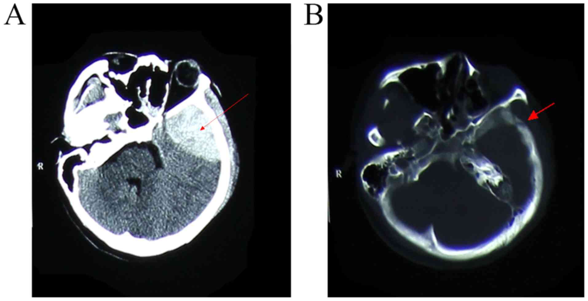
was 14/15 with no localizing neurological signs. Initial cranial CT
scan revealed an ~17-ml epidural hematoma in the left temporal
region along with left temporal bone fracture (Fig. 1). Since there was no mass effect or
midline shift, the patient was treated conservatively with
hemostatic drugs and mannitol, following which she remained
clinically stable. Repeated CT on the same day and the next day
indicated no increase in the size of the hematoma. After medical
treatment for two weeks, a gradual improvement in the signs and
symptoms of the patient was noted. At 1 month after the head
injury, follow-up by serial brain CT indicated a near-complete
resorption of the hematoma and the patient was discharged from
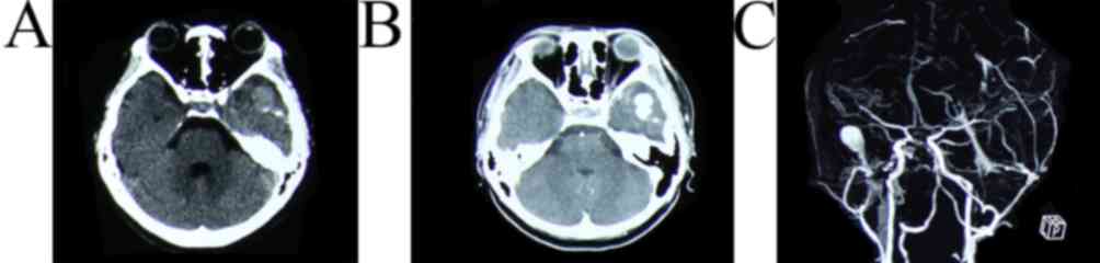
hospital. However, a routine follow-up CT scan performed at 38 days
after the head injury revealed a hyperdense nodule in the left
temporal region (Fig. 2A). CT
revealed a hyperdense nodule with strong homogeneous contrast
enhancement, which was suspected to be vascular in nature (Fig. 2B). Three-dimensional CT angiography
indicated a traumatic pseudoaneurysm of the left external carotid
artery with no communication with the main intracranial arteries
(Fig. 2C). The patient underwent
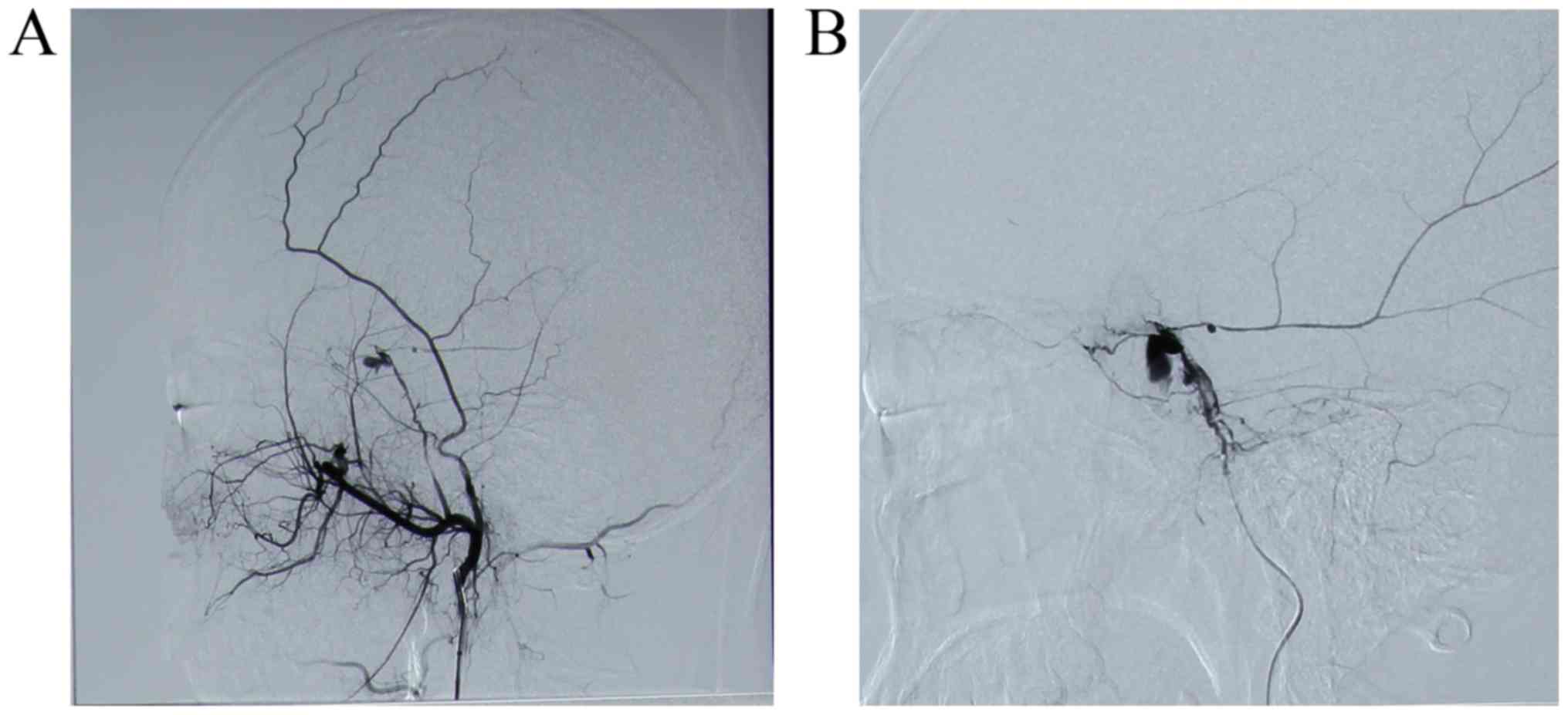
digital subtraction angiography (DSA) three days later. The DSA
confirmed the diagnosis and revealed two pseudoaneurysms arising
from the posterior branch of the left MMA (Fig. 3). The patient and her family members
were reluctant to accept surgery at the local hospital; therefore,
the patient was referred to Jinling Hospital (School of Medicine,
Nanjing University, Nanjing, China).
After 1 week, cerebral angiography was repeated to
evaluate the pseudoaneurysms and to determine the treatment plan.
The angiogram revealed that, while the proximal pseudoaneurysm had
decreased in size, the distal one had enlarged (Fig. 4A). Due to its progressive nature,
there was a high risk of rupture; therefore, immediate embolization
was planned, and informed consent was obtained.
The procedure was performed under general
anesthesia. Access was obtained via a transfemoral approach by
standard techniques. The left external carotid artery and the
branch of the left MMA were selectively catheterized using an
Echelon™ 10 microcatheter. The microcatheter was flushed with 4 ml
saline and its dead space was then filled with 0.3 ml of dimethyl
sulfoxide (DMSO). Under constant fluoroscopic guidance, ONYX™ 34
was injected to embolize the posterior branch of the MMA as
previously reported (Fig. 4B)
(12).
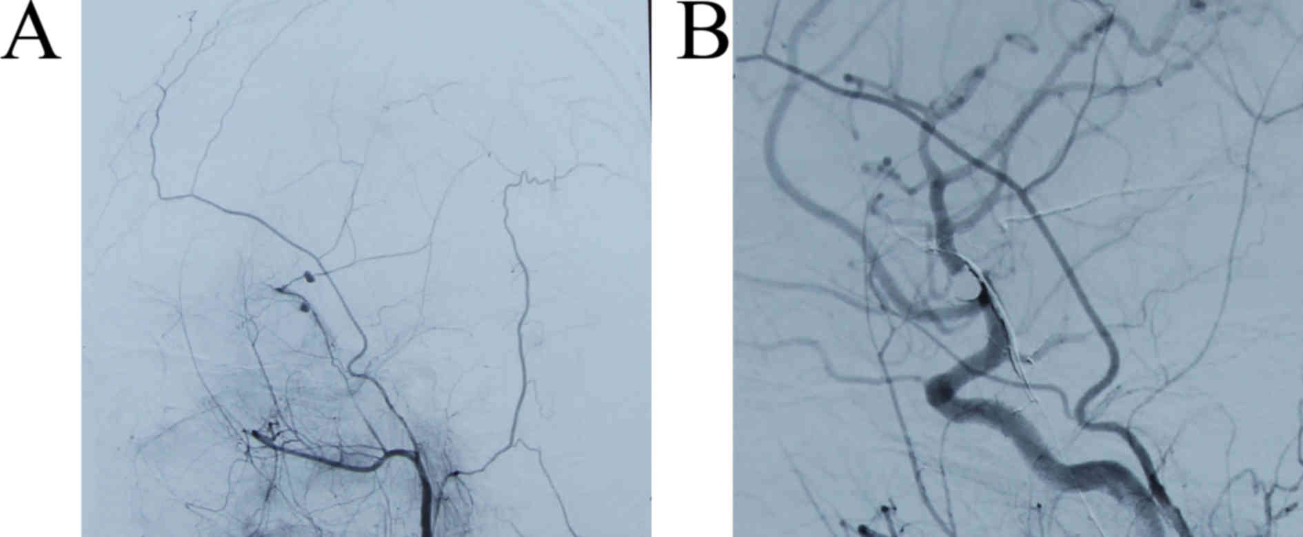
After embolization, selective angiography indicated
that the left external carotid artery and the left MMA were normal
except for the posterior branch, and the pseudoaneurysm was no
longer visible. Repeated CT performed on the next day revealed that
the hyperdense nodule, which was present pre-operatively, had
disappeared and there were no signs of any new hemorrhage. The
patient was discharged from hospital on the third post-operative
day with no neurological deficit. Follow-up DSA performed six
months following the head injury revealed no signs of recurrence of
the pseudoaneurysm (Fig. 5).
Discussion
Traumatic pseudoaneurysm is a rare entity,
accounting for ~1% of all aneurysms (1). In most cases, pseudoaneurysms are
formed due to a tear in the arterial wall after head injury
(13). Approximately 85% of all
traumatic pseudoaneurysms are located in the temporal region and
the remaining ones occur either in the occipital or the frontal
region (14). Traumatic
pseudoaneurysm of the MMA was first described by Schulze in 1957
(15). An estimated 70–90% of
traumatic pseudoaneurysms of the MMA arise in association with a
fracture of the skull bone overlying the temporal part of the MMA
(16). In the current case, an
anterior fracture line passing through the temporal part of the MMA
was clearly discernible and two pseudoaneurysms occurred in the
posterior branch of the left MMA. Besides direct trauma to the
arterial wall, traction of the MMA may occur during closed head
trauma. Cases of pseudoaneurysms of the MMA ostensibly caused by
traction of the MMA have been reported in patients with no
documented bone fracture on the same side (3,13,17).
Almost all MMA pseudoaneurysms reportedly rupture
within 30 days of formation, resulting in high mortality and
morbidity (17,18). Therefore, the requirement for early
diagnosis and timely management of pseudoaneurysms cannot be
overemphasized.
Prior to the availability of DSA, no specific
imaging modalities for diagnosis of traumatic pseudoaneurysms were
available. Wang et al (19)
suggested four indicators for the diagnosis of traumatic
pseudoaneurysm of the MMA based on CT findings: i) Basal skull
fracture in the temporal region on the sequential bone window; ii)
hypodense nodule within an acute hematoma; iii) a well-organized
and encapsulated hematoma with a hypodense nodule; and iv) strong
and homogenous enhancement of the hypodense nodule within the
organized and encapsulated hematoma. However, none of the above
indications were observed in the serial CT scans of the present
case. In contrast to the results of Wang et al (19), in the present case, the
pseudoaneurysm appeared as a hyperdense nodule in the left temporal
part on non-contrast CT, and this nodule exhibited a strong and
homogenous enhancement on contrast CT. The hyperdense nodule was
confirmed to be a pseudoaneurysm by three-dimensional CT
angiography, and later by DSA. Thus, strong and homogenous
enhancement of hyperdense nodule in a hematoma may be an indicator
of pseudoaneurysm. It has also been suggested that enlarging
parenchymal hematoma and atypical hematoma evolution on CT scan
should lead to angiographic validation (20). Several other studies suggested that
CT angiography is a useful and effective tool to confirm the
diagnosis of pseudoaneurysm (4,19).
Digital subtraction angiography is usually
considered the gold standard for the diagnosis of traumatic
pseudoaneurysm. In the case of the present study, the specific
characteristics of traumatic pseudoaneurysm were evidenced by DSA.
The typical characteristics of traumatic pseudoaneurysm observed on
angiography are as follows: Elliptical or irregular-shaped
aneurysmal sac without neck, peripheral location at a distance from
a branching point, and delayed and slow filling and emptying of
contrast agent during the procedure (18). In the present case, initial
angiography at the patient's local hospital revealed the
pseudoaneurysm of MMA; however, the patient was reluctant to
receive open surgery to clip the pseudoaneurysm, and therefore,
conservative therapy was adopted. However, repeat angiography
performed one week later revealed an increase in the size of the
pseudoaneurysm. Similar pseudoaneurysm progressions have been
reported in other studies (5,21). One
of the possible mechanisms of the progression of pseudoaneurysm is
that the initial tear in the meningeal artery is small and tends to
get sealed off by clot and subsequently, the size of clot increases
due to further leakage of blood, resulting in recanalization and
formation of a false lumen. Thus, the pseudoaneurysm gradually
aggravates and may rupture spontaneously (5,21).
A review of literature revealed gaps in the current
knowledge of the pathophysiology of pseudoaneurysms compared with
that of other vascular malformations in the brain (3,22,23). In
addition, there are no definitive treatment guidelines for the
management of affected patients (5,6). Certain
studies have documented spontaneous resolution of aneurysms without
treatment, which suggests that pseudoaneurysm may be followed up
conservatively with periodic conventional angiography (6,24). In
the present case, conservative treatment and observation were
performed for one week; however, repeat DSA revealed that one of
the two pseudoaneurysms had worsened, while the other had slightly
reduced. In another similar report, Lee et al (5) presented a case with progressive
traumatic pseudoaneurysm arising from the MMA, and the lesion was
finally treated with endovascular embolization. Furthermore, the
friable pseudoaneurysmal walls are prone to rupture, and may result
in mortality. It is estimated that >80% of traumatic
pseudoaneurysms rupture and the mortality rate may reach up to 20%
(13,18,25).
Given the high mortality rate following rupture, it is strongly
recommended that the pseudoaneurysms should be managed in a timely
and safe manner.
Open surgery has been the traditional choice of
treatment for pseudoaneurysm, and several reports have established
its success (2,18,22,26).
During open surgery, the pseudoaneurysm may be excised, clipped or
trapped, and the surrounding hematoma may also be evacuated at the
same time. However, surgery may be challenging given the friable
nature of the aneurysm, which often lacks a satisfactory aneurysmal
neck for clip placement; therefore, open surgery for pseudoaneurysm
is associated with a higher risk of intra-operative rupture and
surgical morbidity compared with surgery of a common saccular
aneurysm (27–29).
With advances in technology and materials,
endovascular surgery including coil embolization and parent vessel
occlusion is a reasonable alternative treatment option for
pseudoaneurysms. Kawaguchi et al (16) reported the successful embolization of
a left MMA pseudoaneurysm with four fibered platinum coils. Jussen
et al (23) reported on the
embolization of two MMA pseudoaneurysms by coils and suggested that
endovascular embolization represents a safe treatment option for
traumatic MMA pseudoaneurysms. However, the small size and
irregular morphology of pseudoaneurysms may not allow for the
accommodation of standard-sized coils. Furthermore, any
manipulation of the pseudoaneurysm wall, whether with the
microcatheter or during coil placement, increases the risk of
intraprocedural rupture (10,27). On
the other hand, it is frequently difficult to completely occlude
the aneurysm-bearing vessels with coils. For these reasons,
complete embolization using liquid embolic agents such as Onyx™ may
be a safer and more effective management option.
Onyx™ is an ethylenevinyl alcohol co-polymer
dissolved in DMSO with added micronized tantalum powder to provide
radiopacity (30). A major advantage
of Onyx™ is its non-adherence to the catheter and vessel, which
allows the operator to inject the substance slowly and with more
precision; this facilitates real-time monitoring for reflux and
precludes the concern for catheter adhesion (27). It is thought that Onyx™ allows for
more precise achievement of proximal occlusion than N-butyl
cyanoacrylate or coils (31). Onyx™
embolization of pseudoaneurysms has been described in several
previous case reports (27,32,33). The
present study further demonstrates that Onyx™ treatment is a safe
and effective modality for embolization of pseudoaneurysms.
In conclusion, traumatic pseudoaneurysm of the MMA
is a rare, yet life-threatening condition. Although there are no
typical signs on CT or MRI, meningeal pseudoaneurysm should be
considered as one of the differential diagnoses in cases of
enlarging hematoma or unusual or delayed hematoma evolution on CT,
particularly if a fracture line crosses the middle artery location.
Due to the high rupture rate and mortality associated with
pseudoaneurysm, open surgery remains a traditional choice of
treatment for pseudoaneurysm; however, with the development of
technology and materials, embolization with Onyx™ should be
considered a reasonable treatment option for traumatic
pseudoaneurysm of MMA for patients who are not suitable for open
surgery.
Acknowledgements
Not applicable.
Funding
The author(s) received no financial support for the
research, authorship and/or publication of this article.
Availability of data and materials
All data generated or analyzed during this case are
included in this published article.
Authors' contributions
XL and XZ drafted the manuscript. XZ conceived and
designed the current study and revised the manuscript. All authors
critically reviewed this manuscript and approved the final
version.
Ethical approval and consent to
participate
Not applicable.
Patient consent for publication
Informed consent by the patient was obtained for
publication of this case.
Competing interests
The authors declare that they have no competing
interests.
References
|
1
|
Benoit BG and Wortzman G: Traumatic
cerebral aneurysms. Clinical features and natural history. J Neurol
Neurosurg Psychiatry. 36:127–138. 1973. View Article : Google Scholar : PubMed/NCBI
|
|
2
|
Lim DH, Kim TS, Joo SP and Kim SH:
Intracerebral hematoma caused by ruptured traumatic pseudoaneurysm
of the middle meningeal artery: A case report. J Korean Neurosurg
Soc. 42:416–418. 2007. View Article : Google Scholar : PubMed/NCBI
|
|
3
|
Montanari E, Polonara G, Montalti R,
Vivarelli M, Ricciuti RA, Giorgetti R and Tagliabracci A: Delayed
intracerebral hemorrhage after pseudoaneurysm of middle meningeal
artery rupture: Case report, literature review, and forensic
issues. World Neurosurg. 117:394–410. 2018. View Article : Google Scholar : PubMed/NCBI
|
|
4
|
Paiva WS, Andrade AF, Amorim RL,
Bor-Seng-Shu E, Gattas G, Neville IS, Caldas JG, Figueiredo EG and
Teixeira MJ: Computed tomography angiography for detection of
middle meningeal artery lesions associated with acute epidural
hematomas. Biomed Res Int. 2014:4139162014. View Article : Google Scholar : PubMed/NCBI
|
|
5
|
Lee JY, Lee CY and Kim HW:
Angiographically progressive change of traumatic pseudoaneurysm
arising from the middle meningeal artery. J Korean Neurosurg Soc.
56:423–427. 2014. View Article : Google Scholar : PubMed/NCBI
|
|
6
|
Srinivasan A, Lesiuk H and Goyal M:
Spontaneous resolution of posttraumatic middle meningeal artery
pseudoaneurysm. AJNR Am J Neuroradiol. 27:882–883. 2006.PubMed/NCBI
|
|
7
|
Rahme R, Grande A, Jimenez L, Abruzzo TA
and Ringer AJ: Onyx HD-500 embolization of intracranial aneurysms:
Modified technique using continuous balloon inflation under
conscious sedation. J Clin Neurosci. 21:1383–1387. 2014. View Article : Google Scholar : PubMed/NCBI
|
|
8
|
Ashour R and Ali Aziz-Sultan M: Onyx
HD-500 for embolization of cerebral aneurysms. Neurol Res.
36:363–367. 2014. View Article : Google Scholar : PubMed/NCBI
|
|
9
|
Jittapiromsak P, Ikka L, Benachour N,
Spelle L and Moret J: Transvenous balloon-assisted transarterial
Onyx embolization of transverse-sigmoid dural arteriovenous
malformation. Neuroradiology. 55:345–350. 2013. View Article : Google Scholar : PubMed/NCBI
|
|
10
|
Medel R, Crowley RW, Hamilton DK and
Dumont AS: Endovascular obliteration of an intracranial
pseudoaneurysm: The utility of Onyx. J Neurosurg Pediatr.
4:445–448. 2009. View Article : Google Scholar : PubMed/NCBI
|
|
11
|
La Barge DV III, Ng PP, Stevens EA,
Friedline NK, Kestle JR and Schmidt RH: Extended intracranial
applications for ethylene vinyl alcohol copolymer (Onyx): Mycotic
and dissecting aneurysms. Technical note. J Neurosurg. 111:114–118.
2009. View Article : Google Scholar : PubMed/NCBI
|
|
12
|
van Rooij WJ, Sluzewski M and Beute GN:
Brain AVM embolization with Onyx. AJNR Am J Neuroradiol.
28:172–177. 2007.PubMed/NCBI
|
|
13
|
Wu X, Jin Y and Zhang X: Intraparenchymal
hematoma caused by rupture of the traumatic pseudoaneurysm of
middle meningeal artery. J Craniofac Surg. 25:e111–e113. 2014.
View Article : Google Scholar : PubMed/NCBI
|
|
14
|
Tsutsumi M, Kazekawa K, Tanaka A, Ueno Y,
Nomoto Y, Nii K and Harada H: Traumatic middle meningeal artery
pseudoaneurysm and subsequent fistula formation with the cavernous
sinus: Case report. Surg Neurol. 58:325–328. 2002. View Article : Google Scholar : PubMed/NCBI
|
|
15
|
SCHULZE A: Unusual forms of epidural
hematoma. Zentralbl Neurochir. 17:40–47. 1957.(In German).
PubMed/NCBI
|
|
16
|
Kawaguchi T, Kawano T, Kaneko Y, Ooasa T,
Ooigawa H and Ogasawara S: Traumatic lesions of the bilateral
middle meningeal arteries-case report. Neurol Med Chir (Tokyo).
42:221–223. 2002. View Article : Google Scholar : PubMed/NCBI
|
|
17
|
Bozzetto-Ambrosi P, Andrade G and
Azevedo-Filho H: Traumatic pseudoaneurysm of the middle meningeal
artery and cerebral intraparenchymal hematoma: Case report. Surg
Neurol. 66 Suppl 3:S29–S31. 2006. View Article : Google Scholar : PubMed/NCBI
|
|
18
|
Bruneau M, Gustin T, Zekhnini K and
Gilliard C: Traumatic false aneurysm of the middle meningeal artery
causing an intracerebral hemorrhage: Case report and literature
review. Surg Neurol. 57:174–178. 2002. View Article : Google Scholar : PubMed/NCBI
|
|
19
|
Wang CH, Lee HC and Cho DY: Traumatic
pseudoaneurysm of the middle meningeal artery: Possible indicators
for early diagnosis in the computed tomography era. Surg Neurol.
68:676–681. 2007. View Article : Google Scholar : PubMed/NCBI
|
|
20
|
Brzozowski K, Frankowska E, Piasecki P,
Zięcina P, Zukowski P and Bogusławska-Walecka R: The use of routine
imaging data in diagnosis of cerebral pseudoaneurysm prior to
angiography. Eur J Radiol. 80:e401–e409. 2011. View Article : Google Scholar : PubMed/NCBI
|
|
21
|
Meder JF, Gaston A, Merienne L,
Godon-Hardy S and Fredy D: Traumatic aneurysms of the internal and
external carotid arteries. One case and a review of the literature.
J Neuroradiol. 19:248–255. 1992.(In English and French). PubMed/NCBI
|
|
22
|
Nayil K, Ramzan A, Makhdoomi R, Wani A,
Zargar J and Shaheen F: Incidental traumatic pseudoaneurysm of the
middle meningeal artery: Case report and literature review. Turk
Neurosurg. 22:239–241. 2012.PubMed/NCBI
|
|
23
|
Jussen D, Wiener E, Vajkoczy P and Horn P:
Traumatic middle meningeal artery pseudoaneurysms: Diagnosis and
endovascular treatment of two cases and review of the literature.
Neuroradiology. 54:1133–1136. 2012. View Article : Google Scholar : PubMed/NCBI
|
|
24
|
Shah Q, Friedman J and Mamourian A:
Spontaneous resolution of traumatic pseudoaneurysm of the middle
meningeal artery. AJNR Am J Neuroradiol. 26:2530–2532.
2005.PubMed/NCBI
|
|
25
|
Biondi A: Intracranial aneurysms
associated with other lesions, disorders or anatomic variations.
Neuroimaging Clin N Am. 16:467–482. 2006. View Article : Google Scholar : PubMed/NCBI
|
|
26
|
Kimura T, Sako K, Satoh M, Nakai H,
Yonemasu Y, Takeuchi E and Ishikura H: Posttraumatic pseudoaneurysm
of the middle meningeal artery: A case report. No Shinkei Geka.
23:1021–1025. 1995.(In Japanese). PubMed/NCBI
|
|
27
|
Jadhav AP, Pryor JC and Nogueira RG: Onyx
embolization for the endovascular treatment of infectious and
traumatic aneurysms involving the cranial and cerebral vasculature.
J Neurointerv Surg. 5:562–565. 2013. View Article : Google Scholar : PubMed/NCBI
|
|
28
|
Sim SY, Shin YS and Yoon SH: Endovascular
internal trapping of traumatic pericallosal pseudoaneurysm with
hydrogel-coated self-expandable coil in a child: A case report.
Surg Neurol. 69:418–422. 2008. View Article : Google Scholar : PubMed/NCBI
|
|
29
|
Uzan M, Cantasdemir M, Seckin MS, Hanci M,
Kocer N, Sarioglu AC and Islak C: Traumatic intracranial carotid
tree aneurysms. Neurosurgery. 43:1314–1320. 1998. View Article : Google Scholar : PubMed/NCBI
|
|
30
|
Szajner M, Roman T, Markowicz J and
Szczerbo-Trojanowska M: Onyx(®) in
endovascular treatment of cerebral arteriovenous malformations-a
review. Pol J Radiol. 78:35–41. 2013. View Article : Google Scholar : PubMed/NCBI
|
|
31
|
Choo DM and Shankar JJ: Onyx versus nBCA
and coils in the treatment of intracranial dural arteriovenous
fistulas. Interv Neuroradiol. 22:212–216. 2016. View Article : Google Scholar : PubMed/NCBI
|
|
32
|
Mehta S, Alawi A and Edgell R: E-073
endovascular treatment of a traumatic middle meningeal artery
pseudoaneurysm with Onyx LES: A case report and review of
literature. J Neurointerv Surg. 6:A732014. View Article : Google Scholar
|
|
33
|
Eddleman CS, Surdell D, DiPatri AJ, Tomita
T and Shaibani A: Infectious intracranial aneurysms in the
pediatric population: Endovascular treatment with Onyx. Childs Nerv
Syst. 24:909–915. 2008. View Article : Google Scholar : PubMed/NCBI
|