Introduction
Myocardial infarction (MI), commonly known as heart
attack, can result in the loss of cardiomyocytes, cardiac
dysfunction and heart failure when the unimpaired heart tissue is
unable to compensate for the reduced cardiac output (1,2). For
this reason, MI is a considered a leading cause of morbidity
worldwide (3). To date, cell
transplantation-based regenerative therapy, including bone marrow
(BM)-derived populations, provide a promising approach for MI
(4). Cluster of differentiation
(CD)117 (c-kit), a receptor tyrosine kinase, is an important cell
surface marker used to identify certain types of hematopoietic
progenitors in the BM. Multipotent stem cells derived from the BM
or myocardium can express c-kit (5).
Previous findings indicate that transplantation of
c-kit+ BM precursor cells may lead to recovery of
cardiac function following MI. Although it is clear that
c-kit+ stem cells can exert a beneficial effect on
cardiac repair, the underlying mechanism remains unknown. Notably,
previous studies have suggested that the low rates of cell
engraftment and poor cell survival may hinder BM-derived stem cell
therapy from restoring heart function (6,7).
To overcome the problem of poor cell survival, cells
should be subjected to an environment similar to the cardiac
extracellular matrix (ECM), which may improve their survival and
growth (8,9). To date, biomaterial scaffolds have
provided a promising approach for cell retention and survival.
Cardiac tissue scaffolds are designed primarily on natural and
synthetic biomaterials (10).
Electrospinning has become a popular method for tissue engineering
due to its ability to provide a micron-range network and its
ability to mimic the in vivo ECM structure (11–14).
Electrospun poly(L-lactic acid-co-ε-caprolactone) [P(LLA-CL)]
scaffolds are a copolymer of L-lactic acid and caprolactone that
support the viability and growth of a number of cell types
(15). However, multiple studies
have also indicated that P(LLA-CL) scaffolds have inadequate cell
affinity due to the absence of recognition sites for cell adhesion
(16–18). Silk fibroin (SF) has been widely used
in tissue and cell engineering, including in the construction of
artificial blood vessels, bone and nerves, due to its unique
advantages (good biocompatibility, good oxygen permeability and
controllable morphology) (19,20).
Thus, the blending bioactive SF with the beneficial properties of
P(LLA-CL) to produce a new material may support c-kit+
BM stem cell growth.
Nanofibrous scaffolds in tissue engineering have
attracted interest, predominantly due to their structural
similarity to the natural ECM. Previous studies reported that
blending P(LLA-CL) and SF created electrospun fibrous structures,
which resulted in scaffolds with good mechanical and biological
properties (21,22). However, the function of electrospun
SF/P(LLA-CL) nanofibrous scaffolds in the protection of impaired
heart tissue remains unknown.
In the present study, the contact angle and physical
property of SF/P(LLA-CL) and P(LLA-CL) scaffolds were compared for
the purpose of collecting accurate data using controlled
concentrations of SF and P(LLA-CL). The present study also assisted
to elucidate the accuracy of previously published studies (22,23). The
present study explored the proliferating potential of
c-kit+ BM cells seeded on SF/P(LLA-CL) and P(LLA-CL)
electrospun nanofibrous scaffolds in vitro. The aim of the
present study was to clarify if electrospun SF/P(LLA-CL)
nanofibrous scaffolds seeded with c-kit+ BM cells were
superior to pure SF/P(LLA-CL) electrospun nanofibrous scaffolds
with regard to their effectiveness on cardiac repair following MI
in vivo. To the best of our knowledge, the present study is
the first to demonstrate the effects of the combination of SF and
P(LLA-CL) created electrospun nanofibrous scaffolds with cardiac
repair function compared with P(LLA-CL) scaffolds. The present
findings may be useful for providing a novel treatment of MI.
Materials and methods
Preparation of electrospun
scaffolds
The electrospun scaffolds used in the present study
were prepared according to procedures described previously
(21). Raw silk (Zhejiang Jiaxin
Silk Co., Ltd., Zhejiang, China) was degummed three times using a
0.5% (w/w) Na2CO3 (Sigma-Aldrich; Merck KGaA;
Darmstadt, Germany) solution at 100°C for 30 min and subsequently
washed three times. Degummed silk was dissolved in a ternary
solvent system of CaCl2/H2O/ethanol (mole
ratio: 1/8/2) for 1 h at 70°C. The SF solution was dialyzed using a
cellulose tubular membrane (250–257 µm, Sigma-Aldrich; Merck KGaA)
in distilled water for 3 days at room temperature. The SF solution
was subsequently filtered and lyophilized to obtain regenerated SF
sponges. SF blended with P(LLA-CL) (25:75) and pure P(LLA-CL) were
dissolved in 1,1,1,3,3,3-hexafluoro-2-opropanol solvents (Daikin
Industries Ltd., Osaka, Japan). Subsequently, the blending
solutions were stirred at room temperature for 6 h and the prepared
solutions were loaded into a 2-ml syringe with a blunt-end needle.
The syringe was loaded in a syringe pump (Cole-Parmer Instrument,
LLC, Vernon Hills, IL USA). The flow rate was set to 1 ml/h.
Electrospinning was performed using a high-voltage power supply
(BGG6-358; Bmei Co., Ltd., Beijing, China) and a voltage of 12 kV
was applied across the needle and ground collector. The distance
between the tip of the needle and the collector was 11 cm. The
obtained material films were vacuum-dried for 24 h.
Scanning electron microscope (SEM)
analysis
The morphology of the resultant scaffolds was
observed with a SEM (Jeol, Ltd., Tokyo, Japan). A total of 100
random fibres observed on SEM images were selected and the mean
diameters were subsequently measured using ImageJ software, version
1.8.0 (National Institutes of Health, Bethesda, MD, USA).
Water contact angle determination
The contact angle was measured using a video contact
angle analyzer (DataPhysics Instruments GmbH, Filderstadt,
Germany). Distilled water was used as the reference liquid, which
was dropped randomly in different places of each sample. The
contact angle was measured three times from different positions and
the mean value was calculated.
Determination of mechanical
properties
Mechanical properties were examined using a
materials testing machine (H5K-S; Tinius Olsen TMC, Horsham, PA,
USA). The elongation speed was set at 10 mm/min. At least 6
rectangular-shaped samples (10×10×0.10 mm) were stretched. Each
sample was measured six times.
MI mouse model construction
A total of 36 C57BL/6 mice (male; weight range,
20–25 g) were obtained from Shanghai Laboratory Animal Center,
Chinese Academy of Sciences (SLACCAS, Shanghai, China). All animals
were housed under a 12 h light/dark cycle, with a controlled
temperature range (20-24°C) and relative humidity range (40-70%).
All animals received free access to food and water. Mice were
randomly divided into four groups (each, n= 9) as follows: i) A
sham group; ii) an SF/P(LLA-CL) + c-kit+ group; iii) a pure
SF/P(LLA-CL) group; and iv) an MI group. The study was approved by
the ethics committee of Renji Hospital (Shanghai, China). To induce
MI, mice were anesthetized by inhalation of 3% isoflurane in a
chamber. A rodent ventilator (model 683, Harvard Apparatus, Inc.,
Holliston, MA, USA) was used with 1.5% isoflurane during the
surgical procedure. Depth of anesthesia was assessed as adequate in
the absence of either motor or autonomic responses to nose
pinching. Palpebral reflexes were also monitored continuously and
the disappearance of palpebral reflexes was considered to mean that
animals were fully anesthetized. The mice were kept warm using heat
lamps and heating pads. Rectal temperature was monitored and
maintained between 36.5 and 37.5°C. The chest was opened with a
horizontal incision through the muscle, between the ribs at the
third intercostal space, and ischemia was achieved by ligating the
anterior descending branch of the left coronary artery (LAD) using
a 7-0 nylon suture. Subsequently, the incisions were closed and the
wounds were cleaned and disinfected. Sham-operated mice underwent
the same procedure, with the exception of the LAD ligation.
Clinical observations (respiration, heartbeat and neural reflex)
were performed daily in all mice following the operation.
Cell isolation and flow cytometry
analysis
BM mononuclear cells were isolated from the BM
tissue of mice by density gradient centrifugation 7 days following
MI induction. In brief, femurs and tibias were harvested from
C57BL/6 mice. The BM was collected by repeated washing of the BM
cavity with Hank's balanced salt solution (Biowest, Nuaillé,
France), and loaded on Ficoll solution (Dakewe Biotech Co., Ltd.,
Shenzhen, China). For gradient centrifugation, cells were
centrifuged at 400 × g for 20 min at room temperature.
Subsequently, the cell layer was isolated and combined with three
times the volume of Hanks solution (Biowest, Nuaillé, France). The
mixture was centrifuged at 300 × g for 5 min at room temperature
and cells were resuspended in PBS containing 1% bovine serum
albumin (Biowest, Nuaillé, France). Subsequently, anti-c-kit,
anti-sca-1 and anti-CD34 antibodies (BD Biosciences, San Jose, CA,
USA) were diluted 1:100 in PBS/1% bovine serum albumin (Biowest).
Samples were incubated 30 min in the dark on ice. Finally, cells
were washed with PBS and subjected to a BD Accuri C6 flow
cytometer. Fluorescence was analyzed with CFlow Plus software,
version 1.0.227.4 (BD Biosciences).
c-kit+ BM cell culture and
the cell proliferation assay
c-kit+ cells were positively selected
using a c-kit+ magnetic activated cell sorting kit
(Miltenyi Biotec GmbH, Gladbach, Germany). SF/P(LLA-CL) and
P(LLA-CL) electrospun scaffolds were added to the base of 96-well
plates. Subsequently, c-kit+ BM cells were cultured in
the absence of scaffolds or on the SF/P(LLA-CL) or P(LLA-CL)
electrospun nanofibrous scaffolds, respectively, at concentrations
of 1×10⁵ cells/well. Cells were cultured in Iscove's
modified Dulbecco's medium (Gibco; Thermo Fisher Scientific, Inc.,
Waltham, MA, USA) supplemented with 10% fetal bovine serum
(Biowest).
CellTiter 96® AQueous One Solution
reagent (Promega Corporation, Madison, WI, USA) was added to the
wells according to the manufacturer's instructions. The wells were
incubated at 37°C for 4 h for 0, 1, 3, 5 and 7 days. The absorbance
(OD 490 nm) was measured using a Biotek Synergy™ HT Multi-Mode
Microplate Reader (BioTek Instruments, Inc., Winooski, VT,
USA).
Electrospun scaffold
transplantation
The hearts of mice were exposed for the MI surgery
as described above. Following exposure of the hearts, a 7-0 prolene
was first sutured in advance in the cardiac scaffolds, then, it was
utilized to ligate the left descending artery. Hereafter, the
patches were attached to the surface of the left ventricle. The
incision was closed and the wound was cleaned and disinfected. The
mice were divided into four groups (each, n=9) as follows: i) Sham
group; ii) SF/P(LLA-CL) + c-kit+ group; iii) pure
SF/P(LLA-CL) group; and iv) MI group.
Survival analysis
Survival analysis was performed for 30 days
following transplantation of electrospun scaffolds. The mice
exhibited no cardiorespiratory symptoms until they succumbed to MI.
Any mice that did not succumb to MI by the end of the experiment
were euthanized. Mice were euthanized with carbon dioxide and once
unconscious, were subjected to cervical dislocation. Of note, to
avoid causing pain, the flow rate of carbon dioxide was displaced
at 10–30% of the chamber volume per minute.
Myocardial damage assessment
Myocardial damage was evaluated by measuring the
levels of creatine kinase, MB Form (CK-MB) and lactate
dehydrogenase (LDH) in the blood. A total of 3 days post-MI
induction, blood was collected from the carotid artery and
incubated at room temperature for 30 min. Subsequently, the serum
was separated by centrifugation (400 × g for 20 min at room
temperature) and stored at −70°C. CK-MB (H-197; Nanjing Jiancheng
Bioengineering Institute, Nanjing, China) and LDH (A020-2; Nanjing
Jiancheng Bioengineering Institute) levels were measured using
commercial kits according to the manufacturer's instructions.
Echocardiographic studies
Heart function was assessed by transthoracic
echocardiography using a Vevo 2100 high-resolution imaging system 7
and 28 days following transplantation. The mice were anesthetized
with 3% isoflurane inhalation, and maintained with 1.5% isoflurane
during examinations. Mice were placed in the supine position. The
fur on the chest was removed using a depilatory cream.
Two-dimensional echocardiographic images and M-mode traces were
captured from the parasternal short-axis view at the level of the
papillary muscles. To evaluate ventricular volume changes, left
ventricular end-diastolic volume (EDV) and left ventricular
end-systolic volume (ESV) were measured. Furthermore, the ejection
fraction (EF) was calculated as an indicator of systolic
function.
Infarct size measurement
Infarct size was determined 28 days following
transplantation. Mice were anesthetised with carbon dioxide. When
the mice were insensible, they were sacrificed using cervical
dislocation. Once each assessed mouse was euthanized, the hearts
were carefully removed, sectioned into ~2-mm transverse sections
and placed in 1% 2,3,5-triphenyltetrazolium chloride
(Sigma-Aldrich; Merck KGaA) for 30 min at 37°C. The infarcted areas
of each slice were measured using ImageJ software, version 1.8.0
(National Institutes of Health, Bethesda, MD, USA).
Statistical analysis
Results were presented as the mean ± standard error
of the mean. Each experiment was repeated at least three times.
Differences between two groups were analyzed using the Student's
t-test. Survival differences were compared using the Kaplan-Meier
curve with log-rank analysis and multiple comparisons between
groups were performed using Bonferroni's method. One-way analysis
of variance with Tukey's post-test was used for multiple
comparisons. P<0.05 was used to indicate a statistically
significant difference. SPSS 16.0 software (SPSS Inc., Chicago, IL,
USA) was used for statistical analysis.
Results
Characteristics of SF/P(LLA-CL) and
P(LLA-CL) electrospun scaffolds
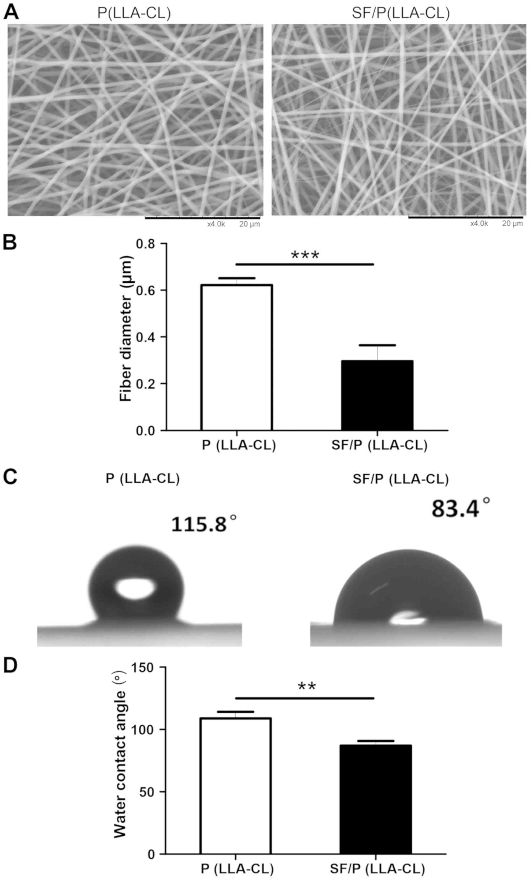
All scaffolds used in the present study were 60–100
µm in thickness and exhibited uniform interconnected pore
structure. Furthermore, SEM observation demonstrated that mixing SF
with P(LLA-CL) changed the morphology of the electrospun scaffolds.
Notably, the diameter of fibers in P(LLA-CL) electrospun scaffolds
(0.6217±0.03007 µm) were significantly thicker when compared with
those blended with SF (0.2966±0.06811 µm; P<0.001; Fig. 1A and B). To evaluate the surface
properties between the two fibrous scaffolds, the wettability was
measured using water contact angle analysis (Fig. 1C). The results indicated that the
addition of SF enhanced the hydrophilicity of the electrospun
sheets when compared with the pure P(LLA-CL) scaffold alone. As
indicated in Fig. 1D, the static
water contact angle was significantly decreased with the addition
of SF content [108.8±5.296° of the P(LLA-CL) group compared with
86.90±3.955° of the SF/P(LLA-CL) group; P<0.01].
Mechanical properties of electrospun
scaffolds
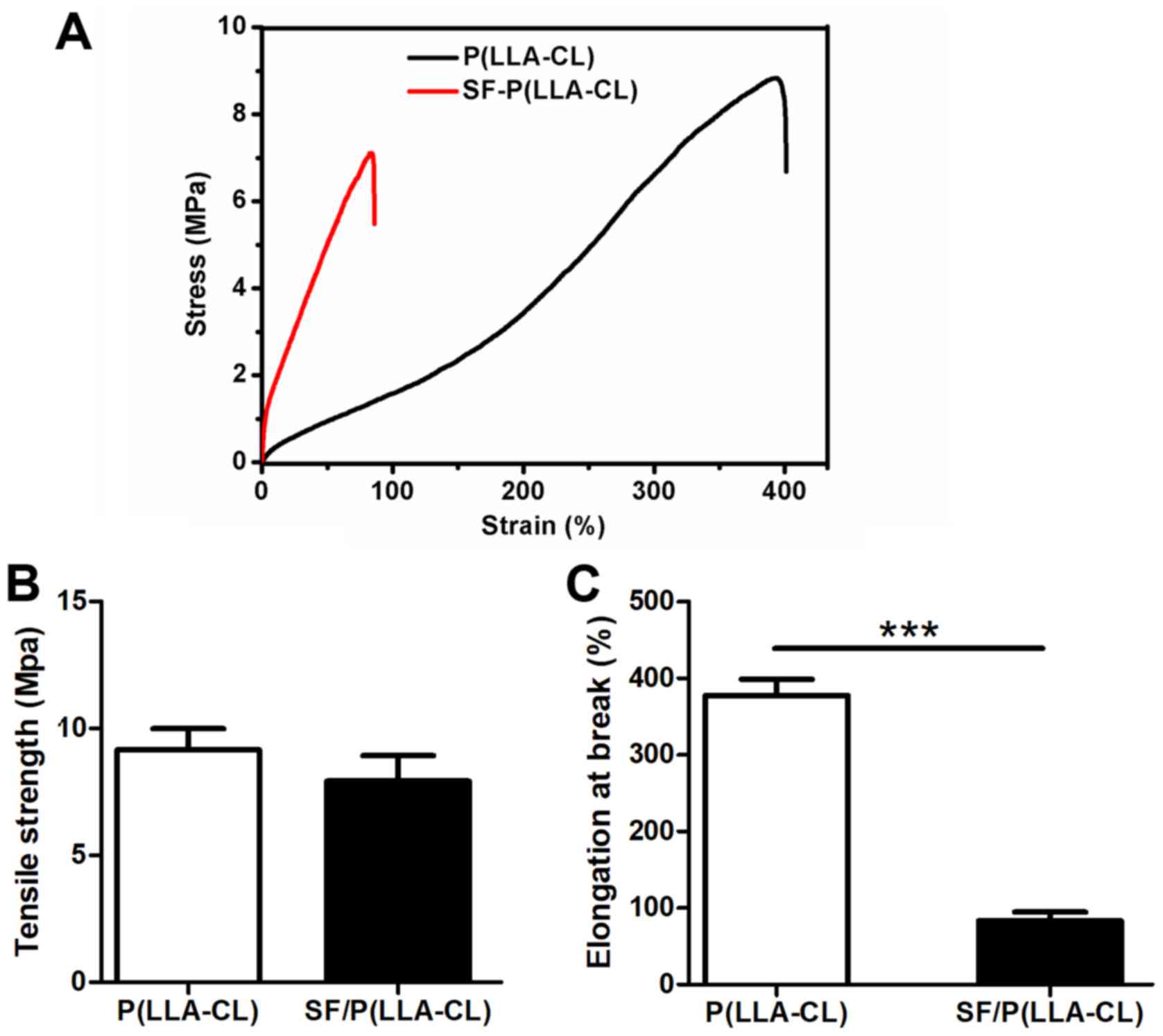
The mechanical properties of P(LLA-CL) and
SF/P(LLA-CL) scaffolds were measured using typical tensile
stress-strain curves (Fig. 2). The
results indicated that the P(LLA-CL) scaffolds transformed from
flexible to brittle when blended with SF. However, no significant
differences were observed between P(LLA-CL) and SF/P(LLA-CL)
electrospun scaffolds, even though tensile strength values of
P(LLA-CL) electrospun scaffolds were increased (9.17±0.82 MPa)
compared with SF/P(LLA-CL) electrospun scaffolds (7.93±1.00 MPa).
In addition, the elongation at break of P(LLA-CL) was significantly
longer (377.90±21.19%) compared with SF/P(LLA-CL) (83.15±11.47%;
P<0.001).
MI increases the expression of
c-kit+ cells in the BM
The stem cell surface markers c-kit, CD34 and sca-1
were evaluated in the present study (Fig. 3A). Notably, the c-kit expression
levels in BM mononuclear cells were significantly upregulated in
response to acute ischemic injury (7.53±0.69%) compared with the
sham group (1.65±0.37%; P<0.001; Fig.
3B). Conversely, the CD34 and sca-1 expression levels were not
significantly altered (Fig. 3C and
D).
 | Figure 3.Flow cytometric analysis and
proliferation assay. (A) Flow cytometric analysis plots of
c-kit+, CD34+ and sca-1+ cells
isolated from MI and sham mice. (B-D) Expression analysis of
c-kit+, CD34+ and sca-1+ in BM
cells. ***P<0.001 as indicated (n=6 for each group). (E)
CellTiter 96® AQueous One Solution reagent was used to
assess c-kit+ BM cells seeded on P(LLA-CL) or
SF/P(LLA-CL) or those cultured without scaffolds at indicated time
points. *P<0.05 (n=6 for each group). SF, silk fibroin;
P(LLA-CL), poly(L-lactic acid-co-ε-caprolactone); MI, myocardial
infarction; BM, bone marrow; CD, cluster of differentiation; CD117,
c-kit; sca-1, stem cell antigen-1; OD, optical density. |
Effect of SF/P(LLA-CL) nanofibrous
scaffolds on the proliferation of c-kit+ BM cells
To evaluate the effect of P(LLA-CL) and SF/P(LLA-CL)
electrospun scaffolds on cell proliferation, c-kit+ BM
cells were seeded on different scaffolds and the cell proliferation
ability was measured using the CellTiter 96® AQueous One
Solution reagent on day 0, 1, 3, 5 and 7. The SF/P(LLA-CL)
electrospun scaffolds were superior in promoting cell proliferation
compared with P(LLA-CL) electrospun scaffolds (P<0.05; day 5 and
7; Fig. 3E). Thus, the SF/P(LLA-CL)
electrospun scaffolds were more suitable for promoting
proliferation in c-kit+ BM cells.
SF/P(LLA-CL) electrospun scaffolds
seeded with c-kit+ BM cells attenuate myocardial damage
and improve survival post-MI
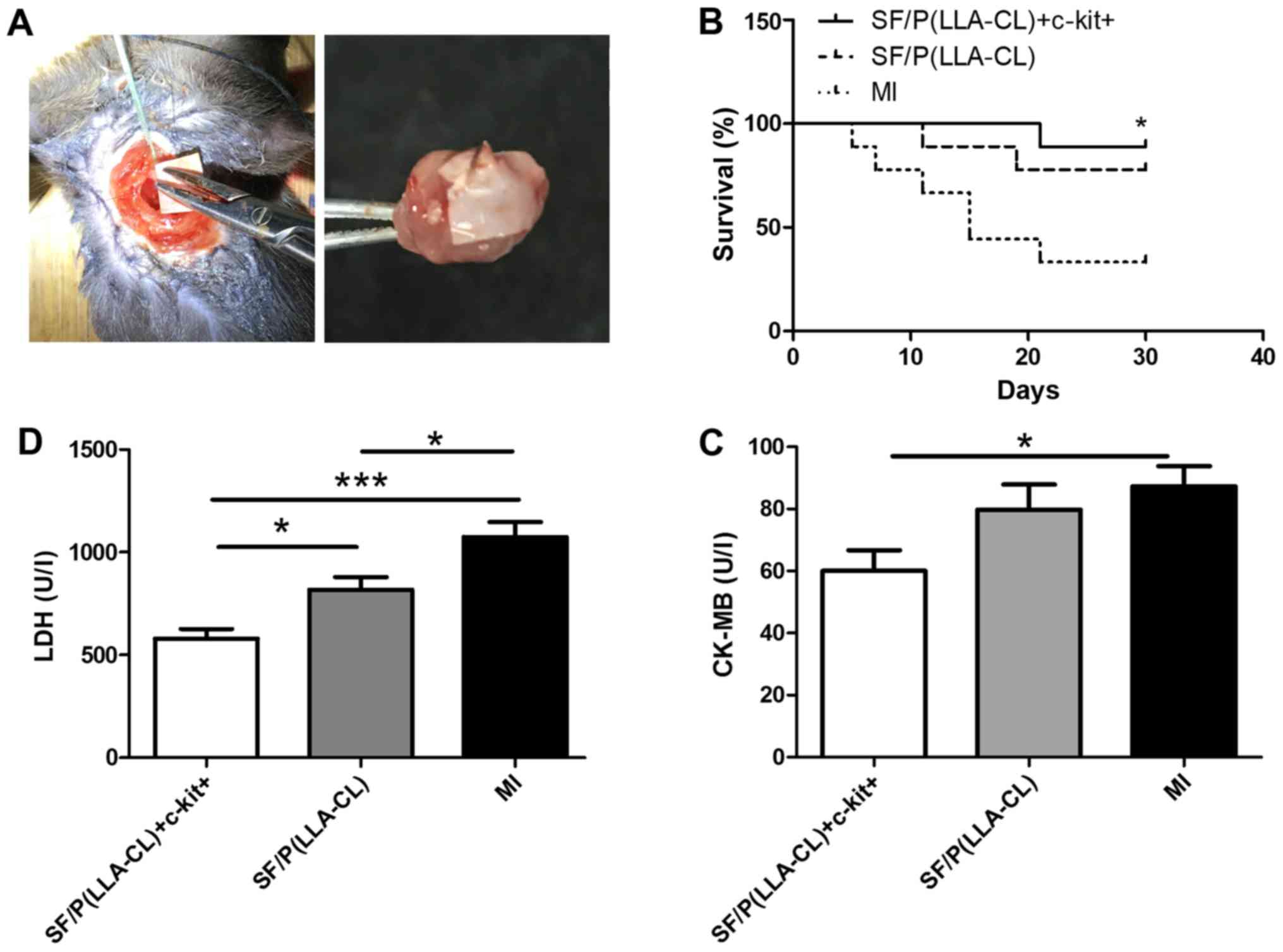
To further assess the role of SF/P(LLA-CL)
electrospun scaffolds in cardiac repair, myocardial damage was
evaluated by measuring the release of several markers in the serum,
including CK-MB and LDH (Fig. 4).
Analysis revealed that SF/P(LLA-CL) + c-kit+ treatment
significantly decreased the serum levels of CK-MB (P<0.05) and
LDH (P<0.001) compared with the MI group. Furthermore, the
survival rates were evaluated following MI induction for up to 30
days in SF/P(LLA-CL) + c-kit+, pure SF/P(LLA-CL) and MI
groups (Fig. 4). The survival rate
of the surgery was consistent with previous research (24–26). The
results demonstrated that the SF/P(LLA-CL) + c-kit+
group exhibited significantly improved survival at 30 days post-MI
compared with the MI group (P<0.05). However, no significant
differences were observed between the pure SF/P(LLA-CL) group and
MI group.
SF/P(LLA-CL) electrospun scaffolds
enhance cardiac repair in vivo
SF/P(LLA-CL) electrospun scaffolds were superior to
P(LLA-CL) scaffolds in terms of promoting cell proliferation in
vitro. To further assess the role of SF/P(LLA-CL) electrospun
scaffolds in cardiac repair, sorted cells were immediately seeded
with SF/P(LLA-CL) electrospun scaffolds and cultured for 5 days
prior to transplantation. Experimental mice were divided into four
groups: i) Sham group; ii) SF/P(LLA-CL) + c-kit+ group;
iii) pure SF/P(LLA-CL); and iv) MI group. Images of the surgical
procedure and a holistic view of the MI heart with scaffold
implantation are presented in Fig.
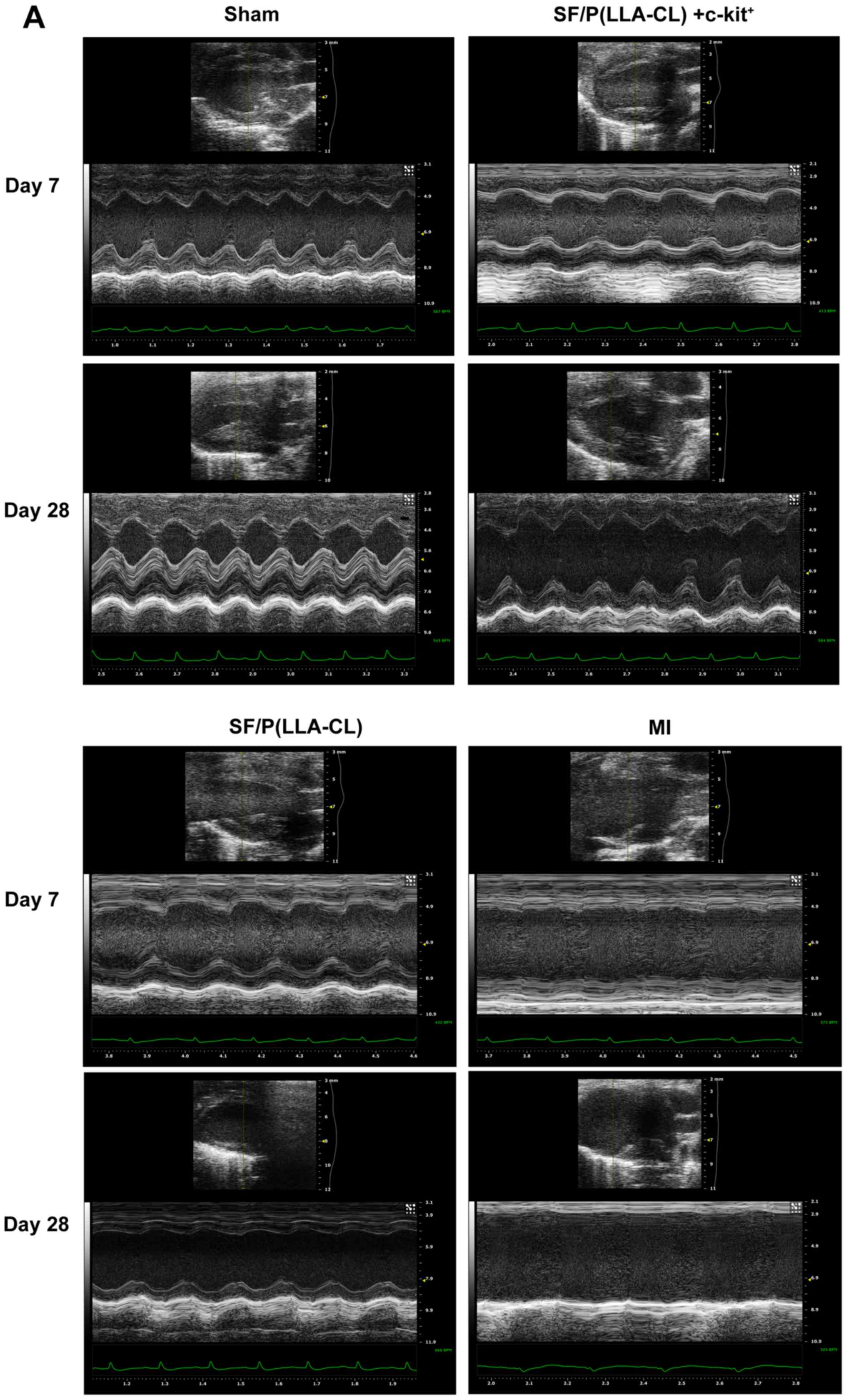
5A. Echocardiography examinations were performed on days 7 and
28 following transplantation. At 7 days post-transplantation, even
though the SF/P(LLA-CL) + c-kit+ electrospun scaffolds group
exhibited significantly improved EF values (Fig. 5B), no significant differences were
observed between the SF/P(LLA-CL) + c-kit+ electrospun scaffolds
group and SF/P(LLA-CL) electrospun scaffolds group regarding EDVs.
Furthermore, the SF/P(LLA-CL) + c-kit+ electrospun
scaffolds group exhibited significantly improved EDVs (P<0.01)
and ESVs (P<0.001) compared with the MI group; (Fig. 5C and D). At 28 days, the reductions
in EDV and ESV were associated with increased EF values in the
SF/P(LLA-CL) + c-kit+ electrospun scaffolds transplantation group.
Mice with SF/P(LLA-CL) + c-kit+ electrospun scaffolds
had significantly decreased EDVs (P<0.01) and ESVs (P<0.001)
compared with the MI group. However, no significant differences
were present between the SF/P(LLA-CL) + c-kit+
electrospun scaffolds and SF/P(LLA-CL) electrospun scaffolds groups
regarding EDV and ESV (Fig. 5B, C and
D). These findings suggested that SF/P(LLA-CL) +
c-kit+ electrospun scaffold transplantation moderately
attenuated post-MI left ventricular function. Pure SF/P(LLA-CL)
electrospun scaffolds were not as effective as SF/P(LLA-CL) +
c-kit+ electrospun scaffolds in protecting cardiac
function post-MI.
Examination of infarct size
All electrospun sheets covered the epicardium of the
left ventricles when the hearts were collected (Fig. 4A). Notably, SF/P(LLA-CL) +
c-kit+ (P<0.001) and pure SF/P(LLA-CL) (P<0.05)
electrospun scaffold transplantation significantly reduced the
infarct size compared with untreated MI (Fig. 6). Furthermore, infarct size in mice
treated with SF/P(LLA-CL) + c-kit+ were reduced more
compared with the pure SF/P(LLA-CL) treated-mice (P<0.05). Thus,
SF/P(LLA-CL) + c-kit+ electrospun scaffolds can reduce
infarct volume to a greater extent compared with pure SF/P(LLA-CL)
scaffolds following transplantation.
Discussion
In the present study, cardiac SF/P(LLA-CL)
electrospun nanofibrous sheets were developed, which served a
protective role in cardiac performance when patched onto infarcted
hearts in mice. SF/P(LLA-CL) electrospun scaffolds and P(LLA-CL)
scaffolds were compared in terms of their mechanical and biotic
characteristics in vitro. SF/P(LLA-CL) + c-kit+
scaffolds and pure SF/P(LLA-CL) scaffolds in cardiac repair were
also examined following MI in vivo. The results demonstrated
that SF/P(LLA-CL) electrospun scaffolds were superior to the
P(LLA-CL) electrospun scaffolds in improving c-kit+ BM
cell survival and proliferation in vitro. Furthermore,
SF/P(LLA-CL) + c-kit+ electrospun scaffolds prevented
cardiac rupture, ameliorated myocardial damage and left ventricular
remodeling and reduced infarct size post-MI induction in
vivo. In conclusion, the data suggested that transplantation of
SF/P(LLA-CL) + c-kit+ electrospun scaffolds may be
useful in MI treatment.
BM cells are easily accessible and grow rapidly
in vitro. Thus, cell therapy using BM-derived stem cells has
been considered in cardiac tissue engineering (1,27).
Cumulative animal studies have revealed that c-kit+
BM-derived stem cell transplantation can improve heart function
following MI (28). However, its
therapeutic efficiency remains low due to poor cell survival
(2,29). Thus, it is critical to establish a
method to improve cell retention and survival rate. In the present
study, it was observed that the c-kit+ BM subpopulation
was significantly increased in response to MI in mice. This
indicated that the c-kit+ BM subpopulation may serve a
role post-MI. Additionally, several studies have revealed that the
nanofibrous architecture of scaffolds may be beneficial to the
proliferation of engrafted cells (30). Therefore, the engraftment of a
c-kit+ BM cell population in P(LLA-CL) and SF/P(LLA-CL)
scaffolds was investigated and it was analyzed whether these
scaffolds enhanced the engrafted c-kit+ cell survival
and proliferation in the present study.
Previous studies have used P(LLA-CL) as a scaffold
in tissue engineering, primarily due to its excellent mechanical
properties and good biocompatibility (6,31).
However, P(LLA-CL) polymers have certain disadvantages, including
inertness, low bioactive properties and hydrophobicity, which
result in limited cell-cell interactions (21,32,33). SF,
a natural protein, has been widely used in tissue engineering due
to its unique properties, including good biocompatibility, good
water vapor permeability and low cost. However, the brittleness of
SF may hinder its use for further study of tissue regeneration
(34,35). In the present study, SF was blended
with P(LLA-CL) to develop a hybrid electrospun nanofibrous scaffold
in order to combine the advantages of SF and P(LLA-CL). It was
revealed that P(LLA-CL) scaffolds transformed from flexible to
brittle when blended with SF. Thus, to maintain the flexibility of
the electrospun scaffolds used in the present study, 25:75 ratios
of SF and P(LLA-CL), respectively, were used. SEM observation
demonstrated that mixing SF with P(LLA-CL) changed the morphology
of electrospun scaffolds, and may therefore increase the cell
affinity. Furthermore, wettability results indicated that the
addition of SF provided electrospun scaffolds with more prominent
hydrophilic properties compared with pure P(LLA-CL).
Good mechanical properties of a scaffold are helpful
for successful cell transplantation (36). In the present study, it was observed
that BM c-kit+ cells exhibited improved proliferation on
SF/P(LLA-CL) and P(LLA-CL) electrospun sheets compared with the
c-kit+ only group. Notably, cell proliferation was the
greatest in the SF/P(LLA-CL) group. The present data demonstrated
that the SF/P(LLA-CL) nanofibrous scaffolds exhibited improved
cytocompatibility, which is a requirement for any biomaterial used
for treatment. In summary, it was revealed that blending SF (25%)
with P(LLA-CL) (75%) resulted in scaffolds with favorable
mechanical and biological properties. However, the mechanisms by
which SF/P(LLA-CL) exerts its effects in stem cell proliferation
are not clear and require further investigation. Thus, SF/P(LLA-CL)
nanofibrous scaffolds were assessed in vivo in the present
study. Providing mechanical support to the infarcted left ventricle
area is a unique superiority of electrospun cardiac scaffolds
(37). It can prevent paradoxical
movement of aneurysms in order to synchronize ventricle movement
and consequently prevent heart failure progression (38,39).
Notably, good flexibility of the cardiac scaffold is required to
withstand repeated left ventricular contractions in vivo.
Previous evidence has indicated that one of the biggest drawbacks
of SF is its brittleness (21,23). In
the present study, 25% SF was blended with 75% P(LLA-CL) to develop
a hybrid electrospun nanofibrous scaffold in order to maintain the
advantages of SF, including its good biocompatibility, water vapor
permeability and cell affinity, in addition to preserving the
flexibility of P(LLA-CL). c-kit+ BM cells have been
demonstrated to support myocardial regeneration in vitro and
in vivo (40). Consequently,
c-kit+ cells were seeded on SF/P(LLA-CL) nanofibrous
scaffolds prior to transplantation in the present study. A key
finding in the present study was that engrafting the SF/P(LLA-CL)
nanofibrous scaffold seeded with c-kit+ BM cells at the
time of coronary ligation attenuated structural remodeling and
improved cardiac function compared with cell-free scaffolds
following MI at days 7 and 28 post-transplantation. In the late
phase of acute myocardial infraction, left ventricle remodelling is
secondary to architectural rearrangements of the surviving
myocardium involving myocyte hypertrophy, interstitial fibrosis,
thinning and dilation of the infarcted myocardial wall (41). Thus, the 28th day was chosen as the
end of the present in vivo experiment due to its match of
the late phase of acute MI in humans (42–44). In
addition, the present study demonstrated that SF/P(LLA-CL)
nanofibrous scaffolds seeded with c-kit+ BM cells
prevented cardiac rupture and attenuated myocardial damage in the
early post-MI phase. Thus, the ECM-mimicking hybrid electrospun
scaffolds seeded with c-kit+ cells may be considered a
viable MI treatment option. Although the present results are
promising, there are still many questions that require answering.
For example, further experiments should be performed to investigate
the regeneration of myocardium tissue in response to
transplantation of the SF/P(LLA-CL) electrospun scaffold. In
addition, more differentiation potential seeds of cells should be
evaluated in cardiac tissue engineering.
In conclusion, in the present study, a new
electrospun nanofibrous scaffold was developed. In vitro, it
was demonstrated that cultured c-kit+ BM cells with
SF/P(LLA-CL) electrospun scaffolds are superior in terms of their
proliferative capacity compared with P(LLA-CL) electrospun
scaffolds or c-kit+ BM cells alone. Furthermore, the
present study provided evidence that the SF/P(LLA-CL) electrospun
scaffold serves as an ECM to support c-kit+ BM cells
survival and retention in the early phase of MI. In the late phase
post-MI, SF/P(LLA-CL) electrospun scaffold are able to support the
infarcted left ventricular area and prevent paradoxical movement of
aneurysms in order to synchronize ventricle movement, ultimately
improving overall heart function and reducing infarct size in a MI
mouse model. The present findings suggest that SF/P(LLA-CL)
electrospun scaffolds may support the brittle and thinning infarct
wall, improve c-kit+ BM cell survival and support
cardiomyocyte performance following MI. Thus, the use of
SF/P(LLA-CL) electrospun sheets may be a novel approach to MI
treatment.
Acknowledgements
Not applicable.
Funding
The present study was supported by grants from the
Medicine-Engineering Cross-Research Foundation of Shanghai Jiao
Tong University (grant no. YG2014ZD02).
Availability of data and materials
The datasets used and/or analyzed during the current
study are available from the corresponding author on reasonable
request.
Authors' contributions
MD performed the histological examination of the
hearts and was a major contributor in writing the manuscript. JG
performed transthoracic electrocardiography. YM, JW and YX analyzed
the data. SX constructed the MI mouse model. XM made the
electrospun scaffolds used in the present study and was a major
contributor in drafting the manuscript.
Ethics approval and consent to
participate
The study was approved by the ethics committee of
Renji Hospital.
Patient consent for publication
Not applicable.
Competing interests
The authors declare that they have no competing
interests.
References
|
1
|
Du M, Schmull S, Zhang W, Wang C, Lian F,
Chen Y and Xue S: c-kit(+)AT2R(+) bone marrow mononuclear cell
subset is a superior subset for cardiac protection after myocardial
infarction. Stem Cells Int. 2016:49135152016. View Article : Google Scholar : PubMed/NCBI
|
|
2
|
Duran JM, Makarewich CA, Sharp TE,
Starosta T, Zhu F, Hoffman NE, Chiba Y, Madesh M, Berretta RM, Kubo
H and Houser SR: Bone-derived stem cells repair the heart after
myocardial infarction through transdifferentiation and paracrine
signaling mechanisms. Circ Res. 113:539–552. 2013. View Article : Google Scholar : PubMed/NCBI
|
|
3
|
Jessup M and Brozena S: Heart failure. N
Engl J Med. 348:2007–2018. 2003. View Article : Google Scholar : PubMed/NCBI
|
|
4
|
Lehtinen M, Pätilä T, Kankuri E, Lauerma
K, Sinisalo J, Laine M, Kupari M, Vento A and Harjula A; Helsinki
BMMC Collaboration, : Intramyocardial bone marrow mononuclear cell
transplantation in ischemic heart failure: Long-term follow-up. J
Heart Lung Transplant. 34:899–905. 2015. View Article : Google Scholar : PubMed/NCBI
|
|
5
|
Gude NA and Sussman MA: Chasing c-Kit
through the heart: Taking a broader view. Pharmacol Res.
127:110–115. 2018. View Article : Google Scholar : PubMed/NCBI
|
|
6
|
Lagostena L, Avitabile D, De Falco E,
Orlandi A, Grassi F, Iachininoto MG, Ragone G, Fucile S, Pompilio
G, Eusebi F, et al: Electrophysiological properties of mouse bone
marrow c-kit+ cells co-cultured onto neonatal cardiac myocytes.
Cardiovasc Res. 66:482–492. 2005. View Article : Google Scholar : PubMed/NCBI
|
|
7
|
Liu J, Wu P, Wang H, Wang Y, Du Y, Cheng
W, Xu Z, Zhou N, Wang L and Yang Z: Necroptosis induced by Ad-HGF
activates endogenous C-Kit+ cardiac stem cells and promotes
cardiomyocyte proliferation and angiogenesis in the infarcted aged
heart. Cell Physiol Biochem. 40:847–860. 2016. View Article : Google Scholar : PubMed/NCBI
|
|
8
|
Awada HK, Long DW, Wang Z, Hwang MP, Kim K
and Wang Y: A single injection of protein-loaded coacervate-gel
significantly improves cardiac function post infarction.
Biomaterials. 125:65–80. 2017. View Article : Google Scholar : PubMed/NCBI
|
|
9
|
Kai D, Prabhakaran MP, Jin G and
Ramakrishna S: Polypyrrole-contained electrospun conductive
nanofibrous membranes for cardiac tissue engineering. J Biomed
Mater Res A. 99:376–385. 2011. View Article : Google Scholar : PubMed/NCBI
|
|
10
|
Godier-Furnemont AF, Martens TP, Koeckert
MS, Wan L, Parks J, Arai K, Zhang G, Hudson B, Homma S and
Vunjak-Novakovic G: Composite scaffold provides a cell delivery
platform for cardiovascular repair. Proc Natl Acad Sci USA.
108:7974–7979. 2011. View Article : Google Scholar : PubMed/NCBI
|
|
11
|
Dhand C, Ong ST, Dwivedi N, Diaz SM,
Venugopal JR, Navaneethan B, Fazil MH, Liu S, Seitz V, Wintermantel
E, et al: Bio-inspired in situ crosslinking and
mineralization of electrospun collagen scaffolds for bone tissue
engineering. Biomaterials. 104:323–338. 2016. View Article : Google Scholar : PubMed/NCBI
|
|
12
|
Mo Y, Guo R, Zhang Y, Xue W and Cheng B:
Controlled dual delivery of angiogenin and curcumin by electrospun
nanofibers for skin regeneration. Tissue Eng Part A. 27:2017.
|
|
13
|
Prabhakaran MP, Kai D, Ghasemi-Mobarakeh L
and Ramakrishna S: Electrospun biocomposite nanofibrous patch for
cardiac tissue engineering. Biomed Mater. 6:1748–6041. 2011.
View Article : Google Scholar
|
|
14
|
Kai D, Prabhakaran MP, Jin G and
Ramakrishna S: Guided orientation of cardiomyocytes on electrospun
aligned nanofibers for cardiac tissue engineering. J Biomed Mater
Res B Appl Biomater. 98:379–386. 2011. View Article : Google Scholar : PubMed/NCBI
|
|
15
|
Wang Z, Lin M, Xie Q, Sun H, Huang Y,
Zhang D, Yu Z, Bi X, Chen J, Wang J, et al: Electrospun silk
fibroin/poly(lactide-co-epsilon-caprolactone) nanofibrous scaffolds
for bone regeneration. Int J Nanomedicine. 11:1483–1500.
2016.PubMed/NCBI
|
|
16
|
Zhang D, Ni N, Chen J, Yao Q, Shen B,
Zhang Y, Zhu M, Wang Z, Ruan J, Wang J, et al: Electrospun SF/PLCL
nanofibrous membrane: A potential scaffold for retinal progenitor
cell proliferation and differentiation. Sci Rep. 5:143262015.
View Article : Google Scholar : PubMed/NCBI
|
|
17
|
Wang CY, Zhang KH, Fan CY, Mo XM, Ruan HJ
and Li FF: Aligned natural-synthetic polyblend nanofibers for
peripheral nerve regeneration. Acta Biomater. 7:634–643. 2011.
View Article : Google Scholar : PubMed/NCBI
|
|
18
|
Chen J, Yan C, Zhu M, Yao Q, Shao C, Lu W,
Wang J, Mo X, Gu P, Fu Y and Fan X: Electrospun nanofibrous
SF/P(LLA-CL) membrane: A potential substratum for endothelial
keratoplasty. Int J Nanomedicine. 10:3337–3350. 2015.PubMed/NCBI
|
|
19
|
Xiang P, Wu KC, Zhu Y, Xiang L, Li C, Chen
DL, Chen F, Xu G, Wang A, Li M and Jin ZB: A novel Bruch's
membrane-mimetic electrospun substrate scaffold for human retinal
pigment epithelium cells. Biomaterials. 35:9777–9788. 2014.
View Article : Google Scholar : PubMed/NCBI
|
|
20
|
Zhang K, Mo X, Huang C, He C and Wang H:
Electrospun scaffolds from silk fibroin and their cellular
compatibility. J Biomed Mater Res A. 93:976–983. 2010.PubMed/NCBI
|
|
21
|
Kuihua Z, Chunyang W, Cunyi F and Xiumei
M: Aligned SF/P(LLA-CL)-blended nanofibers encapsulating nerve
growth factor for peripheral nerve regeneration. J Biomed Mater Res
A. 102:2680–2691. 2014. View Article : Google Scholar : PubMed/NCBI
|
|
22
|
Zhang K, Wang H, Huang C, Su Y, Mo X and
Ikada Y: Fabrication of silk fibroin blended P(LLA-CL) nanofibrous
scaffolds for tissue engineering. J Biomed Mater Res A. 93:984–993.
2010.PubMed/NCBI
|
|
23
|
Aznar-Cervantes S, Pagán A, Martínez JG,
Bernabeu-Esclapez A, Otero TF, Meseguer-Olmo L, Paredes JI and
Cenis JL: Electrospun silk fibroin scaffolds coated with reduced
graphene promote neurite outgrowth of PC-12 cells under electrical
stimulation. Mater Sci Eng C Mater Biol Appl. 79:315–325. 2017.
View Article : Google Scholar : PubMed/NCBI
|
|
24
|
Du J, Zhang L, Wang Z, Yano N, Zhao YT,
Wei L, Dubielecka-Szczerba P, Liu PY, Zhuang S, Qin G and Zhao TC:
Exendin-4 induces myocardial protection through MKK3 and Akt-1 in
infarcted hearts. Am J Physiol Cell Physiol. 310:C270–C283. 2016.
View Article : Google Scholar : PubMed/NCBI
|
|
25
|
Brooks AC, DeMartino AM, Brainard RE,
Brittian KR, Bhatnagar A and Jones SP: Induction of activating
transcription factor 3 limits survival following infarct-induced
heart failure in mice. Am J Physiol Heart Circ Physiol.
309:H1326–H1335. 2015. View Article : Google Scholar : PubMed/NCBI
|
|
26
|
Nishikido T, Oyama J, Shiraki A, Komoda H
and Node K: Deletion of apoptosis inhibitor of macrophage
(AIM)/CD5L attenuates the inflammatory response and infarct size in
acute myocardial infarction. J Am Heart Assoc. 5:0028632016.
View Article : Google Scholar
|
|
27
|
Hu X, Huang X, Yang Q, Wang L, Sun J, Zhan
H, Lin J, Pu Z, Jiang J, Sun Y, et al: Safety and efficacy of
intracoronary hypoxia-preconditioned bone marrow mononuclear cell
administration for acute myocardial infarction patients: The
CHINA-AMI randomized controlled trial. Int J Cardiol. 184:446–451.
2015. View Article : Google Scholar : PubMed/NCBI
|
|
28
|
Bao L, Meng Q, Li Y, Deng S, Yu Z, Liu Z,
Zhang L and Fan H: C-Kit positive cardiac stem cells and bone
marrow-derived mesenchymal stem cells synergistically enhance
angiogenesis and improve cardiac function after myocardial
infarction in a paracrine manner. J Card Fail. 23:403–415. 2017.
View Article : Google Scholar : PubMed/NCBI
|
|
29
|
Fazel S, Cimini M, Chen L, Li S,
Angoulvant D, Fedak P, Verma S, Weisel RD, Keating A and Li RK:
Cardioprotective c-kit+ cells are from the bone marrow and regulate
the myocardial balance of angiogenic cytokines. J Clin Invest.
116:1865–1877. 2006. View Article : Google Scholar : PubMed/NCBI
|
|
30
|
Yao Q, Cosme JG, Xu T, Miszuk JM, Picciani
PH, Fong H and Sun H: Three dimensional electrospun PCL/PLA blend
nanofibrous scaffolds with significantly improved stem cells
osteogenic differentiation and cranial bone formation.
Biomaterials. 115:115–127. 2017. View Article : Google Scholar : PubMed/NCBI
|
|
31
|
Li H, Wu T, Zheng Y, El-Hamshary H,
Al-Deyab SS and Mo X: Fabrication and characterization of
Mg/P(LLA-CL)-blended nanofiber scaffold. J Biomater Sci Polym Ed.
25:1013–1027. 2014. View Article : Google Scholar : PubMed/NCBI
|
|
32
|
Zhang K, Fu Q, Yoo J, Chen X, Chandra P,
Mo X, Song L, Atala A and Zhao W: 3D bioprinting of urethra with
PCL/PLCL blend and dual autologous cells in fibrin hydrogel: An in
vitro evaluation of biomimetic mechanical property and cell growth
environment. Acta Biomater. 50:154–164. 2017. View Article : Google Scholar : PubMed/NCBI
|
|
33
|
Fluke LM, Restrepo RD, Patel S, Hoagland
BD, Krevetski LM and Stephenson JT: Strength and histology of a
nanofiber scaffold in rats. J Surg Res. 205:432–439. 2016.
View Article : Google Scholar : PubMed/NCBI
|
|
34
|
Johari N, Madaah Hosseini HR and
Samadikuchaksaraei A: Optimized composition of nanocomposite
scaffolds formed from silk fibroin and nano-TiO2 for bone tissue
engineering. Mater Sci Eng C Mater Biol Appl. 79:783–792. 2017.
View Article : Google Scholar : PubMed/NCBI
|
|
35
|
Naskar D, Ghosh AK, Mandal M, Das P, Nandi
SK and Kundu SC: Dual growth factor loaded nonmulberry silk
fibroin/carbon nanofiber composite 3D scaffolds for in vitro and in
vivo bone regeneration. Biomaterials. 136:67–85. 2017. View Article : Google Scholar : PubMed/NCBI
|
|
36
|
Yin A, Luo R, Li J, Mo X, Wang Y and Zhang
X: Coaxial electrospinning multicomponent functional
controlled-release vascular graft: Optimization of graft
properties. Colloids Surf B Biointerfaces. 152:432–439. 2017.
View Article : Google Scholar : PubMed/NCBI
|
|
37
|
Liu Y, Xu Y, Wang Z, Wen D, Zhang W,
Schmull S, Li H, Chen Y and Xue S: Electrospun nanofibrous sheets
of collagen/elastin/polycaprolactone improve cardiac repair after
myocardial infarction. Am J Transl Res. 8:1678–1694.
2016.PubMed/NCBI
|
|
38
|
Kai D, Wang QL, Wang HJ, Prabhakaran MP,
Zhang Y, Tan YZ and Ramakrishna S: Stem cell-loaded nanofibrous
patch promotes the regeneration of infarcted myocardium with
functional improvement in rat model. Acta Biomater. 10:2727–2738.
2014. View Article : Google Scholar : PubMed/NCBI
|
|
39
|
Stevens KR, Kreutziger KL, Dupras SK,
Korte FS, Regnier M, Muskheli V, Nourse MB, Bendixen K, Reinecke H
and Murry CE: Physiological function and transplantation of
scaffold-free and vascularized human cardiac muscle tissue. Proc
Natl Acad Sci USA. 106:16568–16573. 2009. View Article : Google Scholar : PubMed/NCBI
|
|
40
|
Beltrami AP, Barlucchi L, Torella D, Baker
M, Limana F, Chimenti S, Kasahara H, Rota M, Musso E, Urbanek K, et
al: Adult cardiac stem cells are multipotent and support myocardial
regeneration. Cell. 114:763–776. 2003. View Article : Google Scholar : PubMed/NCBI
|
|
41
|
Hao H, Hu S, Chen H, Bu D, Zhu L, Xu C,
Chu F, Huo X, Tang Y, Sun X, et al: Loss of endothelial CXCR7
impairs vascular homeostasis and cardiac remodeling after
myocardial infarction: Implications for cardiovascular drug
discovery. Circulation. 135:1253–1264. 2017. View Article : Google Scholar : PubMed/NCBI
|
|
42
|
Pinet F, Cuvelliez M, Kelder T, Amouyel P,
Radonjic M and Bauters C: Integrative network analysis reveals
time-dependent molecular events underlying left ventricular
remodeling in post-myocardial infarction patients. Biochim Biophys
Acta Mol Basis Dis. 1863:1445–1453. 2017. View Article : Google Scholar : PubMed/NCBI
|
|
43
|
Olivier A, Girerd N, Michel JB,
Ketelslegers JM, Fay R, Vincent J, Bramlage P, Pitt B, Zannad F and
Rossignol P; EPHESUS Investigators, : Combined baseline and
one-month changes in big endothelin-1 and brain natriuretic peptide
plasma concentrations predict clinical outcomes in patients with
left ventricular dysfunction after acute myocardial infarction:
Insights from the Eplerenone Post-Acute Myocardial Infarction Heart
Failure Efficacy and Survival Study (EPHESUS) study. Int J Cardiol.
241:344–350. 2017. View Article : Google Scholar : PubMed/NCBI
|
|
44
|
Frey A, Saxon VM, Popp S, Lehmann M,
Mathes D, Pachel C, Hofmann U, Ertl G, Lesch KP and Frantz S: Early
citalopram treatment increases mortality due to left ventricular
rupture in mice after myocardial infarction. J Mol Cell Cardiol.
98:28–36. 2016. View Article : Google Scholar : PubMed/NCBI
|