Introduction
Atherosclerotic disease is systemic and, with the
same pathogenesis, atherosclerosis of the coronary, carotid and
cerebral arteries usually exists at the same time with clinical
relevance (1–4). Cardiovascular and cerebrovascular
diseases are the primary cause of mortality in China, particularly
in the developed area (5).
Screening, diagnosis and evaluation are the most important means to
reduce the threat of mortality in patients with atherosclerotic
disease (5). Compared with gold
standard digital subtraction angiography, computed tomography
angiography (CTA) is a non-invasive and high-powered tool that can
assess atherosclerosis and provide accurate information on arterial
anatomy (2,6), via CTA (2), digital subtraction angiography
(3), ultrasound (4) and magnetic resonance angiography
(7).
However, the CT scan protocol is designed for
cardiovascular and cerebrovascular imaging, and therefore
considerations must be made in regard to the radiation dose, iodine
load and image quality (2). Due to
the risks of radiological examination, the radiologists and
engineers must consider the physical characteristics of CT
scanners. The 128-slice dual-source CT (DSCT) has ~2 vertical tube
detector systems, a rotation time of up to 280 msec and a
table-moving speed of up to 45.8 cm/sec. These physical
characteristics can significantly reduce the radiation dose and the
amount of contrast agent required (8,9). High
temporal resolution can also maintain or improve image quality for
CT coronary angiography (CTCA) (8,9). In
previous studies, cardiovascular and cerebrovascular imaging has
utilized the 128-slice DSCT, obtaining a reliable image quality
with a low-dose length product (DLP; 224.83–256.30 mGy·cm) and
contrast agent (60–80 ml) (10,11).
However, these studies were conservative with a high tube voltage
(100 or 120 kV) and high concentration contrast agent (370 mg/ml),
and did not utilize the advantages of 128-slice DSCT, particularly
by combining the iterative algorithm and ultra-low tube voltage to
reduce the radiation dose for the patients of standard body type.
Cardiovascular and cerebrovascular imaging was also performed with
256-slice CT and first-generation DSCT, with a higher radiation
dose and amount of the contrast agent when compared with those of
the second-generation DSCT (12,13).
In addition, several studies have shown that CTCA
using a high-pitch scanner can maintain the sensitivity and
negative predictive value, with a radiation dose <0.2 mSv and
the contrast agent (370 mgI/ml) as low as 30 ml (14–17). By
combining a low voltage setting and the iterative algorithm, it may
be possible that a low volume and low concentration of the contrast
agent could be used for cardiovascular and cerebrovascular
high-pitch scanning (17,18). As indicated in the present study,
this reliable and safe tool for atherosclerosis assessment should
be considered for use in clinical practice (14,15,17,18).
The aim of the present study was to combine
high-pitch scanning, low tube voltage, iterative algorithm and
low-concentration contrast agent to evaluate an ultra-low dose
(ULD) one-step CTA for coronary, carotid and cerebral arteries.
Materials and methods
Patients
The present prospective study was approved by the
Ethics Committee of Inner Mongolia Medical University Affiliated
Hospital (Inner Mongolia, China), and all recruited patients signed
informed consent forms. A total of 80 patients who were suspected
to have cardiovascular and cerebrovascular diseases were enrolled
in the present study between January 2016 and May 2017. The
inclusion criteria were as follows: i) Heart rate (HR) ≤70 beats
per minute (bpm), or HR could be reduced to <70 bpm by using
oral metoprolol; ii) HR variability (HRv) ≤5 bpm; iii) body weight
(BW) ≤70 kg; and iv) body mass index (BMI) ≤28 kg/m2.
The exclusion criteria included: i) poor breathing; ii)
extravasation of the contrast agent; iii) previous iodine allergy;
and iv) underactive renal function (serum creatinine ≥120 µmol/l).
All patients received nitroglycerin sublingually 5 min prior to the
examination.
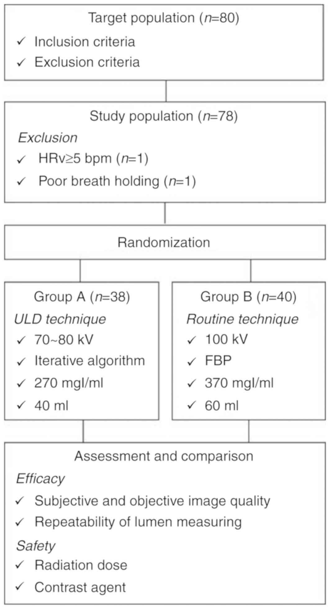
According to the treatment plan, all patients were
randomly divided into two groups (groups A and B), among which 2
patients (all in Group A) were excluded from the study population,
as they had either a HRv of ≥5 bpm (n=1) or poor breathing (n=1).
Finally, a total of 38 patients in group A and 40 patients in group
B were enrolled. The median age was 58 years old (range: 25–80
years old), including 34 males and 44 females. The data of the two
groups is presented in Table I, and
a flow chart of the present study is presented in Fig. 1.
 | Table I.Baseline data. |
Table I.
Baseline data.
| Parameters | Group A (n=38) | Group B (n=40) | Statistics | P-values |
|---|
| Age (years) | 60.47±10.69 | 55.60±9.47 | 2.134a | 0.036 |
| Female (%) | 71.05 | 42.50 | 6.461b | 0.013 |
| Body height
(cm) | 162.13±5.45 | 163.80±6.06 | −1.275a | 0.206 |
| BW (kg) | 61.55±7.46 | 64.20±5.45 | −1.796a | 0.076 |
| Body mass index
(kg/m2) | 23.41±2.58 | 23.97±2.12 | −1.039a | 0.302 |
| Heart rate (beats
per second) | 57.61±7.11 | 60.20±8.49 | −1.459a | 0.149 |
| Scan length
(cm) | 55.30±4.43 | 56.64±3.39 | −1.507a | 0.136 |
| Scan mode | High-pitch
scan | High-pitch
scan | – | – |
| Tube voltage | 70 kVp for BW≤60
kg | 100 kVp | – | – |
|
| 80 kVp for 60
kg<BW≤70 kg |
|
|
|
| Tube current | 320 mAs | 320 mAs | – | – |
| Bolus tracking | Left atrium
trigger | Aortic root
trigger | – | – |
| Contrast agent | 5.0 ml/sec for 40
ml (270 mgI/ml) | 5.0 ml/sec for 60
ml (370 mgI/ml) | – | – |
| Algorithm of image
reconstruction | Iterative algorithm
(SAFIRE 3) | Filter back
projection | – | – |
Scanning and contrast agent injection
technology
The elbow vein was catheterized with an 18-gauge
catheter (BD Intima II 18G; BD Biosciences; Becton, Dickinson and
Company, Franklin Lakes, NJ, USA), and the indwelling needle was
tested with 20 ml physiological saline prior to the contrast agent
injection. All CTA examinations were performed using a
second-generation 128-slice DSCT system (Somatom Definition Flash;
Siemens Healthineers, Erlangen, Bavaria, Germany) with a Stellar
Photon Detector. The acquisition parameters were as follows: i)
Detector collimation was 2×64×0.6 mm; ii) gantry rotation time of
280 msec; iii) a pitch of 3.4 for high-pitch scan; iv) the tube
current for both groups was set to 320 mAs, and the automated tube
current modulation (ATCM; CAREDose 4D; Siemens Healthineers) was
enabled; v) tube voltage was set to 70 or 80 kVp for group A
(experience group) and 100 kVp for group B (control group).
For the bolus-tracking technique, a region of
interest (ROI) was placed on the left atrium and image acquisition
was initiated at 5 sec until the attenuation of left atrium was
>50 HU in group A. In group B, a conventional setting with a ROI
on the aortic root (above 100 HU) and a delay time of 5 sec was
used. The phase to be triggered was set at 55% of the R-R interval.
The low concentration contrast agent (iodixanol injection 270
mgI/ml; GE Healthcare, Chicago, IL, USA) was used for group A. The
high-concentration contrast agent (iopromide 370 mgI/ml; Bayer,
Shanghai, China) was used for group B. Following this, 40 ml of
saline was injected with an injection rate of 5.0 ml/sec for the
two groups. All patients were scanned from 2 cm below the diaphragm
to the top of the head. All CTA technique parameters are presented
in Table I.
Image reconstruction and
processing
The section thickness was reconstructed with 0.75
mm. A medium smooth convolution kernel (I26f) and an iterative
algorithm with SAFIRE 3 (a medium strength level of 3, strength
1–5) were used for group A. Filter back projection with a medium
smooth convolution kernel (B26f) was used for group B. Image
processing and storage were performed with an advanced
three-dimensional workstation (AW version 4.4; GE Healthcare). The
curved planar reconstruction (CPR) technique was applied for the
evaluation of the three types of coronary arteries, and image
quality was evaluated on CPR as well as axial images of vessels and
segments.
Radiation dose and contrast agent
Volume CT dose index (CTDIvol) and DLP
were recorded for each patient. The effective dose (ED) was
calculated with the following formula: ED=DLP × k. In the
present study, the k values for chest and head-neck were
0.014 and 0.0013 mSv/mGy·cm, respectively (19). The injection volume was recorded by
the operators. The mean total iodine content was calculated
according to the product of the concentration and volume.
Image quality
Subjective evaluation of image
quality
Coronary artery
The coronary artery was divided into 15 segments
according to the standards of The American Heart Association
(20): The right coronary artery
(RCA) was divided into segments 1–4, left main (LM) was assigned
segment 5, left anterior descending (LAD) was divided into segments
6–10, and the left circumflex (LCX) was divided into segments
11–15.
Carotid and cerebral arteries
The carotid artery was divided into the following
segments: The common carotid, extracranial internal carotid,
extracranial vertebral and subclavian arteries. The cerebral artery
segments analyzed included the intracranial internal carotid,
intracranial vertebral, anterior cerebral, middle cerebral,
posterior cerebral and basilar arteries.
Rule of image quality scale
Arterial diameters of <1.5 mm were not evaluated.
The image quality of the arteries was assessed by 4 scores:
4=excellent, with no artifacts; 3=good, with mild artifacts and
unrestricted evaluation; 2=assessable, with moderate artifacts, but
still evaluable; and 1=unacceptable (21). All images were evaluated by two
independent readers (A and B, with 5 and 4 years' experience in
cardiovascular diagnosis, respectively) on the AW4.4
workstation.
Objective evaluation of image
quality
Coronary artery
The mean attenuation and standard deviation of the
aortic root, LM, proximal of the LAD, proximal of the LCX, and
proximal of the RCA were measured in all patients by placing ROIs.
The standard deviation was measured as the image noise, and the
mean attenuation of the background was measured in the adipose
tissue around the aortic root.
Carotid and cerebral arteries
Measuring targets on a display length of 1/2 place
were used for various vessels, including the common carotid,
extracranial internal carotid and extracranial vertebral arteries,
as well as the subclavian artery for the carotid artery; the
cerebral arteries included the intracranial internal carotid,
intracranial vertebral, middle cerebral and basilar arteries. The
attenuation of the background for the carotid and cerebral arteries
was measured on the adipose tissue from the right lower jaw space
and temporal fossa.
Rule of density measuring
The artifacts, vessel wall and plaques were avoided
when setting ROIs. The signal-to-noise ratio (SNR) and
contrast-to-noise ratio (CNR) were calculated with the following
formulae: SNR=mean attenuation/noise, and CNR=(mean attenuation of
artery-mean attenuation of adipose tissue)/noise.
Repeatability of lumen measuring for
the experimental group
There were various measuring targets labeled on the
coronary, carotid and cerebral arteries for group A, including the
1/2 display length of the following vessels: LM, LAD, LCX, RCA, and
the common carotid, extracranial internal carotid, extracranial
vertebral, subclavian, intracranial internal carotid, intracranial
vertebral, middle cerebral and basilar arteries. Firstly, according
to the targets, the axial image of the lumen was reconstructed and
then stored by an operator (C). Secondly, the lumen diameter was
measured and recorded by two readers (A and B). Thirdly, the second
measurement was completed by assessor A after 30 days. The vessel
wall and plaques were not measured. Lumen diameter was obtained
using the average value of the maximal and minimal diameters
recorded by the assessors.
Statistical analysis
SPSS version 13 software (SPSS Inc., Chicago, IL,
USA) was used for statistical analysis. Quantitative variables were
expressed as the mean ± standard deviation, and the categorical
variables were expressed as frequencies or percentages. The
independent samples t-test and Mann-Whitney U-test were employed to
compare quantitative variables. The paired t-test was used for
paired data comparisons. Categorical variables were compared with
the χ2 test. Inter-reader variability between the two
readers in regard to image quality scoring was evaluated with
κ statistics. A κ value of <0.20 indicated a poor
agreement; a κ value of 0.21–0.40 indicated a fair
agreement; a κ value of 0.41–0.60 indicated a moderate
agreement; a κ value of 0.61–0.80 indicated a good
agreement; and a κ value of 0.81–1.00 indicated a very good
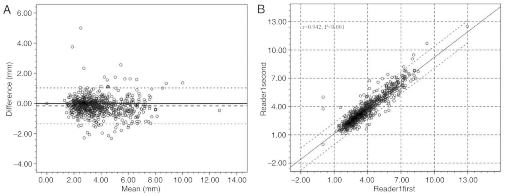
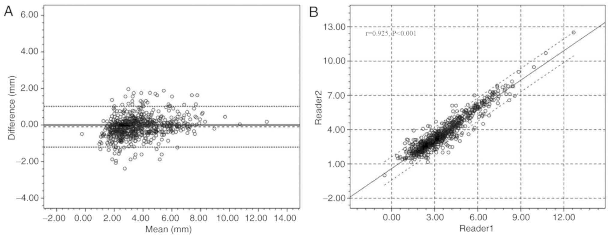
agreement. In the experimental group, measurement correlations
between the intra-reader and inter-reader were demonstrated by a
scatter plot, and measurement consistency between the intra-reader
and inter-reader were indicated using the Bland-Altman plot. The
Bland-Altman plot revealed the average difference, and ±1.96
standard deviation as the consistency of the limits. P<0.05 was
considered to indicate a statistically significant difference.
Results
Patient data
CTA for all patients (n=78) in the two groups was
successfully completed. None of the patients had serious adverse
events. The patient data are presented in Table I.
Radiation dose and contrast agent
The CTDIvol was 1.23±0.41 mGy for Group A
and 3.19±1.05 mGy for Group B. Notably, the difference was
statistically significant (t=−10.811, P<0.001). The DLP was
62.95±21.54 mGy·cm for Group A, and 160.15±15.13 mGy·cm for Group
B, which was decreased by 61% (t=−23.157, P<0.001). The ED was
0.32±0.11 mSv for Group A and 0.79±0.08 mSv for Group B, which
indicated a decrease by 59% in Group A (t=−22.173, P<0.001). The
contrast agent volume was 40±0 and 60±0 ml for Groups A and B,
respectively. The mean total iodine content was 10.8±0 mg for Group
A and 22.2±0 mg for Group B, which indicated a decrease by 51% in
Group A. The comparisons of radiation dose and contrast agent in
each group were presented in Table
II.
| Table II.Comparisons of both groups for
radiation dose and contrast agent. |
Table II.
Comparisons of both groups for
radiation dose and contrast agent.
| Parameters | Group A (n=38) | Group B (n=40) | t-values | P-values |
|---|
| CTDIvol
(mGy) | 1.23±0.41 | 3.19±1.05 | −10.811 | <0.001 |
| DLP (mGy·cm) | 62.95±21.54 | 160.15±15.13 | −23.157 | <0.001 |
| ED (mSv) | 0.32±0.11 | 0.79±0.08 | −22.173 | <0.001 |
| Contrast agent
volume (ml) | 40±0 | 60±0 | – | <0.001 |
| Total iodine
content (mg) | 10.8±0 | 22.2±0 | – | <0.001 |
Image quality
Subjective evaluation of image
quality
There were 2,468 segments evaluated by the two
readers, the frequency of which is presented in Table III. Following the Kappa test,
κ=0.702, P<0.001, in consideration of good agreement between two
readers, any observation was deemed valid. In the present study,
the outcomes recorded by reader A were used.
| Table III.Frequency table for Readers to
evaluate agreement |
Table III.
Frequency table for Readers to
evaluate agreement
|
| Reader A |
|---|
|
|
|
|---|
|
| Readers/Scores | Score 4 | Score 3 | Score 2 | Score 1 | Total |
|---|
| Reader B | Score 4 | 1,889 | 67 | 0 | 0 | 1,956 |
|
| Score 3 | 90 | 263 | 27 | 0 | 380 |
|
| Score 2 | 0 | 65 | 53 | 0 | 118 |
|
| Score 1 | 0 | 0 | 0 | 14 | 14 |
|
| Total | 1,979 | 395 | 80 | 14 | 2,468 |
There were 1,979 segments with a score of 4, 395
with a score of 3, 80 with a score of 2 and 14 with a score of 1.
On a per-segment and -patient basis, differences in assessable
rates between group A and B were not statistically significant
(99.42% vs. 99.45% for per-segment; 84.21% vs. 87.50% for
per-patient; data not shown). Notably, patients who had any segment
of the arteries that was not assessable would be judged as failure.
The mean scores were slightly lower in group A compared with group
B (3.74±0.55 vs. 3.78±0.51, Z=−1.589, P=0.112); however, the
difference was not statistically significant.
In total, 99.19% (489/493) and 99.40% (501/504) of
the coronary arterial segments had assessable image quality for
groups A and B, respectively; the difference was not statistically
significant (χ2=0.167, P=0.683). The mean image scores
for the two groups was similar (3.70±0.58 vs. 3.75±0.53, t=−1.485,
P=0.138). On a per-patient basis, the difference of the proportion
of the two groups was not statistically significant [92.11% (35/38)
vs. 95.00% (38/40), χ2=0.272, P=0.602].
In total, 99.58% (719/722) of the carotid and
cerebral arteries could be assessed for group A and 99.47%
(755/759) for group B; the difference was not statistically
significant (χ2=0.098, P=0.755). Per-patient the
proportion of assessable image quality was 92.11% (35/38) for group
A, and 92.50% (37/40) for group B; the difference was not
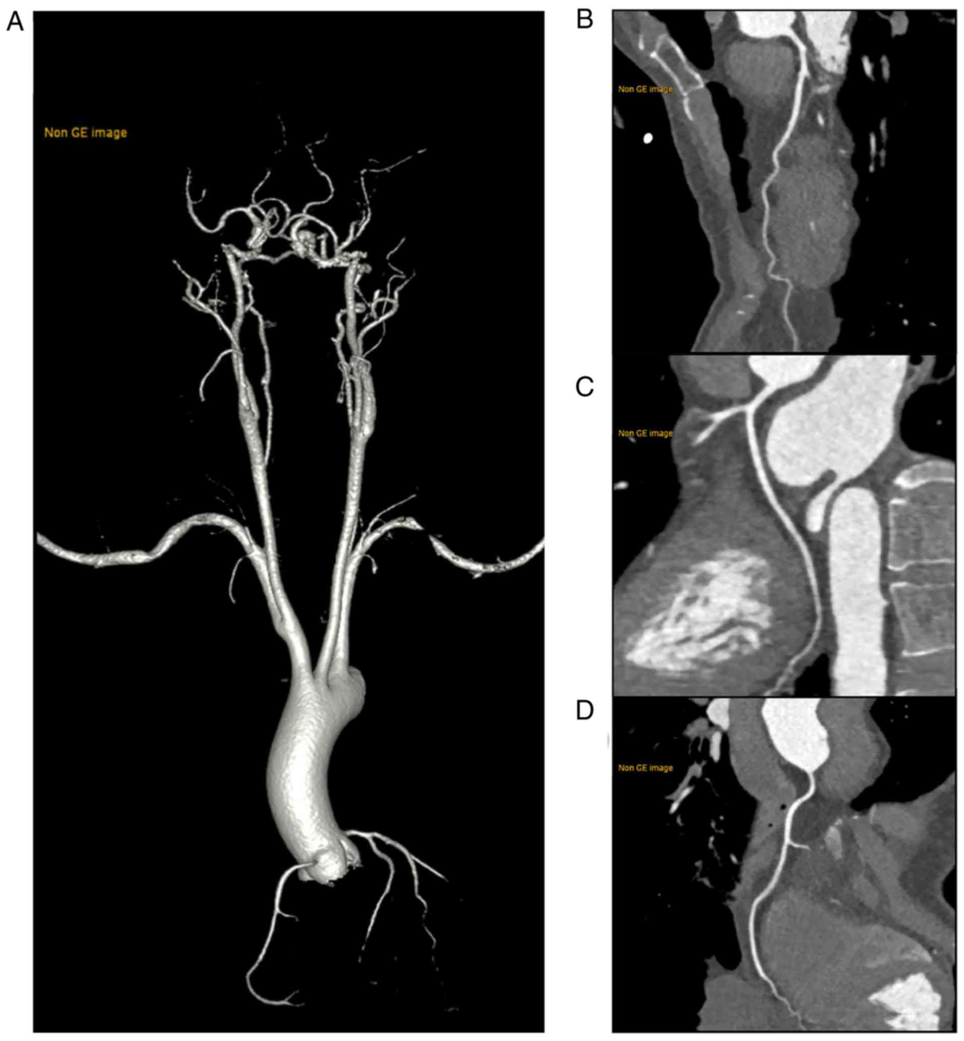
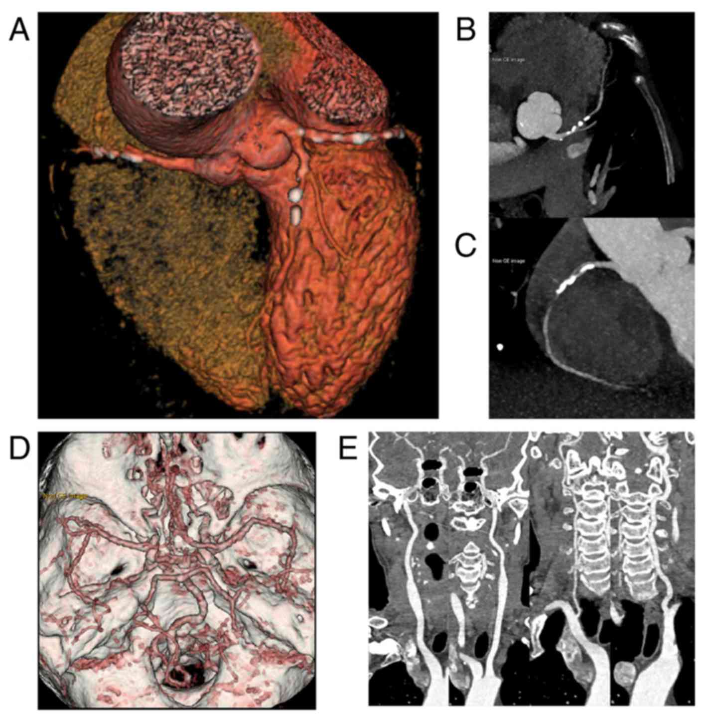
statistically significant (χ2=0.004, P=0.948). Fig. 2 presents high quality images of a
normal patient who received an ultra-low radiation dose and iodine
load. Fig. 3 presents images
obtained by ultra-low dose CTA where coronary artery lesions were
clearly displayed. The images included in Figs. 2 and 3
were all obtained from patients in group A.
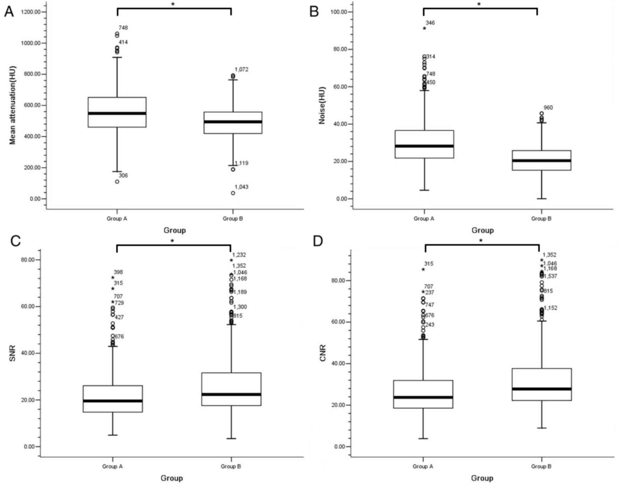
Objective evaluation of image
quality
The mean attenuation and noise of all arteries in
group A was significantly increased compared with that observed in
group B (567.15±145.47 vs. 490.37±107.35 HU; and 30.00±11.93 vs.
20.92±7.68 HU, respectively; P<0.001 for both). The SNR and CNR
were slightly lower in group A compared with in group B (21.58±9.86
vs. 25.54±13.80; and 26.32±11.97 vs. 31.87±17.05, respectively;
P<0.001 for both). The mean attenuation and CNR for each artery
were presented in Fig. 4.
Sub-group analysis
With a median BW of 63.00 kg as the dividing point,
group A was divided into two groups: <63 and ≥63 kg. The mean
attenuation and SNR for the patients <63 kg was higher when
compared with the patients who were ≥63 kg (635.96±118.62 vs.
516.24±104.94; t=3.287, P=0.002 for mean attenuation; 22.51±2.87
vs.19.98±3.07; t=2.579, P=0.014 for SNR). There was no significant
difference between the two sub-groups in regard to noise and CNR
(P=0.293 and P=0.050, respectively). The comparisons of sub-groups
for objective image quality were presented in Table IV.
| Table IV.Comparisons of sub-groups for
objective image quality. |
Table IV.
Comparisons of sub-groups for
objective image quality.
| Parameters | <63 kg | ≥63 kg | t-values | P-values |
|---|
| Attenuation
(HU) | 635.96±118.62 | 516.24±104.94 | 3.287 | 0.002 |
| Noise (HU) | 31.18±5.37 | 29.14±6.08 | 1.069 | 0.292 |
| SNR | 22.51±2.87 | 19.98±3.07 | 2.579 | 0.014 |
| CNR | 27.60±3.07 | 23.39±3.48 | 2.028 | 0.050 |
There were negative correlations between BW, and
attenuation, SNR and CNR, and the r values were −0.535,
−0.404 and −0.322, respectively (all P<0.05). There was no
correlation between BW and noise (r=−0.241, P=0.145).
Per-patient, the mean scores of the coronary artery,
and of the carotid and cerebral arteries in group A were 3.70±0.17
and 3.77±0.11, respectively. The difference was statistically
significant (paired t-test: t=−3.149, P=0.003); however, there was
no significant difference in the assessable rate (489/493 vs.
719/722, χ2=0.002, P=0.962).
Repeatability of lumen measuring for
the experimental group
The difference of the intra-reader was −0.16±0.47 mm
[95% confidence interval (CI): −1.07, 0.77]. There were 96%
(693/722) of the measuring targets inside the 95% limits of
agreement [intraclass correlation coefficient (ICC)=0.954; Fig. 5]. The difference of the inter-reader
was −0.16±0.61 mm (95% CI: −1.35, 1.03). In total, 95% (686/722) of
the measuring targets were inside the 95% limits of agreement
(ICC=0.961; Fig. 6).
Discussion
The aim of the present study was to reduce the
radiation dose and contrast agent to a very low level for
cardiovascular and cerebrovascular CTA. Under the premise of
maintaining image assessment, the present results proved that the
ED and volume of the contrast agent could be limited to <0.5 mSv
and 40 ml, respectively, via a novel scanning protocol in patients
with a BW ≤70 kg and a BMI ≤28 kg/m2. Compared with the
control group, the ED was decreased by 59% and the amount of
contrast agent (total iodine content) was decreased by 51%. The
image assessment and attenuation were not affected by the novel
scanning protocol. The reliability for lumen measuring was also
verified by quantitative evaluation.
Atherosclerosis-associated correlations between
coronary, carotid and cerebral arteries have been reported in
various studies (1–4), and it was necessary to use
cardiovascular and cerebrovascular CTA for preoperative assessment
of the coronary artery via pass-grafting (22). Due to the more accurate anatomical
information and non-invasive operation (2,7),
cardiovascular and cerebrovascular CTA has the potential to replace
other morphological imaging techniques. However, with the potential
risks of medical examinations, the use of CTA has been limited in
clinical practices, particularly in regard to multiple targets of
CT data acquisition, which significantly increase the radiation
exposure and the iodine load (2).
The present study provided a suitable and repeatable strategy to
adapt for clinical requirements. High-pitch scanning has a very
fast table-moving speed, which is due to the double tube-detector
systems with gaps (8,9). In the present study, only ~700 msec was
required for scanning (10,11), which is the primary reason to reduce
the injection time of the contrast agent. However, a low
concentration and volume of the contrast agent were used for CTA,
resulting in a decreasing trend in the time density curve (TDC),
which affected the arterial enhancement. Therefore, based on
previous studies on CTCA (14–18), the
present study used 40 ml low concentration contrast agent for the
designed protocol. Firstly, the ultra-short scanning time was
utilized to provide a sufficient window to identify the peak time.
Secondly, increasing the amount of contrast agent could shift the
TDC to the right and up (23). In
addition, a fast injection rate of contrast agent was used to make
up for the inadequate enhancement. Increasing the injection rate
could also shift the TDC to the left and up. According to the above
adjustments, the present study improved the arterial attenuation
when compared with those observed in previous studies (10–13); the
contrast agent volume was reduced significantly in the present
study.
Coronary arterial motion is intense and complicated
(24); following the high temporal
resolution, DSCT can offer assessable image quality without motion
artifacts within a single cardiac cycle (25). Based on the width of several cardiac
cycles prior to acquisition, high-pitch scanning requires a
suitable acquisition time in order to prospectively predict the
correct trigger exposure (10).
Calculations revealed that the CTA requires two cardiac cycles for
imaging. The scan direction of the conventional CTCA was from head
to foot, including a combination of aortic and coronary artery CTA
(26). Considering the position of
the coronary artery, the present study changed the scanning
direction from foot to head in order to allow part of the coronary
artery to be triggered in the first cardiac cycle. This could
reduce the transient disturbance of electrocardiograms caused by
the fast table-movement, in order to avoid error triggering.
The present study did not include patients with a
high HR (>70 bpm), and Goetti et al (8) demonstrated that high-pitch scanning had
a high failure rate of up to 30% (per patient) in those with a high
HR. The first cases using ULD CTCA were reported in several recent
studies (14,15,17,18),
particularly that of Zhang et al (14,15),
where excellent image quality was obtained. The contrast agent in
the present study was consistent with that used in these studies;
however, a CTA with wide coverage was performed in the present
study. The primary difference was that the present study used
manual bolus tracking by monitoring the left atrium for scanning.
According to our previous study (17) and similar literature (27), it is more accurate in regard to
timing to monitor the left atrium with such a low volume of
contrast agent.
Low-energy X-ray photons can produce a photoelectric
effect and increase the arterial signal (11–18,21,27).
Tube voltage with low kVp settings could produce an attenuation of
>400 HU in the arterial system (14–18).
However, this tube voltage reduction could also significantly
reduce the radiation dose (8–11,14–18,28),
particularly in the improved 128-slice DSCT with a Stellar Photon
Detector, which allows for the lowest tube voltage to be set to 70
kVp. Compared with 80 and 100 kVp, 70 kVp could reduce the
radiation dose to 75 and 56%, respectively (15). Due to the improved equipment, the
radiation dose of the one-step CTA in the present study was
significantly lower than that used in previous studies by Sun et
al (10) and Wang et al
(11). One-step CTA has been
pioneered and demonstrated in previous studies (10–11), but
the present study has improved the low dose technique with the help
of advantageous equipment. A Stellar Photon Detector can improve
the efficiency of photoelectric conversion, and when combined with
an iterative algorithm, the low dose technique can reduce the
radiation dose and contrast agent while still maintaining the image
quality (8,9,29,30).
In the sub-group analysis, the objective image
quality was revealed to be affected by BW, including attenuation,
SNR and CNR, but had no significant effect on noise. In the
equivalent mass injection, there was a negative correlation between
BW and attenuation (23), which was
also the primary reason for the influence of BW on objective image
quality. It was suggested that the injection volume should be
linearly adjusted according to BW. Sub-group analysis also
demonstrated that BW had no effect on noise, which may provide the
stability of ATCM based on patient body type (31). In addition, the image quality of the
carotid and cerebral arteries was improved when compared with that
of the coronary artery. The may be due to the intense movement of
the coronary artery (24). The
single phase acquisition of high-pitch scanning also limits further
improvements in the image quality (8,9).
There were some limitations in the present study.
Firstly, the low sample size. The present study was a feasibility
study that analyzed a small sample size, which may have reduced the
statistical effectiveness. Secondly, the effectiveness of ULD CTA
requires the verification of invasive angiography; however, the
present study validated the reproducibility of the quantitative
evaluation and identified that the measurements of lumen diameter
using ULD technique could maintain acceptable reliability. It has
been indicated that the lumen information obtained by this new
technique records reliable measurements. Thirdly, SNR and CNR in
the experimental group decreased by ~15 and 17% when compared with
the control group, and the subjective score also decreased
significantly; however, the image assessment and subjective scores
between the two groups was not significantly different. In
addition, the present study was limited to a cohort of ≤70 kg
patients and excluded other types of CT scanners, which may
diminish the transferability of the present results in obese
patients and in studies using other types CT scanners. However, it
can be practiced and improved using third-generation DSCT, which
can offer higher image quality and lower radiation dose compared
with second-generation DSCT (32).
In conclusion, the coronary, carotid and cerebral
arteries visualized through one-step high-pitch CTA using
second-generation DSCT can provide images of evaluable quality, and
minimize the radiation dose and contrast agent.
Acknowledgements
Not applicable.
Funding
The present study was supported by the Scientific
Research Fund of the Inner Mongolia Autonomous Region Health and
Family Planning Commission (grant no. 201301050), Natural Science
Foundation of Inner Mongolia (CN) (grant nos. 2017MS0893 and
2017MS0895) and Scientific Research Fund of Inner Mongolia Medical
University Affiliated Hospital (grant no. NYFY YB035).
Availability of data and materials
The datasets used and/or analyzed during the present
study are available from the corresponding author on reasonable
request.
Authors' contributions
LZ, JB and AL conceived and designed the study. XY,
TL, FH, ZW and ZY performed the experiments. LZ, JB and AL wrote
the paper. YG and JL analyzed and interpreted the data, and revised
the paper critically for important intellectual content. All
authors read and approved the manuscript.
Ethics approval and consent to
participate
The present prospective study was approved by the
Ethics Committee of Inner Mongolia Medical University Affiliated
Hospital, and all patients had signed the informed consent
form.
Patient consent for publication
All patients provided consent for publication of
data.
Competing interests
The authors declare that they have no competing
interests.
Glossary
Abbreviations
Abbreviations:
|
ATCM
|
automated tube current modulation
|
|
BMI
|
body mass index
|
|
bpm
|
beats per minute
|
|
BW
|
body weight
|
|
CNR
|
contrast-to-noise ratio
|
|
CPR
|
curved planar reconstruction
|
|
CTA
|
CT angiography
|
|
CTCA
|
CT coronary angiography
|
|
CTDI
|
volume CT dose index
|
|
DLP
|
dose length product
|
|
DSCT
|
dual-source CT
|
|
ED
|
effective dose
|
|
HR
|
heart rate
|
|
HRv
|
heart rate variability
|
|
LAD
|
left anterior descending
|
|
LCX
|
left circumflex
|
|
LM
|
left main
|
|
RCA
|
right coronary artery
|
|
ROI
|
region of interest
|
|
SNR
|
signal-to-noise ratio
|
|
TDC
|
time density curve
|
|
ULD
|
ultra-low dose
|
References
|
1
|
Madisetty MK, Kumaraswami K, Katkam S,
Saumya K, Satyanarayana Raju Y, Chandra N, Jyotsna M, Patnaik S and
Kutala VK: Assessment of oxidative stress markers and carotid
artery intima-media thickness in elderly patients without and with
coronary artery disease. Indian J Clin Biochem. 31:278–285. 2016.
View Article : Google Scholar : PubMed/NCBI
|
|
2
|
Hamirani YS, Larijani V, Isma'eel H,
Pagali SR, Bach P, Karlsberg RP and Budoff MJ: Association of
plaque in thecarotid and coronary arteries: Using MDCT angiography.
Atherosclerosis. 211:141–145. 2010. View Article : Google Scholar : PubMed/NCBI
|
|
3
|
Li AH, Chu YT, Yang LH, Chen KC and Chu
SH: More coronary artery stenosis, more cerebral artery stenosis? A
simultaneous angiographic study discloses their strong correlation.
Heart Vessels. 22:297–302. 2007. View Article : Google Scholar : PubMed/NCBI
|
|
4
|
Coskun U, Yildiz A, Esen OB, Baskurt M,
Cakar MA, Kilickesmez KO, Orhan LA and Yildiz S: Relationship
between carotid intima-media thickness and coronary angiographic
findings: A prospective study. Cardiovasc Ultrasound. 7:592009.
View Article : Google Scholar : PubMed/NCBI
|
|
5
|
Zhou M, Wang H, Zhu J, Chen W, Wang L, Liu
S, Li Y, Wang L, Liu Y, Yin P, et al: Cause-specific mortality for
240 causes in China during 1990–2013: A systematic subnational
analysis for the Global Burden of Disease Study 2013. Lancet.
387:251–272. 2016. View Article : Google Scholar : PubMed/NCBI
|
|
6
|
Min JK, Edwardes M, Lin FY, Labounty T,
Weinsaft JW, Choi JH, Delago A, Shaw LJ, Berman DS and Budoff MJ:
Relationship of coronary artery plaque composition to coronary
artery stenosis severity: Results from the prospective multi center
ACCURACY trial. Atherosclerosis. 219:573–578. 2011. View Article : Google Scholar : PubMed/NCBI
|
|
7
|
Wang Q, Zeng Y, Wang Y, Cai J, Cai Y, Ma L
and Xu X: Comparison of carotid arterial morphology and plaque
composition between patients with acute coronary syndrome andstable
coronary artery disease: A high-resolution magnetic resonance
imaging study. Int J Cardiovasc Imaging. 27:715–726. 2011.
View Article : Google Scholar : PubMed/NCBI
|
|
8
|
Goetti R, Feuchtner G, Stolzmann P,
Desbiolles L, Fischer MA, Karlo C, Baumueller S, Scheffel H,
Alkadhi H and Leschka S: High-pitch dual-source CT coronary
angiography: Systolic data acquisition at high heart rates. Eur
Radiol. 20:2565–2571. 2010. View Article : Google Scholar : PubMed/NCBI
|
|
9
|
Leschka S, Stolzmann P, Desbiolles L,
Baumueller S, Goetti R, Schertler T, Scheffel H, Plass A, Falk V,
Feuchtner G, et al: Diagnostic accuracy of high pitch dual-source
CT for the assessment of coronary stenoses: First experience. Eur
Radiol. 19:2896–2903. 2009. View Article : Google Scholar : PubMed/NCBI
|
|
10
|
Sun K, Li K, Han R, Li W, Chen N, Yang Q,
Du X, Wang C, Liu G, Li Y, et al: Evaluation of high-pitch
dual-source CT angiography for evaluation of coronary and
carotid-cerebrovascular arteries. Eur J Radiol. 84:398–406. 2015.
View Article : Google Scholar : PubMed/NCBI
|
|
11
|
Wang Z, Chen Y, Wang Y, Xue H, Jin Z, Kong
L, Cao J and Li S: Feasibility of low-dose contrast medium high
pitch CT angiography for the combined evaluation of coronary, head
and neck arteries. PLoS One. 9:e902682014. View Article : Google Scholar : PubMed/NCBI
|
|
12
|
Zhang JL, Liu BL, Zhao YM, Liang HW, Wang
GK, Wan Y, Huang YH and Shen BZ: Combining coronary with carotid
and cerebrovascular angiography using prospective ECG gating and
iterative reconstruction with 256-slice CT. Echocardiography.
32:1291–1298. 2015. View Article : Google Scholar : PubMed/NCBI
|
|
13
|
Tognolini A, Arellano CS, Marfori W,
Heidari G, Sayre JW, Krishnam MS and Ruehm SG: Comprehensive
low-dose imaging of carotid and coronary arteries with a
single-injection dualsource CT angiography protocol. Clin Radiol.
69:246–253. 2014. View Article : Google Scholar : PubMed/NCBI
|
|
14
|
Zhang LJ, Qi L, Wang J, Tang CX, Zhou CS,
Ji XM, Spearman JV, De Cecco CN, Meinel FG, Schoepf UJ and Lu GM:
Feasibility of prospectively ECG-triggered high-pitch coronary CT
angiography with 30 mL iodinated contrast agent at 70 kVp: Initial
experience. Eur Radiol. 24:1537–1546. 2014. View Article : Google Scholar : PubMed/NCBI
|
|
15
|
Zhang LJ, Qi L, De Cecco CN, Zhou CS,
Spearman JV, Schoepf UJ and Lu GM: High-pitch coronary CT
angiography at 70 kVp with low contrast medium volume comparison of
80 and 100 kVp high-pitch protocols. Medicine (Baltimore).
93:e922014. View Article : Google Scholar : PubMed/NCBI
|
|
16
|
Schuhbaeck A, Achenbach S, Layritz C,
Eisentopf J, Hecker F, Pflederer T, Gauss S, Rixe J, Kalender W,
Daniel WG, et al: Image quality of ultra-low radiation exposure
coronary CT angiography with an effective dose <0.1 mSv using
high-pitch spiral acquisition and raw data-based iterative
reconstruction. Eur Radiol. 23:597–606. 2013. View Article : Google Scholar : PubMed/NCBI
|
|
17
|
Zhao L, Liu A and Guo Y: Ultra-low-dose CT
coronary angiography using 128-slice dual source CT with low
concentration contrast agent: Initial experience. Jpn J Radiol.
35:724–732. 2017. View Article : Google Scholar : PubMed/NCBI
|
|
18
|
Zheng M, Wu Y, Wei M, Liu Y, Zhao H and Li
J: Low-concentration contrast medium for 128-slice dual-source CT
coronary angiography at a very low radiation dose using
prospectively ECG-triggered high-pitch spiral acquisition. Acad
Radiol. 22:195–202. 2015. View Article : Google Scholar : PubMed/NCBI
|
|
19
|
Halliburton SS, Abbara S, Chen MY, Gentry
R, Mahesh M, Raff GL, Shaw LJ and Hausleiter J; Society of
Cardiovascular Computed Tomography, : SCCT guidelines on radiation
dose and dose-optimization strategies in cardiovascular CT. J
Cardiovasc Comput Tomogr. 5:198–224. 2011. View Article : Google Scholar : PubMed/NCBI
|
|
20
|
Austen WG, Edwards JE, Frye RL, Gensini
GG, Gott VL, Griffith LS, McGoon DC, Murphy ML and Roe BB: A
reporting system on patients evaluated for coronary artery disease.
Report of the Ad hoc committee for grading of coronary artery
disease, council on cardiovascular surgery, american heart
association. Circulation. 51 (4 Suppl):S5–S40. 1975. View Article : Google Scholar
|
|
21
|
Ghadri JR, Küest SM, Goetti R, Fiechter M,
Pazhenkottil AP, Nkoulou RN, Kuhn FP, Pietsch C, von Schulthess P,
Gaemperli O, et al: Image quality and radiation dose comparison of
prospectively triggered low-dose CTCA: 128-slice dual-source
high-pitch spiral versus 64-slice single-source sequential
acquisition. Int J Cardiovasc Imaging. 28:1217–1225. 2011.
View Article : Google Scholar : PubMed/NCBI
|
|
22
|
Masabni K, Sabik JF III, Raza S, Carnes T,
Koduri H, Idrees JJ, Beach J, Riaz H, Shishehbor MH, Gornik HL and
Blackstone EH: Nonselective carotid artery ultrasound screening in
patients undergoing coronary artery bypass grafting: Is it
necessary? J Thorac Cardiovasc Surg. 151:402–408. 2016. View Article : Google Scholar : PubMed/NCBI
|
|
23
|
Bae KT, Seeck BA, Hildebolt CF, Tao C, Zhu
F, Kanematsu M and Woodard PK: Contrast enhancement in
cardiovascular MDCT: Effect of body weight, height, body surface
area, body mass index, and obesity. AJR Am J Roentgenol.
190:777–784. 2008. View Article : Google Scholar : PubMed/NCBI
|
|
24
|
Lu B, Mao SS, Zhuang N, Bakhsheshi H,
Yamamoto H, Takasu J, Liu SC and Budoff MJ: Coronary artery motion
during the cardiac cycle and optimal ECG triggering for coronary
artery imaging. Invest Radiol. 36:250–256. 2001. View Article : Google Scholar : PubMed/NCBI
|
|
25
|
Adler G, Meille L, Rohnean A,
Sigal-Cinqualbre A, Capderou A and Paul JF: Robustness of
end-systolic reconstructions in coronary dual-source CT angiography
for high heart rate patients. Eur Radiol. 20:1118–1123. 2010.
View Article : Google Scholar : PubMed/NCBI
|
|
26
|
Goetti R, Baumüller S, Feuchtner G,
Stolzmann P, Karlo C, Alkadhi H and Leschka S: High-pitch
dual-source CT angiography of the thoracic and abdominal aorta: Is
simultaneous coronary artery assessment possible? AJR Am J
Roentgenol. 194:938–944. 2010. View Article : Google Scholar : PubMed/NCBI
|
|
27
|
Lembcke A, Schwenke C, Hein PA, Knobloch
G, Durmus T, Hamm B and Huppertz A: High-pitch dual-source CT
coronary angiography with low volumes of contrast medium. Eur
Radiol. 24:120–127. 2014. View Article : Google Scholar : PubMed/NCBI
|
|
28
|
Ertl-Wagner BB, Hoffmann RT, Bruning R,
Herrmann K, Snyder B, Blume JD and Reiser MF: Multi-detector row CT
angiography of the brain at various kilovoltage settings.
Radiology. 231:528–535. 2004. View Article : Google Scholar : PubMed/NCBI
|
|
29
|
Renker M, Nance JW Jr, Schoepf UJ, O'Brien
TX, Zwerner PL, Meyer M, Kerl JM, Bauer RW, Fink C, Vogl TJ and
Henzler T: Evaluation of heavily calcified vessels with coronary CT
angiography: Comparison of iterative and filtered back projection
image reconstruction. Radiology. 260:390–399. 2011. View Article : Google Scholar : PubMed/NCBI
|
|
30
|
Niesten JM, van der Schaaf IC, Vos PC,
Willemink MJ and Velthuis BK: Improving head and neck CTA with
hybrid and model-based iterative reconstruction techniques. Clin
Radiol. 70:1252–1259. 2015. View Article : Google Scholar : PubMed/NCBI
|
|
31
|
Beeres M, Williams K, Bauer RW, Scholtz J,
Kaup M, Gruber-Rouh T, Lee C, Wichmann JL, Frellesen C, Nour-Eldin
NE, et al: First clinical evaluation of high-pitch
dual-sourcecomputed tomographic angiography comparing automatedtube
potential selection with automated tube current modulation. J
Comput Assist Tomogr. 39:624–628. 2015. View Article : Google Scholar : PubMed/NCBI
|
|
32
|
Meyersohn NM, Szilveszter B, Staziaki PV,
Scholtz JE, Takx RAP, Hoffmann U and Ghoshhajra BB: Coronary CT
angiography in the emergency department utilizing second and third
generation dual source CT. J Cardiovasc Comput Tomogr. 11:249–257.
2017. View Article : Google Scholar : PubMed/NCBI
|