Introduction
Diabetic retinopathy, also referred to as diabetic
eye disease, is a type of damage to the retina caused by the
high-glucose environment in patients with diabetes mellitus
(1). Clinical studies have indicated
that diabetic retinopathy is a leading cause of blindness in
different populations all over the world (2). An estimated 10–50% of patients with
diabetes eventually develop diabetic retinopathy during the course
of the disease (3). With the
increasing incidence rate of diabetes mellitus (4,5), the
prevalence of diabetic retinopathy is expected to further increase
in the near future. In spite of efforts to improve the treatment
strategies for diabetic retinopathy, blindness inevitably occurs in
certain cases (6,7). However, the underlying mechanisms
remain largely elusive. At present, early detection followed by
active treatment remains key in the management of diabetic
retinopathy.
Long non-coding RNAs (lncRNAs) is a group of
non-coding RNAs comprising >200 nucleotides. A growing body of
evidence has revealed that certain lncRNAs are key factors in
various human diseases (8,9). However, the involvement of lncRNAs in
diabetic retinopathy has remained largely unexplored. LncRNA B-Raf
proto-oncogene, serine/threonine kinase-activated non-protein
coding RNA (BANCR) is a well-characterized lncRNA in human cancers
(10,11). Of note, lncRNA BANCR was recently
proved to participate in retinoblastoma, indicating its potential
involvement in other eye diseases (12). In the present study, it was
demonstrated that lncRNA BANCR participated in the development of
retinopathy among diabetic patients, possibly through its
regulatory role in cell apoptosis.
Materials and methods
Cell line and patients
The human retinal pigment epithelial (RPE) cell line
ARPE-19 was purchased from the American Type Culture Collection
(ATCC; Manassas, VA, USA). ARPE-19 cells were with normal karyology
and form polarized epithelial monolayers on porous filter supports.
ARPE-19 cells have structural and functional properties resembling
those of RPE cells in vivo (13). Cells were cultivated with
ATCC-formulated Dulbecco's modified Eagle's medium (DMEM)/F12
(30–2006™; ATCC) containing 10% fetal bovine serum in an incubator
(37°C, 5% CO2).
A total of 244 patients with type II diabetes were
enrolled in the present study. Those patients were diagnosed and
treated at Lanzhou University Second Hospital (Lanzhou, China)
between March 2008 to March 2010 and subsequently treated. The
following diagnostic criteria were used: i) Random plasma glucose
≥11.1 mmol/l with diabetic symptoms (polyuria, fatigue, polydipsia
or weight loss); and ii) 2-h post-load glucose ≥11.1 mmol/l
following 75 g oral glucose uptake. The inclusion criteria were as
follows: i) Patients diagnosed with diabetes for the first time;
ii) patients without any obvious diabetic complications; iii)
patients completed 8-year follow-up; iv) patients developed a
single diabetic complication. The exclusion criteria were as
follows: i) Patients complicated with multiple diseases; ii)
patients had already developed diabetic complications at the
time-point of diagnosis; iii) patients who were treated within 90
days prior to admission; iv) patients died during follow-up; v)
patients developed multiple diabetic complications. At the same
time, the present study also included 102 healthy volunteers to
serve as a control group. All healthy volunteers exhibited all
physiological parameters within normal range following systemic
physiological examinations. The patient group included 133 males
and 111 females, and the age ranged from 30 to 67 years, with an
average age of 46.4±5.5 years. The control group included 60 males
and 52 females, and the age ranged from 28 to 69 years, with an
average age of 48.1±6.2 years. The patient and control groups had
similar age and gender distributions. The present study passed the
review of the Ethics Committee of Lanzhou University Second
Hospital (Lanzhou, China). All participants provided written
informed consent.
Follow-up and plasma samples
Blood was extracted from each participant on the day
of admission. Patients were followed-up for 8 years to record the
occurrence of any diabetic complications. Patients were followed up
every 6 months and blood was extracted on each visit. Plasma was
prepared using conventional methods. All specimens were stored in
liquid nitrogen prior to analysis.
Reverse transcription-quantitative
polymerase chain reaction (RT-qPCR)
RNAzol® RT RNA Isolation Reagent
(Genecopoeia, Guangzhou, China) was used to extract total RNA.
After RT using the RevertAid RT Reverse Transcription kit (cat. no.
K1691; Thermo Fisher Scientific, Inc., Waltham, MA, USA), PCR
mixtures were prepared using the SensiFAST™ Real-Time PCR kit (cat.
no. BIO-86005; Bioline, London, UK). The reaction conditions were
45 sec at 95°C, followed by 40 cycles of 28 sec at 95°C and 36 sec
at 56°C. Sequences of primers used for PCR were as follows: lncRNA
BANCR forward, 5′-ACAGGACTCCATGGCAAACG-3′ and reverse,
5′-ATGAAGAAAGCCTGGTGCAGT-3′; β-actin forward,
5′-GACCTCTATGCCAACACAGT-3′ and reverse, 5′-AGTACTTGCGCTCAGGAGGA-3′.
Quantitative expression values were determined using the
2−ΔΔCq method (14).
Cell transfection
pIRES2 vectors expressing lncRNA BANCR and empty
pIRES2 vectors were purchased from Genecopoeia. LncRNA BANCR small
interfering (si)RNA and negative control siRNA were designed and
synthesized by Sangon Biotech Co., Ltd. (Shanghai, China).
Transfection was performed using Lipofectamine 2000™ reagent
(Invitrogen; Thermo Fisher Scientific, Inc.) with the respective
vectors at a dose of 15 nM. Untransfected cells were used as
control cells and cells transfected with empty vector were used as
negative control cells. The cells were subjected to the respective
assays at 24 h after transfection.
Cell apoptosis assay
Cells with an lncRNA BANCR overexpression rate of
>200% and their controls were subjected to the apoptosis assay.
In brief, cell suspensions (6×10⁴ cells/ml) were
prepared using serum-free medium. Cells were cultivated in a 6-well
plate with 10 ml cell suspension in each well, followed by the
addition of 5, 10, 20 or 40 mM d-glucose. After the cells were
cultivated for 48 h (15), digestion
with 0.25% trypsin was performed, and the cells were collected and
suspended in DMEM medium. subsequently, Annexin V-fluorescein
isothiocyanate (FITC; Dojindo, Kumamoto, Japan) and propidium
iodide (PI) staining was performed and apoptotic cells were
detected by flow cytometry using WOLF Cell Sorter (NanoCellect
Biomedical, Inc., San Diego, CA, USA).
Statistical analysis
All experiments were performed in triplicate and
values are expressed as the mean ± standard deviation. The
diagnostic value of the plasma levels of lncRNA BANCR for diabetic
retinopathy was evaluated by receiver operating characteristic
(ROC) curve analysis. Student's t-test was used for comparisons
between two groups. One-way analysis of variance followed by
Tukey's test was used for comparisons among multiple groups.
P<0.05 was considered to indicate statistical significance.
Results
Occurrence of diabetic complications
during 8-year follow-up
During the 8-year follow-up, diabetic retinopathy
occurred in 38 cases (occurred 18–96 months following admission;
median, 60.7±22.3 months), diabetic cardiomyopathy in 23 cases,
diabetic neuropathy in 56 cases and other complications in 28
cases. No obvious complications were detected in the 99 remaining
cases. The basic information of the patients on the day of
admission is provided in Table I. No
significant differences in the baseline data were detected among
patients with different complications.
 | Table I.Baseline information of patients with
different diabetic complications. |
Table I.
Baseline information of patients with
different diabetic complications.
| Parameter | Diabetic retinopathy
(n=38) | Diabetic
cardiomyopathy (n=23) | Diabetic neuropathy
(n=56) | Other complications
(n=28) | No complications
(n=99) |
|---|
| Age on admission
(years) | 45.6±4.1 | 46.1±6.5 | 47.2±6.8 | 45.4±5.1 | 46.0±5.3 |
| Gender, n (%) |
|
|
|
|
|
| Male | 22 (56.4%) | 13 (56.5%) | 33 (58.9%) | 12 (42.9%) | 53 (53.5%) |
|
Female | 17 (43.6%) | 10 (43.5%) | 23 (41.1%) | 16 (57.1%) | 46 (46.5%) |
| Hemoglobin A1c | 11.07±3.49 | 10.82±4.11 | 11.05±3.88 | 10.75±4.01 | 10.68±3.82 |
| BMI | 23.22±1.97 | 23.53±2.11 | 23.36± 2.08 | 23.79±2.33 | 23.52±1.78 |
| Lifestyle habits |
|
|
|
|
|
|
Smoking | 16 (42.1) | 11 (47.8) | 23 (41.1) | 14 (50.0) | 44 (44.4) |
|
Drinking | 21 (55.3) | 13 (56.5) | 33 (58.9) | 17 (60.1) | 56 (56.6) |
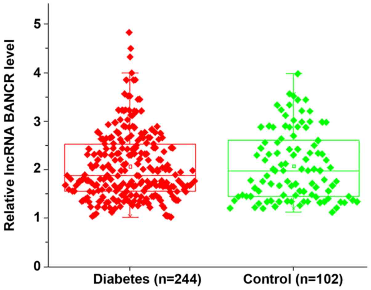
LncRNA BANCR was not differentially
expressed in diabetic patients and healthy controls on the day of
admission
RT-qPCR analysis indicated no significant
differences in the plasma levels of lncRNA BANCR between diabetic
patients and healthy controls on the day of admission, indicating
that lncRNA BANCR is unlikely involved in the development of
diabetes (Fig. 1).
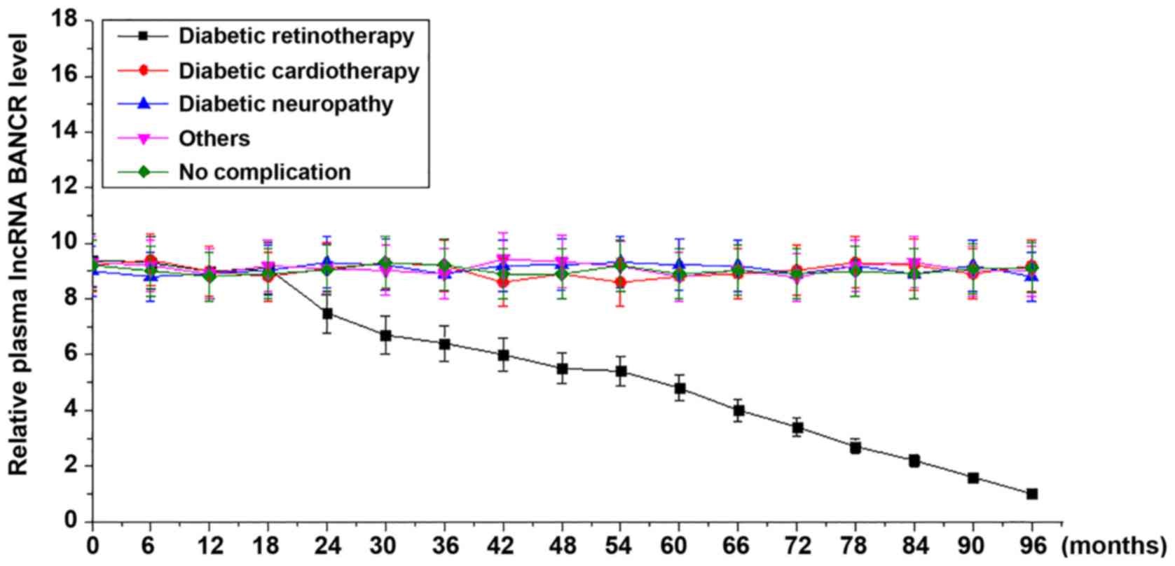
Plasma levels of lncRNA BANCR increase
in diabetic patients developing retinopathy but not in diabetic
patients developing other or no complications during follow-up
During the 8-year follow-up, continuous decreases
were observed in the plasma levels of lncRNA BANCR among patients
who developed diabetic retinopathy. However, no significant changes
in the plasma levels of lncRNA BANCR were identified among patients
who developed diabetic cardiomyopathy, diabetic neuropathy, other
complications or no complications (Fig.
2).
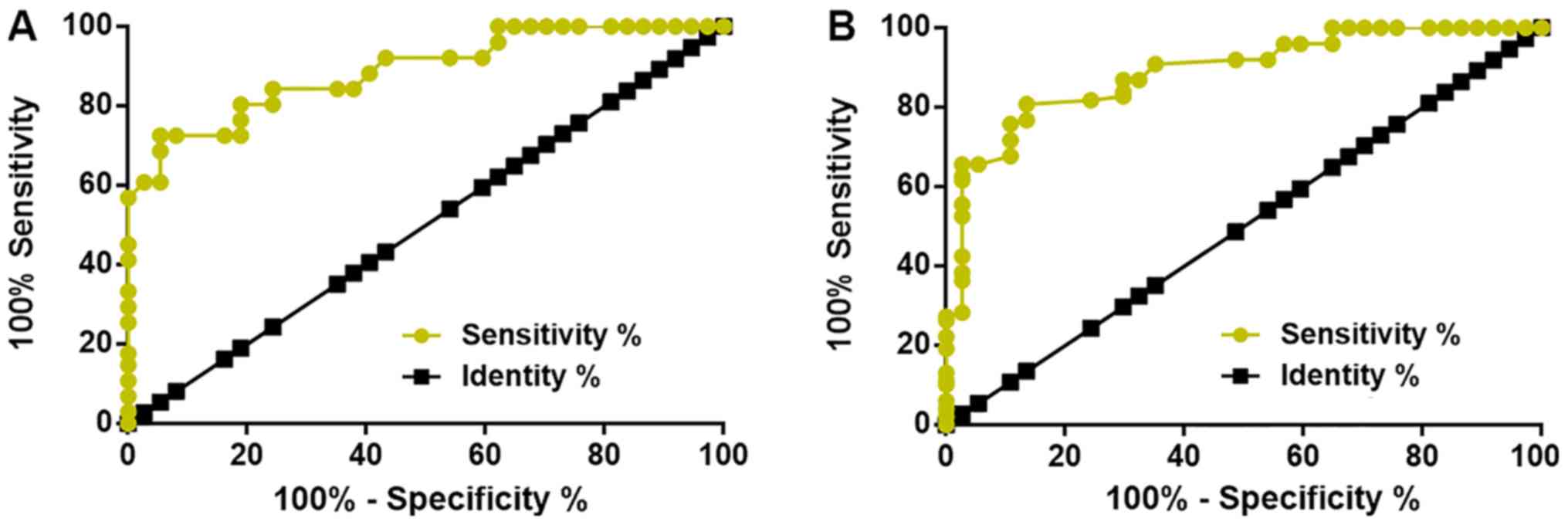
Plasma levels of lncRNA BANCR at 12
months prior to diagnosis is able to distinguish patients who will
develop diabetic retinopathy from healthy controls and diabetic
patients without obvious complications
The diagnostic value of plasma lncRNA BANCR for
diabetic retinopathy was evaluated by ROC curve analysis, which was
performed for healthy controls or diabetic patients without any
obvious complications as true-negative cases and diabetic
retinopathy patients as true-positive cases. It was revealed that
the plasma levels of lncRNA BANCR at 12 months prior to the
diagnosis of diabetic retinopathy are able to sufficiently
distinguish patients who will develop diabetic retinopathy from
healthy controls (Fig. 3A) and
diabetic patients without any obvious complications (Fig. 3B). The plasma levels of lncRNA BANCR
at earlier time-points failed to predict diabetic retinopathy. With
the healthy controls as a reference, the area under the curve was
0.8887, with a standard error of 0.02713 and a 95% confidence
interval of 0.8355–0.9419 (Fig. 3A).
With diabetic patients without obvious complications as a
reference, the area under the curve was 0.8896, with a standard
error of 0.02958 and a 95% confidence interval of 0.8316–0.9476
(Fig. 3B). In addition, plasma
levels of lncRNA BANCR at 12 months prior to diagnosis is also able
to distinguish patients who will develop diabetic retinopathy from
diabetic patients who will develop other complications (data not
shown).
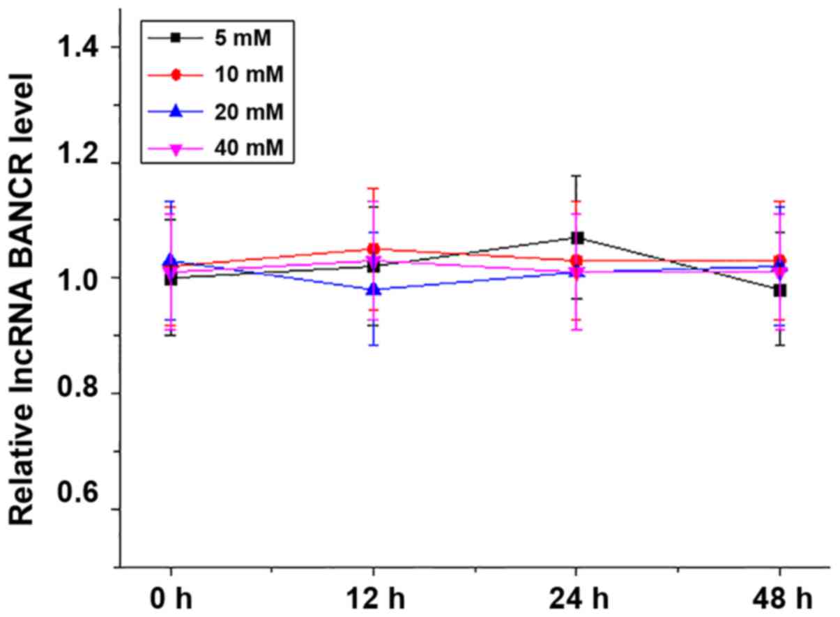
High glucose treatment failed to alter
lncRNA BANCR expression in the ARPE-19 human RPE cell line
The ARPE-19 human RPE cell line was treated with 5,
10, 20 or 40 mM d-glucose for 12, 24 or 48 h, and the expression of
lncRNA BANCR in those cells was detected by RT-qPCR. As presented
in Fig. 4, high-glucose treatment
failed to alter lncRNA BANCR expression in the RPE cells within 48
h.
LncRNA BANCR overexpression inhibits
apoptosis of ARPE-19 cells under high-glucose conditions
LncRNA BANCR overexpression and siRNA-mediated
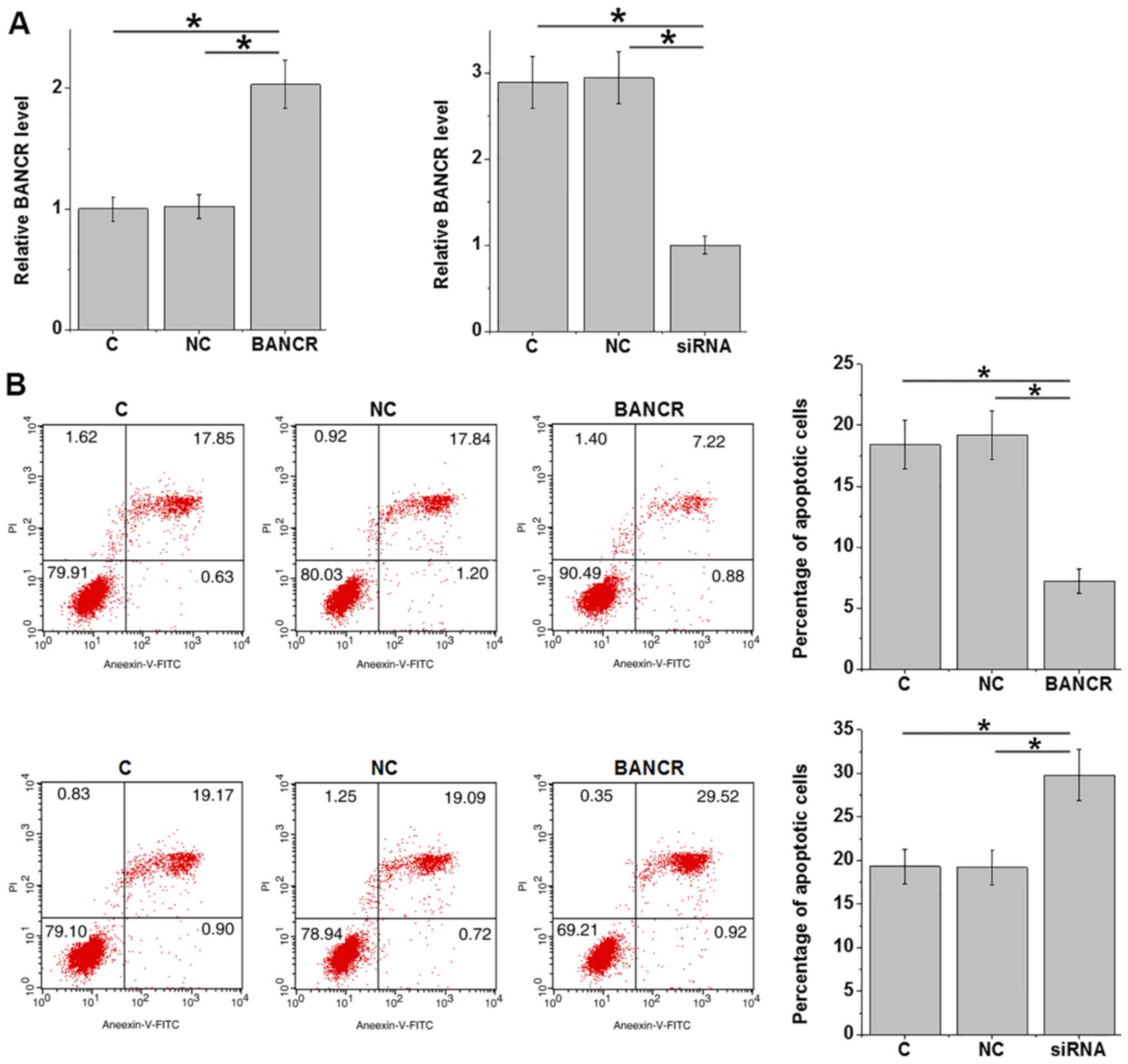
silencing in the ARPE-19 human RPE cell line was achieved after
transfection (P<0.05; Fig. 5A).
ARPE-19 cells were cultured in medium containing 20 mM d-glucose
for 24 h and cell apoptosis was detected. Compared with that in the
control group and the negative control group, lncRNA BANCR
overexpression inhibited, while lncRNA BANCR silencing promoted the
apoptosis of ARPE-19 cells (P<0.05; Fig. 5B).
Discussion
The functions of lncRNA BANCR have been well
characterized in cancer biology (10,11),
while its involvement in diabetic complications has remained
elusive. The key result of the present study is that lncRNA BANCR
is downregulated in diabetic retinopathy and ectopic overexpression
of lncRNA inhibits the apoptosis of RPE cells under high-glucose
conditions.
Previous studies have indicated that the development
of diabetic retinopathy globally affects the expression of lncRNAs,
indicating the involvement of lncRNAs in this disease (16,17).
However, studies on the involvement of lncRNAs in diabetic
retinopathy are rare. In a recent study, Zhang et al
(18) reported that lncRNA
maternally expressed 3 (MEG3) was downregulated in diabetic
retinopathy and overexpression of lncRNA MEG3 may improve the
condition of this disease by regulating the expression of
transforming growth factor-β1 and vascular endothelial growth
factor. In another study, Biswas et al (19) indicated that lncRNA
metastasis-associated lung adenocarcinoma transcript 1 and HOX
transcript antisense RNA are the key epigenetic regulators in
diabetic retinopathy-associated pathological changes. In the
present study, it was demonstrated that lncRNA BANCR was
downregulated specifically in patients with diabetic retinopathy
but not in patients with other diabetic complications. In
vitro cell experiments using the ARPE-19 human RPE cell line
also suggested that lncRNA BANCR expression was not affected by a
high-glucose environment, whereas BANCR overexpression inhibited
the apoptosis of ARPE-19 cells under high glucose treatment. The
present authors speculate that BANCR may be affected by long-term,
but not short-term, high glucose treatment, or BANCR downregulation
is caused by the formation of lesions in the eyes. Nevertheless,
the present ROC curve analysis data suggested that lncRNA BANCR may
be a specific biomarker for diabetic retinopathy due to the
observation that altered BANCR distinguished diabetic retinopathy
patients from healthy controls, diabetic patients without obvious
complications and diabetic patients with other complications.
Blindness inevitably occurs in certain patients with
diabetic retinopathy. For the efficient management of diabetic
retinopathy, early detection and prediction of risks are critical
(20). In the present study, it was
demonstrated that the plasma levels of lncRNA BANCR at 12 months
prior to the diagnosis of diabetic retinopathy are able to
sufficiently distinguish patients who will develop diabetic
retinopathy from healthy controls and diabetic patients without any
obvious complications. Therefore, circulating lncRNA BANCR may be
of predictive value for diabetic retinopathy.
Accelerated apoptosis of human RPE cells is a major
pathological change in eyes of patients with diabetic retinopathy
and inhibition of human RPE cell apoptosis is considered as a
promising therapeutic strategy for diabetic retinopathy (21). In the present study, lncRNA BANCR
overexpression inhibited the apoptosis of human RPE cells in a
high-glucose environment. Therefore, lncRNA BANCR overexpression
may be a potential strategy for the treatment of diabetic
retinopathy. However, the molecular mechanisms of the regulatory
role of lncRNA BANCR in the apoptosis of human RPE cells remain
elusive.
It is worth noting that the present study failed to
detect the expression of lncRNA BANCR in aqueous humor or vitreous
specimens. Future studies by our group will aim at performing this
analysis. In addition, the present study also failed to explore the
molecular mechanisms of BANCR in diabetic retinopathy. Future
studies by our group will investigate the molecular mechanisms of
the action of BANCR in diabetic retinopathy. However, the present
study suggests that BANCR may serve as a potential therapeutic
target for diabetic retinopathy, which provides a novel approach
for the clinical treatment of this disease. In the present study,
only Annexin V-FITC and PI to were used to detect cell apoptosis.
However, apoptotic modulation at the molecular level should be
validated by further approaches, including the detection of
cleavage of caspases and poly(ADP ribose) polymerase and DNA
laddering, and the presence of apoptosis should be screened by
microscopic view of nuclear condensation. The present study failed
to perform these analyses due to limited resources. In future
studies, these experiments may be performed to further confirm the
present conclusions.
In conclusion, the present study revealed that
lncRNA BANCR was downregulated in diabetic retinopathy and that
overexpression of lncRNA BANCR to inhibit cell apoptosis may be a
novel therapeutic approach to improve diabetic retinopathy.
Acknowledgements
Not applicable.
Funding
No funding received.
Availability of data and materials
All data generated or analyzed during the present
study are included in this published article.
Author's contributions
XZh, XZo, YL and YW were responsible for the
conception and design of the study. XZh, XZo and YL performed the
experiments. XZh, XZo and YL analyzed and interpreted the data. XZh
and YW drafted the manuscript. XZh, XZo, YL and YW were responsible
for the revision of the manuscript.
Ethics approval and consent to
participate
The protocol of the present study was approved by
the Ethics Review Committee of Lanzhou University Second Hospital
(Lanzhou, China). All participants provided written informed
consent.
Patient consent for publication
Not applicable.
Competing interests
The authors declare that they have no competing
interests.
References
|
1
|
Semeraro F, Cancarini A, dell'Omo R,
Rezzola S, Romano MR and Costagliola C: Diabetic retinopathy:
Vascular and inflammatory disease. J Diabetes Res. 2015:5820602015.
View Article : Google Scholar : PubMed/NCBI
|
|
2
|
Lee R, Wong TY and Sabanayagam C:
Epidemiology of diabetic retinopathy, diabetic macular edema and
related vision loss. Eye Vis (Lond). 2:172015. View Article : Google Scholar : PubMed/NCBI
|
|
3
|
Ting DS, Cheung GC and Wong TY: Diabetic
retinopathy: Global prevalence, major risk factors, screening
practices and public health challenges: A review. Clin Exp
Ophthalmol. 44:260–277. 2016. View Article : Google Scholar : PubMed/NCBI
|
|
4
|
Wang C, Li J, Xue H, Li Y, Huang J, Mai J,
Chen J, Cao J, Wu X, Guo D, et al: Type 2 diabetes mellitus
incidence in Chinese: Contributions of overweight and obesity.
Diabetes Res Clin Pract. 107:424–432. 2015. View Article : Google Scholar : PubMed/NCBI
|
|
5
|
Guariguata L, Whiting DR, Hambleton I,
Beagley J, Linnenkamp U and Shaw JE: Global estimates of diabetes
prevalence for 2013 and projections for 2035. Diabetes Res Clin
Pract. 103:137–149. 2014. View Article : Google Scholar : PubMed/NCBI
|
|
6
|
Simó R and Hernández C: Novel approaches
for treating diabetic retinopathy based on recent pathogenic
evidence. Prog Retin Eye Res. 48:160–180. 2015. View Article : Google Scholar : PubMed/NCBI
|
|
7
|
Leasher JL, Bourne RRA, Flaxman SR, Jonas
JB, Keeffe J, Naidoo N, Pesudovs K, Price H, White RA, Wong TY, et
al: Global estimates on the number of people blind or visually
impaired by diabetic retinopathy: A meta-analysis from 1990 to
2010. Diabetes Care. 39:1643–1649. 2016. View Article : Google Scholar : PubMed/NCBI
|
|
8
|
Shi X, Sun M, Liu H, Yao Y and Song Y:
Long non-coding RNAs: A new frontier in the study of human
diseases. Cancer Lett. 339:159–166. 2013. View Article : Google Scholar : PubMed/NCBI
|
|
9
|
Esteller M: Non-coding RNAs in human
disease. Nat Rev Genet. 12:861–874. 2011. View Article : Google Scholar : PubMed/NCBI
|
|
10
|
Li L, Zhang L, Zhang Y and Zhou F:
Increased expression of LncRNA BANCR is associated with clinical
progression and poor prognosis in gastric cancer. Biomed
Pharmacother. 72:109–112. 2015. View Article : Google Scholar : PubMed/NCBI
|
|
11
|
Wang D, Wang D, Wang N, Long Z and Ren X:
Long non-coding RNA BANCR promotes endometrial cancer cell
proliferation and invasion by regulating MMP2 and MMP1 via ERK/MAPK
signaling pathway. Cell Physiol Biochem. 40:644–656. 2016.
View Article : Google Scholar : PubMed/NCBI
|
|
12
|
Su S, Gao J, Wang T, Wang J, Li H and Wang
Z: Long non-coding RNA BANCR regulates growth and metastasis and is
associated with poor prognosis in retinoblastoma. Tumuor Biol.
36:7205–7211. 2015. View Article : Google Scholar
|
|
13
|
Dunn KC, Aotaki-Keen AE, Putkey FR and
Hjelmeland LM: ARPE-19, a human retinal pigment epithelial cell
line with differentiated properties. Exp Eye Res. 62:155–170. 1996.
View Article : Google Scholar : PubMed/NCBI
|
|
14
|
Livak KJ and Schmittgen TD: Analysis of
relative gene expression data using real-time quantitative PCR and
the 2(-Delta Delta C(T)) method. Methods. 25:402–408. 2001.
View Article : Google Scholar : PubMed/NCBI
|
|
15
|
Cregan SP, Smith BP, Brown DL and Mitchel
RE: Two pathways for the induction of apoptosis in human
lymphocytes. Int J Radiat Biol. 75:1069–1086. 1999. View Article : Google Scholar : PubMed/NCBI
|
|
16
|
Yan B, Tao ZF, Li XM, Zhang H, Yao J and
Jiang Q: Aberrant expression of long noncoding RNAs in early
diabetic retinopathy. Invest Ophthalmol Vis Sci. 55:941–951. 2014.
View Article : Google Scholar : PubMed/NCBI
|
|
17
|
Jaé N and Dimmeler S: Long noncoding RNAs
in diabetic retinopathy. Circ Res. 116:1104–1106. 2015. View Article : Google Scholar : PubMed/NCBI
|
|
18
|
Zhang D, Qin H, Leng Y, Li X, Zhang L, Bai
D, Meng Y and Wang J: LncRNA MEG3 overexpression inhibits the
development of diabetic retinopathy by regulating TGF-β1 and VEGF.
Exp Ther Med. 16:2337–2342. 2018.PubMed/NCBI
|
|
19
|
Biswas S, Thomas AA, Feng B, Chen S,
Aref-Eshghi E, Gonder J, Sadikovic B and Chakrabarti S: MALAT1 and
HOTAIR-Key epigenetic regulators in diabetic retinopathy. Diabetes.
67:2402018. View Article : Google Scholar
|
|
20
|
Olafsdottir E, Andersson DKG, Dedorsson I,
Svärdsudd K, Jansson SP and Stefánsson E: Early detection of type 2
diabetes mellitus and screening for retinopathy are associated with
reduced prevalence and severity of retinopathy. Acta Ophthalmol.
94:232–239. 2016. View Article : Google Scholar : PubMed/NCBI
|
|
21
|
Kim DI, Park MJ, Lim SK, Choi JH, Kim JC,
Han HJ, Kundu TK, Park JI, Yoon KC, Park SW, et al:
High-glucose-induced CARM1 expression regulates apoptosis of human
retinal pigment epithelial cells via histone 3 arginine 17
dimethylation: Role in diabetic retinopathy. Arch Biochem Biophys.
560:36–43. 2014. View Article : Google Scholar : PubMed/NCBI
|