Introduction
Diabetes mellitus, or diabetes, is a common
metabolic disorder affecting a considerable portion of the
worldwide population (1). It is
generally considered that the incidence of diabetes will be
significantly increased in the future due to various factors,
including the popularization of western-style diet structures and a
sedentary lifestyle (2,3). Patients suffering from long-term
diabetes usually develop complications in most major organs, an
example being chronic renal failure (4,5). A
previous clinical study demonstrated that >30% of patients with
type 1 diabetes and 25% of patients with type 2 diabetes will
develop nephropathy (6), which
causes high mortality rates (7). At
present, survival rates of those with the condition remain poor
(8).
It has been reported that the activation of
hypoxia-inducible factors (HIF) prevents diabetic nephropathy
(9). In another study, it was
suggested that chronic intermittent hypobaric hypoxia ameliorates
rat models of diabetic nephropathy by activating HIF1 signaling
(10). The current study suggests
that HIF1 signaling may serve as a potential therapeutic target for
diabetic nephropathy. It has been determined that HIF1 signaling
participates in certain pathological changes by interacting with
different long non-coding RNAs (lncRNAs) (11,12). A
recent study reported LINK-A as a novel lncRNA that promotes triple
negative breast cancer by interacting with HIF1α (13). Therefore, it is reasonable to
hypothesize that LINK-A may also interact with HIF1α in diabetic
nephropathy. The current study revealed that diabetic nephropathy
may be improved via the LINK-A IncRNA mediated induction of HIF1α
upregulation.
Materials and methods
Subjects and renal biopsies
A total of 102 diabetic patients with nephropathy
and 466 diabetic patients without complications were treated from
January 2014 to March 2018 at Kunming Medical University (Kunming,
China). Form those patients, a total of 32 patients with diabetic
nephropathy and 28 diabetic patients without obvious complications
were enrolled in the current study. The inclusion criteria were as
follows: i) Patients who had not been treated within 3 months prior
to admission; ii) patients with normal major organ function except
kidney function in patients with diabetic nephropathy and; iii)
patients received renal biopsies to detect potential renal lesions
or if renal lesions were excluded in diabetic patients without
complications. The exclusion criteria were as follows: i) patients
who suffered from other severe diseases or chronic diseases, (63
patients were excluded based on this parameter); ii) patients who
could not fully understand experimental protocol, (6 patients were
excluded based on this parameter). During the same time period
(January 2014 to March 2018), 56 individuals without diabetes also
received renal biopsies to confirm suspected renal lesions, the
results of which revealed that renal lesions were not present in 16
of them. Among these 16 people, 14 were included in the control
group and 2 cases were excluded due to individuals suffering from
additional severe diseases, including one patient with Hepatitis B
and another patient with heart disease. The current study received
ethical approval from the Ethics Committee of Kunming Medical
University (Kunming, China). Renal biopsies used within the
experiment were obtained from the specimen library of Kunming
Medical University (Kunming, China). All patients and their
families provided their written informed consent. See Table I for basic data of 3 groups of
participants.
 | Table I.Basic data of the study groups. |
Table I.
Basic data of the study groups.
|
| Sex |
|---|
|
|
|
|---|
| Groups | Male, n | Female, n | Age range
(years) | Average age
(years) |
|---|
| Diabetic
nephropathy | 17 | 15 | 23–67 | 45.9±5.2 |
| Diabetes | 15 | 13 | 25–69 | 46.4±6.7 |
| Control | 11 | 13 | 24–69 | 46.1±5.5 |
Cell culture and transfection
Mouse podocyte cells (PrimCells, LLC) were used to
perform in vitro experiments. Cells were cultured in mouse
podocyte primary cell culture complete medium with serum
(Celprogen, Inc.) and maintained at 37°C in a 5%
CO2-humidifed incubator. Full-length LINK-A lncRNA and
HIF1α cDNAs were amplified via PCR using the following primer
pairs: LINK-A forward, 5′-TGGAATTCAAGATGTGGGTGAG-3′ and reverse,
5′-AAGGATAATGCATTTTTATTTTAATTGAG-3′; HIF1α forward,
5′-AGTGCACAGTGCTGCCTCGTCTG-3′ and reverse,
5′-CCTGGTCCACAGAAGATGTTTA-3′. PCR amplification was performed using
Phusion High-Fidelity DNA Polymerase (Thermo Fisher Scientific,
Inc.) using the following thermocycling conditions: Initial
denaturation at 95°C for 1 min; 35 cycles at 95°C for 12 sec, 60°C
for 12 sec and 70°C for 2 min. LINK-A lncRNA and HIF1α cDNA were
cloned into a pEGFPC3 vector (Shanghai GenePharma Co., Ltd.) to
generate LINK-A lncRNA and HIF1α expression vectors. LINK-A lncRNA
and HIF1α expression vectors were used to transfect mouse podocyte
cells using Lipofectamine® 2000 reagent (Thermo Fisher
Scientific, Inc.). Untransfected mouse podocyte cells were used as
controls. Transfection with empty vector was used as a negative
control. Following 24-h transfection, cells were collected and
LINK-A lncRNA and HIF1α overexpression was confirmed by reverse
transcription-quantitative (RTq)PCR.
RT-qPCR
Total RNA was extracted from renal biopsies and cell
lines using TRIzol® reagent (Invitrogen; Thermo Fisher
Scientific, Inc.). Total RNA was reverse transcribed into cDNA
using the Reverse Transcriptase AMV kit (Sigma-Aldrich; Merck
KGaA). qPCR was subsequently performed using the SuperScript III
Platinum SYBR Green One-Step qPCR kit (Thermo Fisher Scientific
Inc.). The following primer pairs were used for the qPCR: LINK-A
IncRNA forward, 5′-TTCCCCCATTTTTCCTTTTC-3′ and reverse,
5′-CTCTGGTTGGGTGACTGGTT-3′; β-actin forward,
5′-GACCTCTATGCCAACACAGT-3′ and reverse, 5-AGTACTTGCGCTCAGGAGG-3′.
The following thermocycling conditions were as follows: Initial
denaturation at 95°C for 56 sec; 40 cycles of 95°C for 12 sec and
57.6°C for 40 sec. Data was quantified using the 2−ΔΔCq
method and normalized to the internal reference gene β-actin
(14).
ELISA
Renal biopsies were used to measure levels of HIF1α
using an ELISA kit (cat. no. EHIF1A; Thermo Fisher Scientific,
Inc.), according to the manufacturer's protocol. Levels of HIF1α
were normalized to ng/g.
Cell apoptosis assay
Cell apoptosis under 20 mM D-glucose treatments were
detected via a cell apoptosis assay. Mouse podocyte cell
suspensions (5×10⁴ cells/ml) were prepared in mouse
podocyte cell culture complete medium with serum (Celprogen, Inc.).
Cell suspensions (10 ml) were added to each well of a six-well
plate, followed by the addition of 20 mM D-glucose (Sangon Biotech
Co., Ltd.). Cells were cultured for 24 h at 37°C. Cells were
digested using 0.25% trypsin, collected and mixed with Mouse
Podocyte Cell Culture Complete medium with serum. Following
centrifugation at 1,200 × g for 3 min at 22°C, cells were stained
using Annexin V-FITC (Dojindo Molecular Technologies, Inc.) and
propidium iodide at 22°C for 18 min. Apoptotic cells were detected
using a CytoFLEX LX flow cytometer (Beckman Coulter, Inc.) and
analyzed using FCSalyzer-0.9.15 (sourceforge.net/projects/fcsalyzer/).
Western blotting
Protein was extracted using RIPA Lysis and
Extraction Buffer (Thermo Fisher Scientific, Inc.) and protein
concentration was measured using a BCA assay. After denaturing at
95°C for 10 min, 20 µg protein was subjected to 12% SDS-PAGE gel
electrophoresis. Following gel transfer to PVDF membranes, 5%
skimmed milk was used to block the membranes at room temperature
for 1 h. Membranes were then incubated with rabbit anti-human HIF1α
(1:1,650; cat. no. ab2185; Abcam) and rabbit anti-human GAPDH
(1:1,400; cat. no. ab8255; Abcam) primary antibodies overnight at
4°C. Membranes were incubated the next day with IgG-horseradish
peroxide conjugated secondary antibodies (goat anti-rabbit;
1:1,500; MBS435036; MyBioSource, Inc.) at room temperature for 1 h.
Signals were subsequently developed by ECL (Sigma-Aldrich; Merck
KGaA). Data normalization was performed using ImageJ software
(v.1.60; National Institutes of Health).
Statistical analysis
GraphPad Prism 6 software (GraphPad Software, Inc.)
was used for all statistical analyses. Data were expressed as the
mean ± standard deviation and analyzed using one-way analysis of
variance followed by a least significant difference post-hoc test.
The correlation between LINK-A lncRNA and HIF1α expression was
analyzed using Pearson's correlation coefficient. Diagnostic values
of LINK-A lncRNA for diabetic nephropathy was evaluated by receiver
operating characteristic (ROC) curve analysis. P<0.05 was
considered to indicate a statistically significant difference.
Results
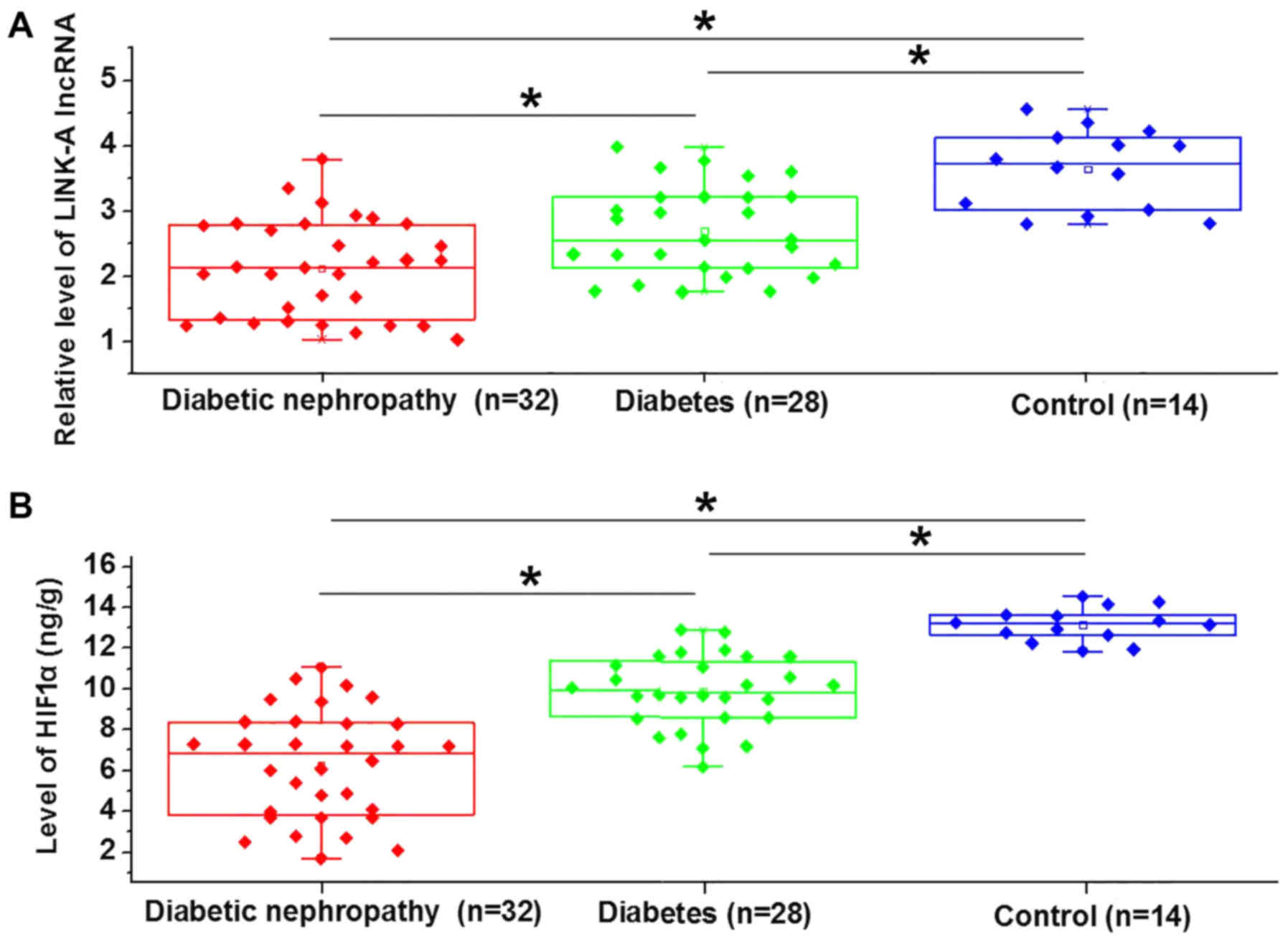
Expression of LINK-A lncRNA and HIF1α
are downregulated in patients with diabetic nephropathy
LINK-A lncRNA (Fig.
1A) and HIF1α (Fig. 1B) were
significantly downregulated in patients with diabetic nephropathy,
with diabetic patients without complications exhibiting an
intermediate expression and healthy controls exhibiting the lowest
expression. This indicates that the inhibition of LINK-A lncRNA and
HIF1α may contribute to diabetic nephropathy.
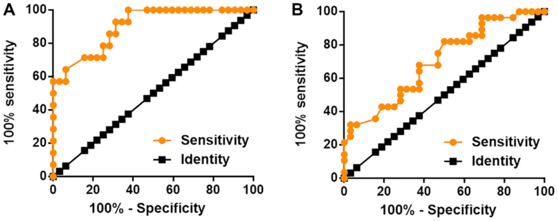
Upregulation of LINK-A lncRNA
distinguished patients with diabetic nephropathy from diabetic
patients without complications as well as from healthy
controls
The diagnostic value of LINK-A lncRNA for diabetic
nephropathy was analyzed via ROC curve analysis. With controls as
references, the area under the curve (AUC) value was 0.9007
(standard error; 0.04579; 95% confidence interval; 0.8109–0.9904;
Fig. 2A). With use of diabetic
patients with no complications as references, the AUC was 0.7031
(standard error; 0.06672; 95% confidence interval; 0.5723–0.8339;
Fig. 2B). These data may therefore
indicate that LINK-A lncRNA may serve as a promising diagnostic
biomarker for diabetic nephropathy.
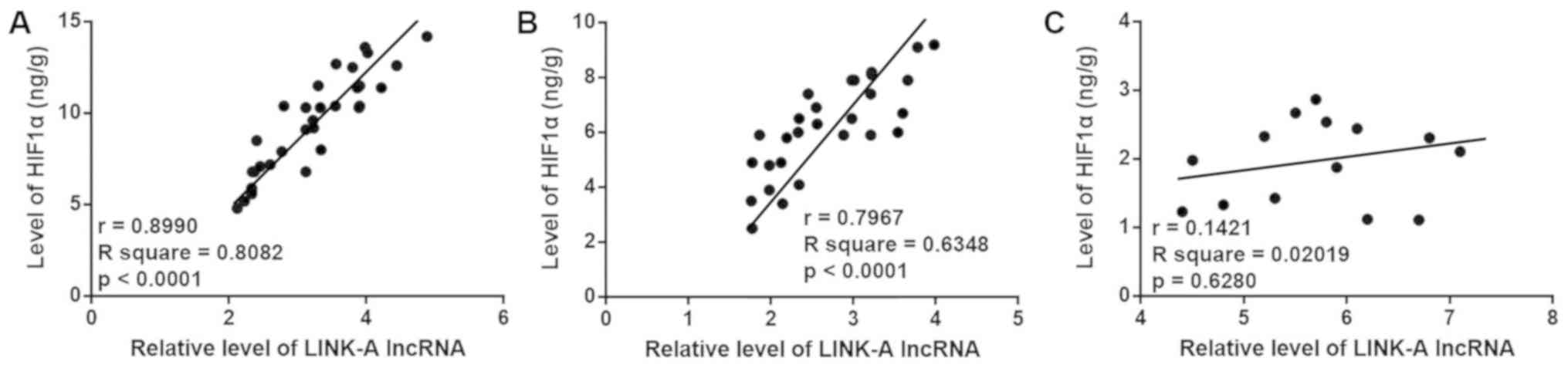
Expression of LINK-A lncRNA and HIF1α
are positively correlated in the 2 patient groups, but not in the
control group
As presented in Fig.
3, Pearson's correlation coefficient revealed that the
expression of LINK-A lncRNA and HIF1α were positively correlated in
patients with diabetic nephropathy (Fig.
3A) and in patients without complications (Fig. 3B) but not in the control group
(Fig. 3C). Additionally, the
positive correlation between LINK-A lncRNA and HIF1α was stronger
in patients with diabetic nephropathy than in diabetic patients
without complications. These data indicate that LINK-A lncRNA and
HIF1α may interact in diabetic nephropathy and in diabetes.
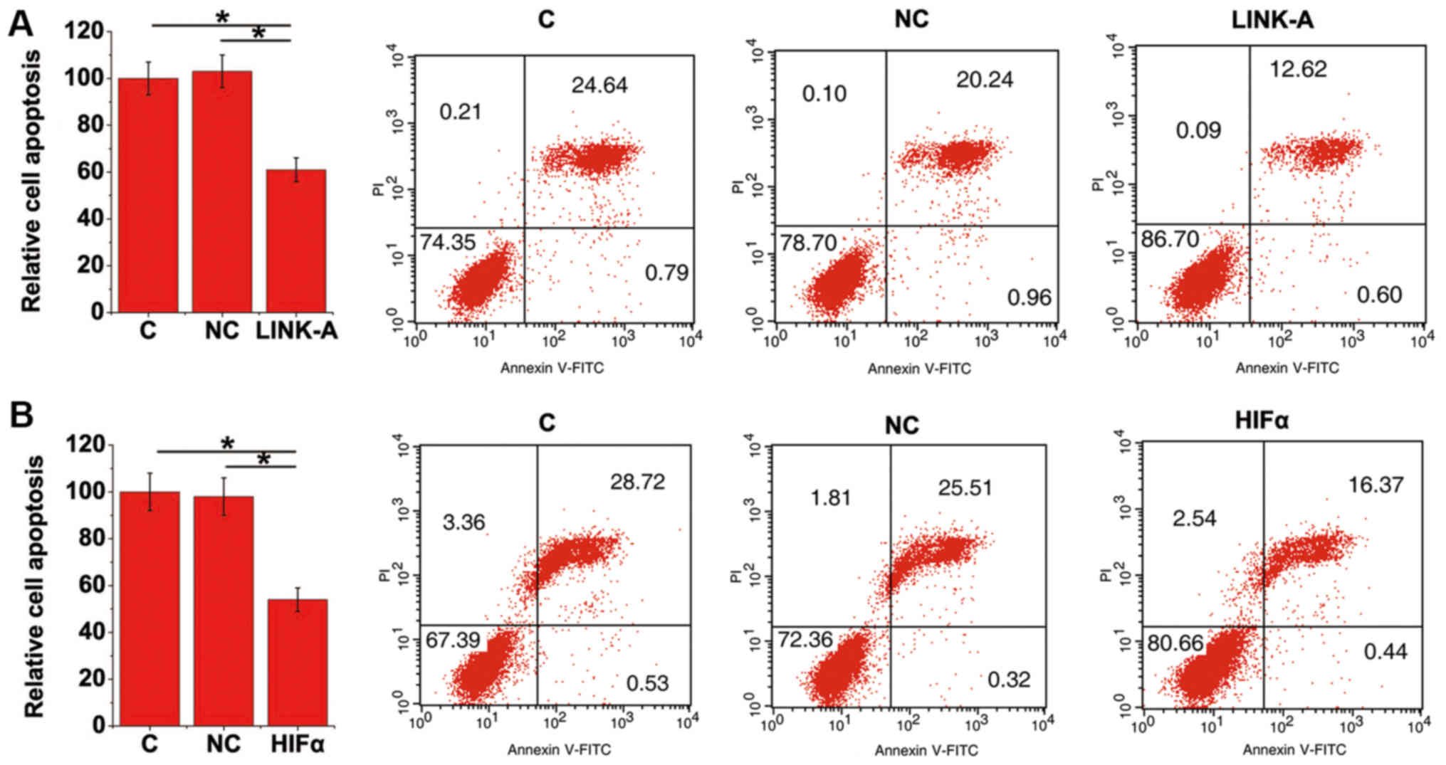
LINK-A lncRNA and HIF1α overexpression
inhibits apoptosis of mouse podocyte cells under high glucose
treatment
Cell apoptosis under 20 mM D-glucose treatment was
detected via a cell apoptosis assay. Compared with control and
negative control cells with LINK-A lncRNA overexpression (Fig. 4A) and HIF1α (Fig. 4B) exhibited significant inhibition of
cell apoptosis. The overexpression of LINK-A lncRNA and HIF1α may
therefore serve as a potential therapeutic target for diabetic
nephropathy.
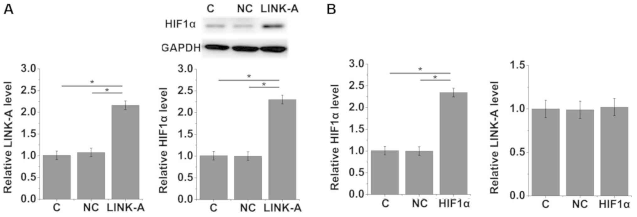
LINK-A lncRNA activates HIF1α in mouse
podocyte cells
To further investigate the interactions between
LINK-A lncRNA and HIF1α, LINK-A lncRNA and HIF1α expression vectors
were transfected into mouse podocyte cells. The expression of
LINK-A lncRNA and HIF1α was detected by RT-qPCR and Western
blotting. As presented in Fig. 5A,
compared with the control and negative control cells with LINK-A
lncRNA overexpression exhibited significantly upregulated HIF1α
expressions. In contrast, the overexpression of HIF1α did not
significantly affect LINK-A lncRNA expression (Fig. 5B). Therefore, the results indicate
that LINK-A lncRNA may be an upstream activator of HIF1α in mouse
podocyte cells.
Discussion
Human materials were used in the current study to
assess the role of LINK-A lncRNA in diabetic nephropathy. The
results of the present study indicated that LINK-A, as a recently
identified lncRNA, may participate in diabetic nephropathy by
interacting with HIF1α. Data from the present study also
demonstrated that LINK-A may be a potential therapeutic target for
the treatment of diabetic nephropathy.
The development of diabetic nephropathy can affect
the expression pattern of a large set of lncRNAs (15). Certain lncRNAs, which serve critical
roles in diabetic nephropathy, are considered to be potential
therapeutic targets. The present study observed the lowest
expression of LINK-A lncRNA in patients with diabetic nephropathy.
However, compared with the control group, significant
downregulation of LINK-A lncRNA was also observed in diabetic
patients without complications. Therefore, the downregulation of
LINK-A lncRNA may occur in diabetes and further downregulation may
be accompanied by the development of nephropathy among patients
with diabetes.
The present study indicated that the downregulation
of LINK-A may be used to distinguish patients with diabetic
nephropathy from healthy controls and patients with diabetes but
without obvious complications. These data indicate that LINK-A may
serve as a potential diagnostic biomarker for the diagnosis of
diabetic nephropathy. LINK-A, as a recently identified lncRNA, has
an unknown expression in triple negative breast cancer (15). The expression of LINK-A may therefore
be affected by other human diseases. Multiple diagnostic markers
should thus be used to improve diagnostic specificity.
The interaction between LINK-A and HIF1α has been
revealed to occur in triple negative breast cancer (16). The current study also observed a
positive correlation between LINK-A and HIF1α in patients with
diabetes but without complications and patients with diabetic
nephropathy. Mouse podocyte cells were used to further confirm the
interaction between LINK-A and HIF1α in the current study. Podocyte
cells are critical for renal function and the number of podocyte
cells have been widely used as biomarkers for renal function
(17). Podocyte cell injuries are
common in patients with diabetes and patients with diabetic
nephropathy (18,19). In addition, podocyte cells are
considered to be therapeutic targets for diabetic nephropathy
(20). In the current study, the
results indicated that LINK-A may be an upstream activator of HIF1α
in podocyte cells. No significant correlation between LINK-A and
HIF1α was observed in the patient control group, while LINK-A
overexpression led to the significant upregulation of HIF1α in
podocyte cells. These data suggest that overexpression techniques
may not be able to fully represent the interactions between LINK-A
and HIF1α in the human body.
The present study only performed in vitro
experiments. The in vivo functions of LINK-A and HIF1α in
diabetic nephropathy are therefore unclear and future studies
should be performed with animal models to support the conclusions
drawn from the current study.
LINK-A overexpression inhibited the apoptosis of
mouse podocyte cells under high glucose treatment in the current
study. Therefore, LINK-A overexpression may serve as a therapeutic
target for diabetic nephropathy. The present study is limited by
the small sample size due to the difficulties in collecting renal
biopsies. In the future, greater sample sizes should be used to
further support results.
In conclusion, the current study revealed that
LINK-A and HIF1α were upregulated in patients with diabetic
nephropathy. LINK-A may also upregulate HIF1α to improve diabetic
nephropathy and therefore may be a useful therapeutic target for
potential treatments and therapies.
Acknowledgements
Not applicable
Funding
No funding was received.
Availability of data and materials
The datasets used and/or analyzed during the present
study are available from the corresponding author on reasonable
request.
Authors' contributions
JY and LL designed the study and performed all of
the experiments. SH, ZZ and WF analyzed data. LL interpreted data
and prepared the manuscript. All authors read and approved the
final manuscript.
Ethics approval and consent to
participate
The present study was approved by the Ethics
Committee of Kunming Medical University (Kunming, China). All
patients and healthy volunteers provided written informed consent
prior to their inclusion within the study.
Ethics approval and consent to
participate
The present study was approved by the Ethics
Committee of Kunming Medical University (Kunming, China). All
patients and healthy volunteers provided written informed consent
prior to their inclusion within the study.
Patient consent for publication
All patients provided informed consent for
publication.
Competing interests
The authors declare that they have no competing
interests.
References
|
1
|
Zimmet P, Alberti KG, Magliano DJ and
Bennett PH: Diabetes mellitus statistics on prevalence and
mortality: facts and fallacies. Nat Rev Endocrinol 12(10). 616–622.
2016. View Article : Google Scholar
|
|
2
|
Rowley WR, Bezold C, Arikan Y, Byrne E and
Krohe S: Diabetes 2030: Insights from yesterday, today, and future
trends. Popul Health Manag. 20:6–12. 2017. View Article : Google Scholar : PubMed/NCBI
|
|
3
|
Rowley WR and Bezold C: Creating public
awareness: State 2025 diabetes forecasts. Popul Health Manag.
15:194–200. 2012. View Article : Google Scholar : PubMed/NCBI
|
|
4
|
Ahlqvist E, Van Zuydam NR, Groop LC and
McCarthy MI: The genetics of diabetic complications. Nat Rev
Nephrol. 11:277–287. 2015. View Article : Google Scholar : PubMed/NCBI
|
|
5
|
American Diabetes Association: Standards
of medical care in diabetes-2014. Diabetes Care 37(Supplement 1).
S14–S80. 2014. View Article : Google Scholar
|
|
6
|
Navarro-González JF, Jarque A, Muros M,
Mora C and García J: Tumor necrosis factor-alpha as a therapeutic
target for diabetic nephropathy. Cytokine Growth Factor Rev.
20:165–173. 2009. View Article : Google Scholar : PubMed/NCBI
|
|
7
|
Lei L, Mao Y, Meng D, Zhang X, Cui L, Huo
Y and Wang Y: Percentage of circulating CD8+ T lymphocytes is
associated with albuminuria in type 2 diabetes mellitus. Exp Clin
Endocrinol Diabetes. 122:27–30. 2014.PubMed/NCBI
|
|
8
|
Fried LF, Emanuele N, Zhang JH, Brophy M,
Conner TA, Duckworth W, Leehey DJ, McCullough PA, O'Connor T,
Palevsky PM, et al: Combined angiotensin inhibition for the
treatment of diabetic nephropathy. N Engl J Med. 369:1892–1903.
2013. View Article : Google Scholar : PubMed/NCBI
|
|
9
|
Nordquist L, Friederich-Persson M,
Fasching A, Liss P, Shoji K, Nangaku M, Hansell P and Palm F:
Activation of hypoxia-inducible factors prevents diabetic
nephropathy. J Am Soc Nephrol. 26:328–338. 2015. View Article : Google Scholar : PubMed/NCBI
|
|
10
|
Tian YM, Guan Y, Li N, Ma HJ, Zhang L,
Wang S and Zhang Y: Chronic intermittent hypobaric hypoxia
ameliorates diabetic nephropathy through enhancing HIF1 signaling
in rats. Diabetes Res Clin Pract. 118:90–97. 2016. View Article : Google Scholar : PubMed/NCBI
|
|
11
|
Hong Q, Li O, Zheng W, Xiao WZ, Zhang L,
Wu D, Cai GY, He JC and Chen XM: LncRNA HOTAIR regulates HIF-1α/AXL
signaling through inhibition of miR-217 in renal cell carcinoma.
Cell Death Dis. 8:e27722017. View Article : Google Scholar : PubMed/NCBI
|
|
12
|
Xiang S, Gu H, Jin L, Thorne RF, Zhang XD
and Wu M: LncRNA IDH1-AS1 links the functions of c-Myc and HIF1α
via IDH1 to regulate the Warburg effect. Proc Natl Acad Sci U S A.
115:E1465–E1474. 2018. View Article : Google Scholar : PubMed/NCBI
|
|
13
|
Lin A, Li C, Xing Z, Hu Q, Liang K, Han L,
Wang C, Hawke DH, Wang S, Zhang Y, et al: The LINK-A lncRNA
activates normoxic HIF1α signalling in triple-negative breast
cancer. Nat Cell Biol. 18:213–224. 2016. View Article : Google Scholar : PubMed/NCBI
|
|
14
|
Livak KJ and Schmittgen TD: Analysis of
relative gene expression data using real-time quantitative PCR and
the 2(-Delta Delta C(T)) method. Mothods. 25:402–408. 2001.
|
|
15
|
Chen S, Dong C, Qian X, Huang S, Feng Y,
Ye X, Miao H, You Q, Lu Y and Ding D: Microarray analysis of long
noncoding RNA expression patterns in diabetic nephropathy. J
Diabetes Complications. 31:569–576. 2017. View Article : Google Scholar : PubMed/NCBI
|
|
16
|
Long J and Danesh FR: Values and
limitations of targeting lncRNAs in diabetic nephropathy. Diabetes.
67:552–553. 2018. View Article : Google Scholar : PubMed/NCBI
|
|
17
|
Wasung ME, Chawla LS and Madero M:
Biomarkers of renal function, which and when? Clin Chim Acta.
438:350–357. 2015. View Article : Google Scholar : PubMed/NCBI
|
|
18
|
Fufaa GD, Weil EJ, Lemley KV, Knowler WC,
Brosius FC 3rd, Yee B, Mauer M and Nelson RG: Structural predictors
of loss of renal function in American Indians with type 2 diabetes.
Clin J Am Soc Nephrol. 11:254–261. 2016. View Article : Google Scholar : PubMed/NCBI
|
|
19
|
Maezawa Y, Takemoto M and Yokote K: Cell
biology of diabetic nephropathy: Roles of endothelial cells,
tubulointerstitial cells and podocytes. J Diabetes Investig.
6:3–15. 2015. View Article : Google Scholar : PubMed/NCBI
|
|
20
|
Chen J, Chen JK and Harris RC: EGF
receptor deletion in podocytes attenuates diabetic nephropathy. J
Am Soc Nephrol. 26:1115–1125. 2015. View Article : Google Scholar : PubMed/NCBI
|