Introduction
Myocardial infarction (MI) has a high morbidity and
mortality worldwide (1).
Percutaneous coronary intervention and thrombolytic therapy are
widely accepted as standard treatments for MI and the mortality
rate of patients decreases gradually in the acute phase (2). However, the mortality rate of coronary
artery disease remains high in the general population (3). Thus, it is necessary to identify novel
biomarkers for the early diagnosis of myocardial ischemic injury,
which may provide valuable benefits to current therapies.
Certain cyclic myocardial injury markers, including
high-sensitivity cardiac troponin (4), natriuretic peptide (5) and cyclic microRNA (miRNA) (6) have garnered a great deal of attention.
However, the number of available markers remains limited. Exosomes
are small cell-derived vesicles, with a diameter of 30–120 nm,
which are located in many, if not all biological fluids (7). These contain mRNA, miRNAs, long
non-coding RNAs, DNA, lipids and other small molecules (7). miRNAs have been studied in many
diseases due to their utility in disease diagnoses, to monitor
therapy and to predict the probability of disease recurrence; a
recent study demonstrated that various exosomal miRNA biomarkers
may be useful for the diagnosis of various diseases, including
nervous system diseases, malignant tumors and respiratory diseases
(8). For example, miR-21, miR-26a
and miR-146a were expressed in bronchoalveolar lavage exosomes,
however the expression of other miRNAs involved in the initiation
and progression of inflammation in sarcoidosis was not observed
(8). In addition, circulating
muscle-specific miRNAs were different between COPD and non-COPD
subjects (8). The aforementioned
study suggested that the plasma level of exosomal miRNA can reflect
changes within the skeletal muscle and so can act as a biomarker of
skeletal muscle dysfunction. In fact, exosomal miRNAs have been
considered a noninvasive diagnostic biomarkers and therapeutic
target in respiratory diseases (8).
However, the diagnostic value of exosomal miRNAs remains unknown in
patients with myocardial ischemic injury. Thus, there is a growing
need for diagnostic markers to improve the outcome of patients with
myocardial ischemic injury.
To the best of our knowledge, the current study is
the first to compare the miRNA profile of dysregulated plasma
exosomes in patients with MI compared with healthy controls.
Furthermore, the present study assessed the possible mechanism that
underlies the regulation of miRNA in myocardial ischemic
injury.
Materials and methods
Collection and preparation of plasma
samples
Plasma samples from 14 patients with MI, 10 patients
with stable angina pectoris and 14 healthy individuals were
collected between January 2016 and December 2016 from the First
Hospital of Shanxi Medical University (Taiyuan, China). The current
study recruited 20 males and 18 females, aged 41–78 years. The mean
age of these subjects was 47.6 years. Following collection, the
samples were stored at −80°C. These patients were diagnosed using
coronary angiograms, electrocardiograms and cardiac troponin tests,
and in accordance with previously published diagnostic criteria
guidelines (9,10). The exclusion criteria were as
follows: i) Patients unable to co-ordinate with doctors, ii)
patients with multiple medical complications or iii) patients who
received drugs treatment associated with angiocardiopathy within
one month of recruitment. Written informed consent was obtained
from all the participants and the present study was approved by the
Ethics Committee of the First Hospital of Shanxi Medical University
(Taiyuan, China).
Exosomes and exosomal RNA
isolation
Plasma from each participant was filtered using a
Millipore Millex-AA syringe (cat. no. SLAA033SB; Merck KGaA,
Darmstadt, Germany) prior to isolation. An exoEasy Maxi kit (Qiagen
GmbH, Hilden, Germany) was used to isolate exosomes from 4 ml of
pre-filtered plasma according to the manufacturer's protocol.
Following exosomal isolation, the remaining plasma was diluted with
PBS (1:1) and utilized as a control. For exosomal RNA extraction,
an exoRNeasy Serum/Plasma Maxi kit (Qiagen GmbH) was used to
isolate total exosome RNA from 4 ml pre-filtered plasma according
to the manufacturer's protocol. Finally, an RNeasy MinElute spin
column was placed into a 1.5 ml collection tube and 14 µl of
RNase-free water (both Qiagen GmbH) was directly added to the
center of the spin column membrane. The lid was closed gently, the
column was left to stand for 1 min and was then centrifuged for 1
min at 5,000 × g and 25°C to elute the RNA. Collected RNA was then
used in the subsequent experiments.
Transmission electron microscopy
Following exosome isolation, extracellular vesicles
were fixed with 4% paraformaldehyde for 30 min at 25°C and treated
as previously described (11). The
morphological characteristics of plasma-derived extracellular
vesicles were observed via transmission electron microscopy at a
magnification of ×30,000 (11).
Microarray analysis
miRNA microarray analysis was performed using the
Human miRNA OneArray® v6 (PhalanxBio, Inc., San Diego,
CA, USA). This contained triplicate 2539 unique miRNA probes from
humans (miRBase release v20; http://www.mirbase.org/), each printed in technical
triplicate, with 114 experimental control probes. The experiment
was performed according to a previous study (12), and fold-change (FC) was calculated.
FC=log2avg (MI)-log2avg (healthy control). The heatmap of
differentially expressed miRNAs was constructed by R software
(version 3.4.2; Vienna University of Economics and Business,
Vienna, Austria).
RNA preparation and reverse
transcription-quantitative polymerase chain reaction
Total RNA was isolated using the Trizol reagent
(Invitrogen; Thermo Fisher Scientific, Inc., Waltham, MA, USA)
according to the manufacturer's protocol and RNA (0.5 µg) was
utilized to synthesize cDNA using Super Scriptase III (Invitrogen;
Thermo Fisher Scientific, Inc.) with stem-loop RT primers
(5′-GTCGTATCCAGTGCAGGGTCCGAGGTATTCGCACTGGATACGACAGTGAA-3′). The
resultant cDNA was used for PCR. The thermocycling conditions were
as follows: Denaturation at 95°C for 3 min, then 40 cycles of 95°C
for 15 sec, 58°C for 30 sec and 72°C for 30 sec. To detect miR-183,
the polymerase a method was utilized, followed by SYBR-Green qPCR
(Thermo Fisher Scientific, Inc.) as described previously (3). Relative gene expression data was
produced using the 2−∆∆Cq method (13). U6 was used as an internal control.
The U6 primers used in the qPCR analysis were as follows: Forward:
5′-GCACTTTATTGGTCCATCATCC-3′; reserve:
5′-GACACCTGGCTCTTTTTGATTC-3′. The miR-183 primers used in the qPCR
analysis were as follows: Forward: 5′-CGCGGTATGGCACTGGTAGA-3′;
reverse: 5′-AGTGCAGGGTCCGAGGTATTC-3′.
Bioinformatic analysis
The target genes of miR-183 were downloaded from the
Targetscan database (http://www.targetscan.org/vert_50/) and all verified
genes were selected. To explore the functional annotation
enrichment of exosomal miR-183 target genes, gene ontology (GO) and
Kyoto Encyclopedia of Genes and Genomes (KEGG) analyses were
performed using the String database (http://string-db.org/). All target genes of miR-183
were then used to construct a protein-protein interaction network
via the String database, with the aim of obtaining a comprehensive
description of cellular mechanisms and functions.
Statistical analysis
Statistical analyses were performed using SPSS 19.0
(IBM Corp., Armonk, NY, USA). miRNA microarray data were analyzed
using a paired sample t-test. The comparison of multiple groups was
determined using one-way analysis of variance followed by an S-N-K
post-hoc test. P<0.05 was considered to indicate a statistically
significant result.
Results
Isolation of plasma-derived
exosomes
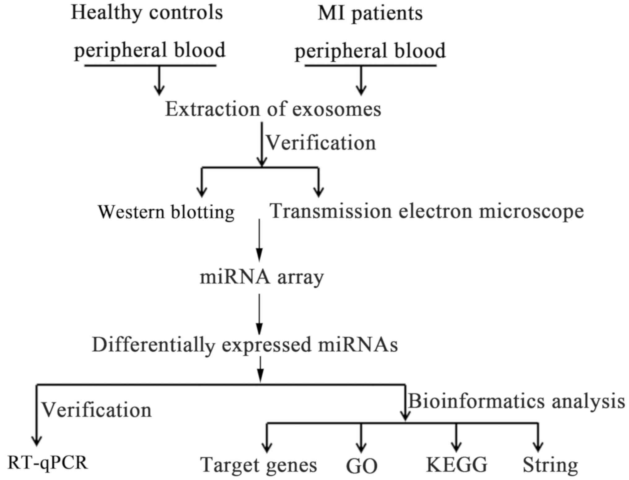
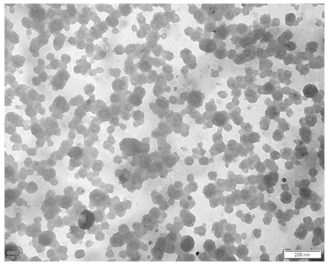
The experimental workflow of the current study is
presented in Fig. 1. The presence of
extracellular vesicles in patient plasma samples was confirmed
using electron microscopy and western blotting. The electron
micrographs of harvested extracellular vesicles revealed circular
membrane-bound structures with diameters varying from 50 to 100 nm
(Fig. 2A). According to these
results, it was confirmed that exosomes were successfully isolated
from human plasma.
Expression level of exosomal miR-183
is upregulated in patients with MI
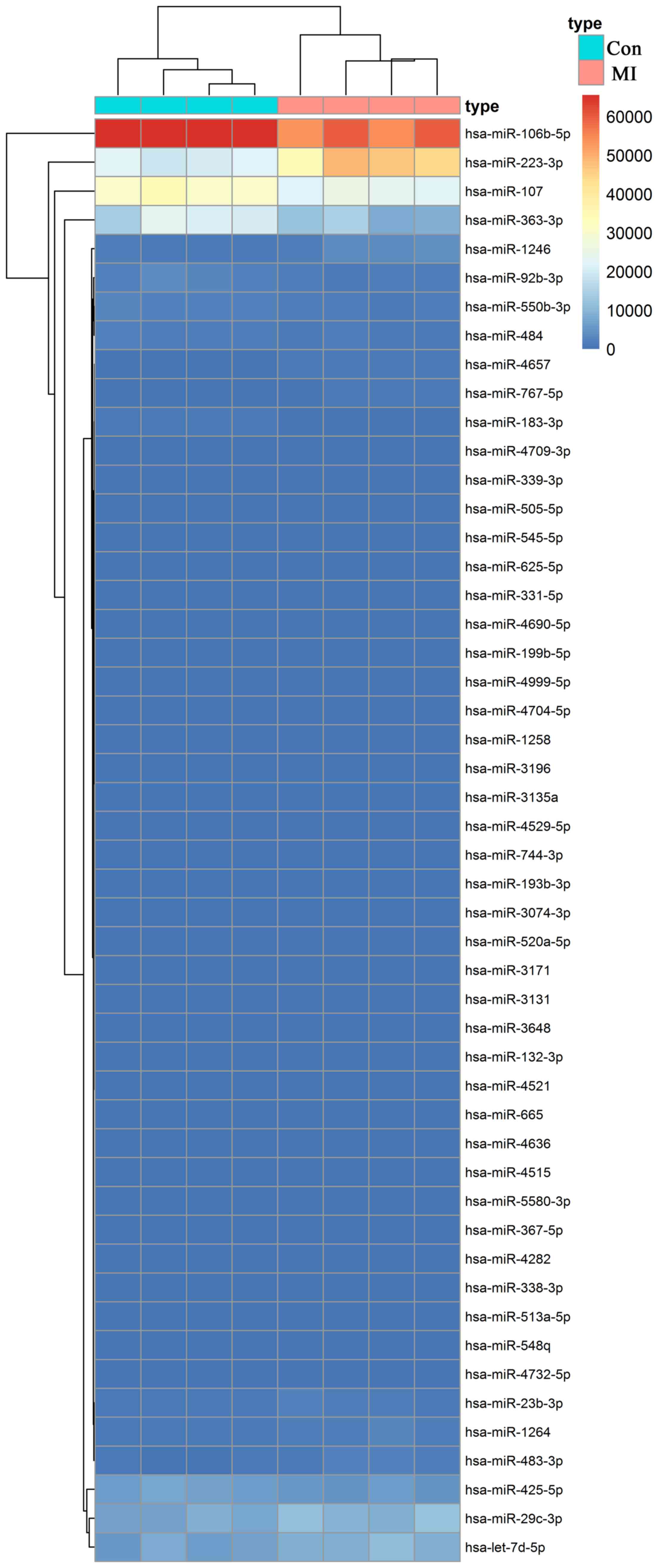
To identify the differentially expressed exosomal
miRNAs associated with the occurrence and development of myocardial
ischemic injury, a miRNA microarray was used in the present study.
The relative expression level of exosomal miRNAs in patients with
MI (n=4) and healthy controls (n=4) were compared using a miRNA
microarray, from which 85 differentially expressed miRNAs were
identified (P<0.05; 1<log2FC<-1; data not
shown). The heatmap of the top 50 differentially expressed miRNAs
is presented in Fig. 3. The
statistical results of 42 significantly different miRNAs
(P<0.01) are presented in Table
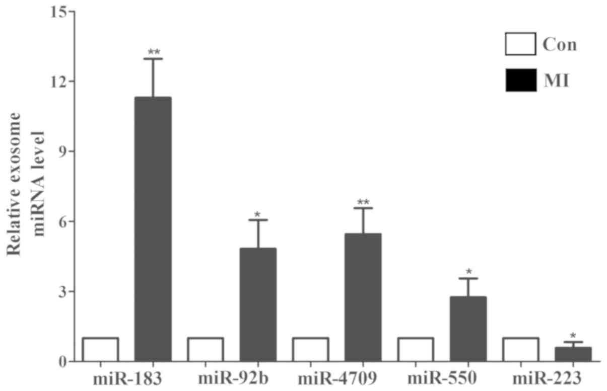
I. The top 10 most markedly dysregulated miRNAs were verified
using RT-qPCR in plasma samples from patients with MI (n=14) and
healthy individuals (n=14). Five miRNAs were consistent with those
of the microarray results: miR-183, miR-92b, miR-4709, miR-550 and
miR-223 (Fig. 4). Furthermore, the
change in miR-183 expression was the most significant
(P<0.01).
 | Table I.Differentially expressed exosomal
miRNAs in patients with myocardial infarction patients and healthy
individuals. |
Table I.
Differentially expressed exosomal
miRNAs in patients with myocardial infarction patients and healthy
individuals.
| No. | miRNA | Fold-change | Average
expression | t-value | P-value |
|---|
| 1 | hsa-miR-183-3p | 30.4197969 | 517.4346016 | 8.137940658 | 0.000104492 |
| 2 | hsa-miR-4657 | −24.922 | 548.13275 | −7.9080916 | 0.000124557 |
| 3 | hsa-miR-223-3p | −22.225 | 32320.3625 | −7.03659641 | 0.000252489 |
| 4 | hsa-miR-331-5p | 7.78202188 | 98.53995781 | 6.170875344 | 0.000547483 |
| 5 | hsa-miR-3610 | 19.66065609 | −16.82911102 | 6.08685269 | 0.000592724 |
| 6 | hsa-miR-548q | 10.4839219 | 169.6091641 | 5.603577385 | 0.000950949 |
| 7 | hsa-miR-4709-3p | 4.8494688 | 308.1036719 | 5.523099852 | 0.001031681 |
| 8 | hsa-miR-4521 | 3.04811797 | 8.192506641 | 5.340919802 | 0.001244448 |
| 9 | hsa-miR-92b-3p | 14.987188 | 1970.016406 | 5.338298419 | 0.001247849 |
| 10 |
hsa-miR-550b-3p | 9.2721875 | 1788.251406 | 5.292485194 | 0.001309002 |
| 11 | hsa-miR-3196 | 4.64892188 | 55.62916875 | 5.164620854 | 0.001498195 |
| 12 |
hsa-miR-106b-5p | 8.55 | 61145.74375 | 5.153722569 | 0.001515686 |
| 13 |
hsa-miR-4999-5p | 3.437765 | 27.2591675 | 4.881966157 | 0.002035064 |
| 14 | hsa-miR-665 | 2.66090297 | 8.935673828 | 4.686898091 | 0.00253041 |
| 15 | hsa-miR-4636 | 20.69043406 | −14.23388922 | 4.615443949 | 0.002744343 |
| 16 | hsa-miR-505-5p | 9.85890625 | 255.3029219 | 4.580595453 | 0.002855913 |
| 17 | hsa-miR-545-5p | 14.4396688 | 225.7731656 | 4.560897844 | 0.0029212 |
| 18 |
hsa-miR-4529-5p | −7.56569 | 16.08826438 | −4.541569 | 0.002986879 |
| 19 | hsa-miR-484 | 7.528 | 1503.836 | 4.504105993 | 0.003118896 |
| 20 | hsa-miR-339-3p | 16.245125 | 293.7521563 | 4.356629784 | 0.003705222 |
| 21 |
hsa-miR-4690-5p | 9.08543781 | 87.77904672 | 4.313023159 | 0.003901248 |
| 22 |
hsa-miR-4732-5p | 17.3946422 | 156.6910852 | 4.260649424 | 0.004151997 |
| 23 |
hsa-miR-199b-5p | −4.177 | 52.0643375 | −4.08801447 | 0.005113256 |
| 24 |
hsa-miR-4704-5p | 2.50220313 | 27.57288906 | 4.0354906 | 0.005452571 |
| 25 | hsa-miR-23b-3p | −7.8964844 | 1120.02457 | −4.0042717 | 0.005665949 |
| 26 | hsa-miR-1258 | 28.15745063 | 34.60022938 | 3.971930513 | 0.005896726 |
| 27 | hsa-miR-625-5p | 12.6166406 | 250.8808047 | 3.941044287 | 0.006126798 |
| 28 | hsa-miR-4515 | 17.91870313 | −30.97231094 | 3.917868717 | 0.006305908 |
| 29 | hsa-miR-1264 | −9.83 | 1324.7825 | −3.89505726 | 0.006487833 |
| 30 |
hsa-miR-193b-3p | −3.86354734 | 6.368044609 | −3.83238497 | 0.007017982 |
| 31 |
hsa-miR-5580-3p | 13.43604625 | −23.61720031 | 3.8175008 | 0.007150752 |
| 32 | hsa-miR-367-5p | −14.87524359 | −15.89417508 | −3.8087925 | 0.007229706 |
| 33 | hsa-miR-363-3p | 9.45 | 15382.3 | 3.778905829 | 0.007508021 |
| 34 | hsa-miR-1246 | −19.130625 | 2229.092188 | −3.74986284 | 0.007789749 |
| 35 | hsa-miR-3131 | 16.80226016 | −0.828861328 | 3.733061229 | 0.007957996 |
| 36 | hsa-miR-3135a | 3.89429531 | 46.65076797 | 3.690694624 | 0.008400146 |
| 37 | hsa-miR-29c-3p | −29.9475 | 8943.44375 | −3.61090831 | 0.00930743 |
| 38 | hsa-let-7d-5p | −26.55 | 7891.1825 | −3.60204751 | 0.009414606 |
| 39 |
hsa-miR-3074-3p | −23.74049453 | 13.19788555 | −3.59284785 | 0.009527305 |
| 40 | hsa-miR-3648 | 18.56568 | −3.532107031 | 3.586636253 | 0.009604231 |
| 41 |
hsa-miR-520a-5p | −9.91408813 | 20.38453563 | −3.57706628 | 0.009724074 |
| 42 | hsa-miR-338-3p | −11.5395641 | 104.142593 | −3.57291933 | 0.00977651 |
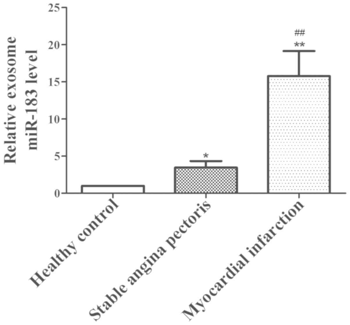
Upregulated miR-183 is associated with
the degree of myocardial ischemic injury
To elucidate the role of miR-183 in myocardial
injury, the expression of miR-183 in different degrees of
myocardial injury was analyzed. The results revealed that exosomal
miR-183 levels were significantly lower in the plasma of the
healthy individuals (P<0.05) compared with the stable angina and
MI groups, and increased accordingly with an increase in severity
of myocardial ischemic injury. Furthermore, the expression of
miR-183 in patients with MI (n=14) was significantly higher than
the levels in patients with stable angina pectoris (n=10;
P<0.01; Fig. 5). These results
indicate that upregulated exosomal miR-183 may be a novel
predictive marker for patients with myocardial ischemic injury.
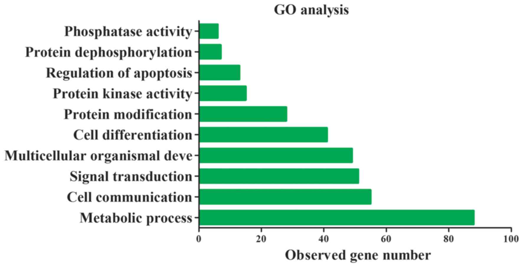
miR-183 is involved in the regulation
of protein kinase activity
miRNAs are closely associated with heart disease
occurrence and progression through the activation of certain
signaling pathways (14). To further
understand the underlying molecular mechanism of miR-183 function,
target genes of miR-193 in GO and KEGG pathways were assessed. The
top 30 target genes of miR-183 are presented in Table II. miR-183 target genes were
primarily involved in 10 significant functions, including cell
communication, signal transduction and the regulation of kinase and
phosphatase activity (Fig. 6). The
results of KEGG indicate that these target genes are also primarily
involved in cardiomyocyte and dopaminergic synapse adrenergic
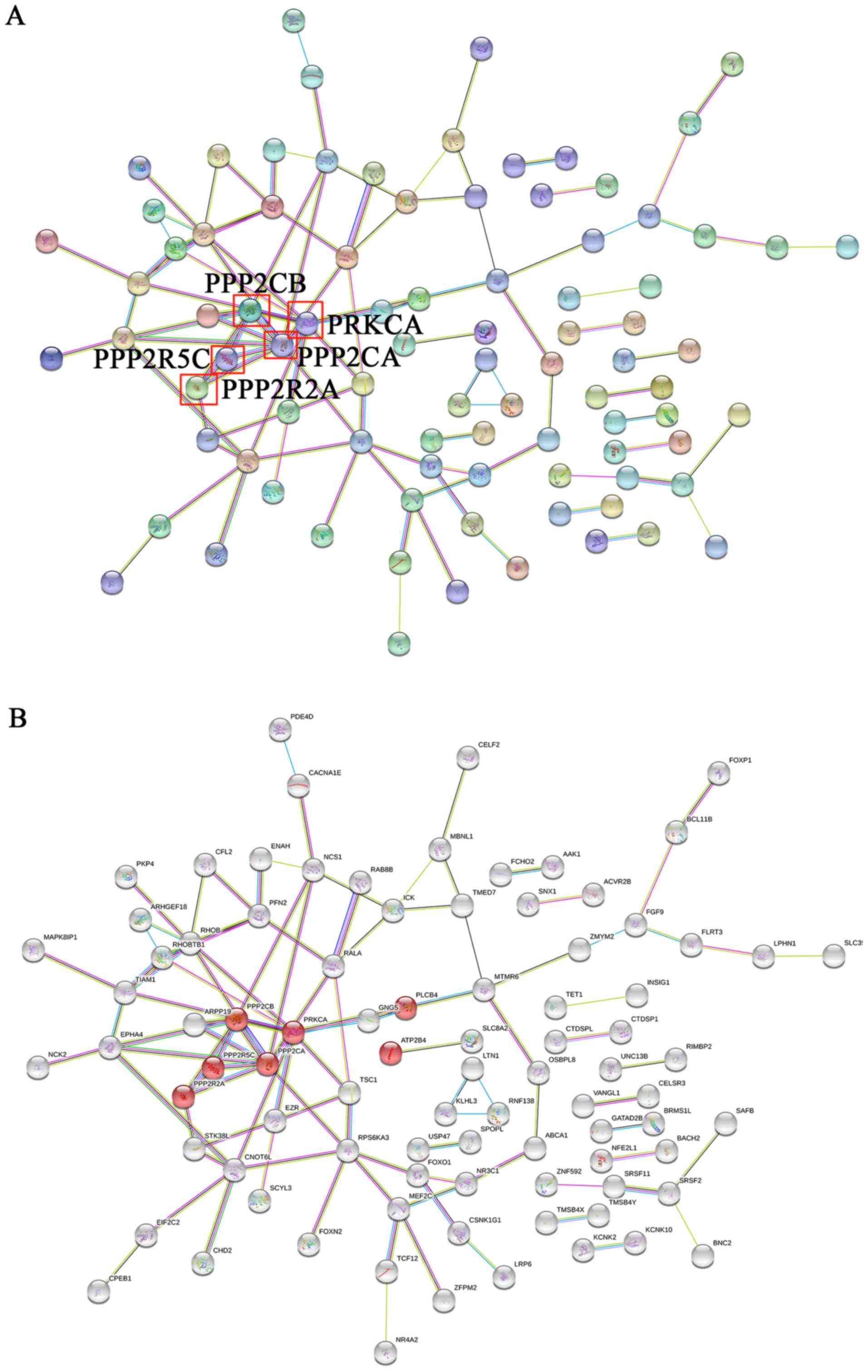
signaling (Table III). A
protein-protein interaction network was constructed using the
String database and the results demonstrated that the core genes of
the network were PPP2CB, PPP2R5C, PPP2R2A, PPP2CA and
PRKCA (Fig. 7A). Furthermore,
these key genes code for proteins of the kinase family and are
involved in adrenergic signaling in cardiomyocytes (Fig. 7B). These results indicate that
exosomal miR-183 is involved in cardiomyocyte adrenergic signaling
via the regulation of protein kinase activity.
| Table II.The top 30 target genes of miR-183
with higher scores in the TargetScan database. |
Table II.
The top 30 target genes of miR-183
with higher scores in the TargetScan database.
| No. | Target gene | Total context
score | Context score of
the aggregate PCT |
|---|
| 1 | SPRY3 | −0.18 | 0.9 |
| 2 | PFN2 | −0.7 | 0.9 |
| 3 | PSEN2 | −0.52 | 0.85 |
| 4 | PPP2R5C | −0.41 | 0.85 |
| 5 | PPP2CA | −0.49 | 0.85 |
| 6 | PDE4D | −0.3 | 0.85 |
| 7 | OSBPL8 | −0.28 | 0.85 |
| 8 | MAL2 | −0.62 | 0.85 |
| 9 | LRP6 | −0.53 | 0.85 |
| 10 | KCNK10 | −0.46 | 0.85 |
| 11 | IDH2 | −0.4 | 0.85 |
| 12 | FCHO2 | −0.39 | 0.85 |
| 13 | EZR | −0.36 | 0.85 |
| 14 | ENAH | −0.41 | 0.85 |
| 15 | DUSP10 | −0.47 | 0.85 |
| 16 | DGCR2 | −0.22 | 0.85 |
| 17 | CTDSP1 | −0.33 | 0.85 |
| 18 | CHD2 | −0.26 | 0.85 |
| 19 | ARHGAP21 | −0.37 | 0.85 |
| 20 | KLHL28 | −0.31 | 0.84 |
| 21 | C16orf72 | −0.43 | 0.83 |
| 22 | PRKCA | −0.1 | 0.77 |
| 23 | PHF15 | −0.4 | 0.77 |
| 24 | PDCD4 | −0.43 | 0.77 |
| 25 | KIAA0368 | −0.22 | 0.77 |
| 26 | KIAA0182 | −0.23 | 0.77 |
| 27 | DAGLA | −0.19 | 0.77 |
| 28 | CDK5R1 | −0.32 | 0.77 |
| 29 | ARPP-19 | −0.5 | 0.77 |
| 30 | AP3M1 | −0.26 | 0.77 |
| Table III.Kyoto Encyclopedia of Genes and
Genomes pathway analysis of miR-183 target genes. |
Table III.
Kyoto Encyclopedia of Genes and
Genomes pathway analysis of miR-183 target genes.
| No. | Pathway
description | False discovery
rate | Matching proteins
in network |
|---|
| 1 | Dopaminergic
synapse | 0.0171 | GNG5, PLCB4,
PPP2CA, PPP2CB, PPP2R2A, PPP2R5C, PRKCA |
| 2 | Adrenergic
signaling in cardiomyocytes | 0.0186 | ATP2B4, PLCB4,
PPP2CA, PPP2CB, PPP2R2A, PPP2R5C, PRKCA |
| 3 | Regulation of actin
cytoskeleton | 0.0186 | CFL2, ENAH, EZR,
FGF9, PFN2, TIAM1, TMSB4X, TMSB4Y |
| 4 | Gastric acid
secretion | 0.0186 | EZR, KCNK10, KCNK2,
PLCB4, PRKCA |
| 5 | AMPK signaling
pathway | 0.0214 | FOXO1, PPP2CA,
PPP2CB, PPP2R2A, PPP2R5C, TSC1 |
| 6 | MAPK signaling
pathway | 0.0428 | CACNA1E, DUSP10,
FGF9, MAP3K4, MAPK8IP1, MEF2C, PRKCA, RPS6KA3 |
| 7 | Long-term
depression | 0.0473 | PLCB4, PPP2CA,
PPP2CB, PRKCA |
Discussion
In 2012, the World Health Organization Global Status
Report on non-communicable diseases revealed that 7.4 million
mortalities were a result of ischemic heart disease (15). Specifically, acute MI has a high
morbidity and mortality (16). It is
thus necessary to elucidate early diagnostic markers. Circulating
miRNAs have become novel biomarkers for the detection of various
diseases, including those of the cardiovascular system (3). Previous studies have demonstrated that
certain circulating miRNAs are involved in acute MI, including
cardiac-specific miRNAs, non-cardiac miRNAs (17) and long non-coding RNA (3). Circulating non-coding RNAs can be used
as novel biomarkers in the diagnosis of cardiovascular diseases and
can assist in cell communication and signal transduction (3,18). Along
with the intensification of exosome research, exosomal miRNAs have
become novel diagnostic markers in various diseases (19–21).
However, the expression of exosomal miRNAs between patients with MI
and healthy individuals is yet to be fully elucidated, and
confirming the expression profile of exosomal miRNAs in MI is
crucial for performing functional research on miRNAs in MI.
Exosomes are 30–120 nm endocytic membrane-derived
vesicles (22) that have been
reported to serve crucial roles in the clinical diagnosis of
certain conditions (22), including
ovarian cancer, lung cancer, cardiovascular disease, colorectal
cancer (23), and type 1 diabetes
(24). Although these exosomes
comprise different RNA species, lipids and proteins (25), miRNA levels are closely associated
with disease pathophysiology and are high in plasma exosomes
(26). A previous study has
demonstrated that damaged cardiac muscle cells release exosomes,
which regulate the expression of sarcomeric and ion channel genes
involved in cardiac conductance, rhythmicity and automaticity via
the release of cardiac-specific miRNAs (27).
The present study demonstrated that 50 exosomal
miRNAs were dysregulated in patients with MI. Among the
dysregulated exosomal miRNAs, miR-183 expression was significantly
upregulated in these patients. Previous studies have revealed that
miR-183 is primarily involved in the regulation of cancer cell
proliferation, invasion and migration in various types of tumor
(28–30). Recently, a study demonstrated that
the angiogenic factor AGGF1, effectively treats cardiac hypertrophy
and heart failure by regulating miR-183 (31). However, the role of miR-183 in MI
diagnosis is still unknown. To the best of our knowledge, the
present study was the first to demonstrate that exosomal miR-183 is
significantly upregulated in patients with MI.
To assess the association between exosomal miR-183
and myocardial ischemic injury, the expression of exosomal miR-183
was measured in healthy individuals, patients with stable angina
pectoris and patients with MI. It was revealed that with the
increase in the severity of myocardial injury, the expression of
exosomal miR-183 also increased. In particular, the expression of
exosomal miR-183 was the highest in patients with MI compared to
that in healthy controls and patients with stable angina pectoris.
To the best of our knowledge, this is the first study to reveal
that exosomal miR-183 levels are positively associated with the
degree of myocardial injury. A previous study have demonstrated
that miR-1, miR-133a and miR-34a are also associated with
myocardial injury severity through the induction of adverse
structural remodeling, which impairs cardiac contractile function
(32). The results of the integrated
bioinformatics analysis of the current study indicated that
exosomal miR-183 is primarily involved in cardiomyocyte adrenergic
signaling via the regulation of protein kinase activity. To the
best of our knowledge, there is no study that assesses the
involvement of miR-183 in cardiomyocyte adrenergic signaling via
the regulation of protein kinase activity. This should therefore be
further assessed in future studies. However, the current study has
certain limitations. For example, the effects and mechanisms of
exosomal miR-183 on cardiac myocyte function need to be further
assessed. In addition, the number of cases was small in the present
study and as such, there is a requirement for large-scale clinical
validation.
In summary, the present study provides an insight
into the dynamic changes in exosomal miR-183 levels observed in the
early phase of MI and confirms that exosomal miR-183 expression is
positively associated with myocardial ischemic injury. Furthermore,
the current study confirmed that exosomal miR-183 is primarily
involved in cardiomyocyte adrenergic signaling via the regulation
of certain protein kinases. Thus, exosomal miR-183 may be a novel
diagnostic biomarker for patients with myocardial ischemic
injury.
Acknowledgements
Not applicable.
Funding
No funding was received.
Availability of data and materials
The datasets used and/or analyzed during the current
study are available from the corresponding author on reasonable
request.
Authors' contributions
XZ and YJ was responsible for drafting the
manuscript, as well as the analysis and interpretation of data. HC
and HY collected the data. WG contributed to design of the current
study. All authors read and approved the final manuscript
Ethics approval and consent to
participate
The present study was approved by the Ethics
Committee of the First Hospital of Shanxi Medical University
(Taiyuan, China). Written informed consent was obtained from each
patient or their relatives prior to Collection of plasma
samples.
Patient consent for publications
All patients agreed with the publication of this
study
Competing interests
The authors declare that they have no competing
interests.
References
|
1
|
Xu W, Yu H, Ma R, Ma L, Liu Q and Shan H,
Wu C, Zhang R, Zhou Y and Shan H: Apelin protects against
myocardial ischemic injury by inhibiting dynamin-related protein 1.
Oncotarget. 8:100034–100044. 2017.PubMed/NCBI
|
|
2
|
Giustino G and Dangas GD:
Ischemia-reperfusion injury and ischemic post-conditioning in acute
myocardial infarction: Lost in translation. Catheter Cardiovasc
Interv. 90:1068–1069. 2017. View Article : Google Scholar : PubMed/NCBI
|
|
3
|
Gao L, Liu Y, Guo S, Yao R, Wu L, Xiao L,
Wang Z, Liu Y and Zhang Y: Circulating long noncoding RNA HOTAIR is
an essential mediator of acute myocardial infarction. Cell Physiol
Biochem. 44:1497–1508. 2017.PubMed/NCBI
|
|
4
|
Willeit P, Welsh P, Evans JDW, Tschiderer
L, Boachie C, Jukema JW, Ford I, Trompet S, Stott DJ, Kearney PM,
et al: High-sensitivity cardiac troponin concentration and risk of
first-ever cardiovascular outcomes in 154,052 participants. J Am
Coll Cardiol. 70:558–568. 2017. View Article : Google Scholar : PubMed/NCBI
|
|
5
|
Shemisa K, Bhatt A, Cheeran D and Neeland
IJ: Novel biomarkers of subclinical cardiac dysfunction in the
general population. Curr Heart Fail Rep. 14:301–310. 2017.
View Article : Google Scholar : PubMed/NCBI
|
|
6
|
Paiva S and Agbulut O: MiRroring the
multiple potentials of MicroRNAs in acute myocardial infarction.
Front Cardiovasc Med. 4:732017. View Article : Google Scholar : PubMed/NCBI
|
|
7
|
Zhou J, Li XL, Chen ZR and Chng WJ:
Tumor-derived exosomes in colorectal cancer progression and their
clinical applications. Oncotarget. 8:100781–100790. 2017.PubMed/NCBI
|
|
8
|
Alipoor SD, Mortaz E, Garssen J,
Movassaghi M, Mirsaeidi M and Adcock IM: Exosomes and exosomal
miRNA in respiratory diseases. Mediators Inflamm. 2016:56284042016.
View Article : Google Scholar : PubMed/NCBI
|
|
9
|
Anderson JL, Adams CD, Antman EM, Bridges
CR, Califf RM, Casey DE Jr, Chavey WE II, Fesmire FM, Hochman JS,
Levin TN, et al: 2012 ACCF/AHA focused update incorporated into the
ACCF/AHA 2007 guidelines for the management of patients with
unstable angina/non-ST-elevation myocardial infarction: A report of
the American College of Cardiology Foundation/American Heart
Association Task Force on Practice Guidelines. J Am Coll Cardiol.
61:e179–e347. 2013. View Article : Google Scholar : PubMed/NCBI
|
|
10
|
O' Flynn N, Timmis A, Henderson R, Rajesh
S and Fenu E; Guideline Development Group, : Management of stable
angina: Summary of NICE guidance. BMJ. 343:d41472011. View Article : Google Scholar : PubMed/NCBI
|
|
11
|
Au Yeung CL, Co NN, Tsuruga T, Yeung TL,
Kwan SY, Leung CS, Li Y, Lu ES, Kwan K, Wong KK, et al: Exosomal
transfer of stroma-derived miR21 confers paclitaxel resistance in
ovarian cancer cells through targeting APAF1. Nat Commun.
7:111502016. View Article : Google Scholar : PubMed/NCBI
|
|
12
|
Wang S, Ruan QF, Xie WG, Chen L, Jiang MJ,
Ruan JJ and Ye ZQ: Differential expression of microRNAs in serum of
severe burn patients and analysis of the signaling pathway at early
stage. Zhonghua Shao Shang Za Zhi. 33:639–643. 2017.(In Chinese).
PubMed/NCBI
|
|
13
|
Livak KJ and Schmittgen TD: Analysis of
relative gene expression data using real-time quantitative PCR and
the 2(-Delta Delta C(T)) method. Methods. 25:402–408. 2001.
View Article : Google Scholar : PubMed/NCBI
|
|
14
|
Verjans R, Peters T, Beaumont FJ, van
Leeuwen R, van Herwaarden T, Verhesen W, Munts C, Bijnen M, Henkens
M, Diez J, et al: MicroRNA-221/222 family counteracts myocardial
fibrosis in pressure overload-induced heart failure. Hypertension.
71:280–288. 2018. View Article : Google Scholar : PubMed/NCBI
|
|
15
|
Maarman GJ, Mendham AE, Lamont K and
George C: Review of a causal role of fructose-containing sugars in
myocardial susceptibility to ischemia/reperfusion injury. Nutr Res.
42:11–19. 2017. View Article : Google Scholar : PubMed/NCBI
|
|
16
|
Tran HV, Lessard D, Tisminetzky MS,
Yarzebski J, Granillo EA, Gore JM and Goldberg R: Trends in length
of hospital stay and the impact on prognosis of early discharge
after a first uncomplicated acute myocardial infarction. Am J
Cardiol. 121:397–402. 2018. View Article : Google Scholar : PubMed/NCBI
|
|
17
|
Wang ZH, Sun XY, Li CL, Sun YM, Li J, Wang
LF and Li ZQ: miRNA-21 expression in the serum of elderly patients
with acute myocardial infarction. Med Sci Monit. 23:5728–5734.
2017. View Article : Google Scholar : PubMed/NCBI
|
|
18
|
Wang F, Long G, Zhao C, Li H, Chaugai S,
Wang Y, Chen C and Wang DW: Atherosclerosis-related circulating
miRNAs as novel and sensitive predictors for acute myocardial
infarction. PLoS One. 9:e1057342014. View Article : Google Scholar : PubMed/NCBI
|
|
19
|
Pan J, Ding M, Xu K, Yang C and Mao LJ:
Exosomes in diagnosis and therapy of prostate cancer. Oncotarget.
8:97693–97700. 2017.PubMed/NCBI
|
|
20
|
Pillay P, Moodley K, Moodley J and Mackraj
I: Placenta-derived exosomes: Potential biomarkers of preeclampsia.
Int J Nanomedicine. 12:8009–8023. 2017. View Article : Google Scholar : PubMed/NCBI
|
|
21
|
Zhang G and Yang P: A novel cell-cell
communication mechanism in the nervous system: Exosomes. J Neurosci
Res. 96:45–52. 2018. View Article : Google Scholar : PubMed/NCBI
|
|
22
|
Lin J, Li J, Huang B, Liu J, Chen X, Chen
XM, Xu YM, Huang LF and Wang XZ: Exosomes: Novel biomarkers for
clinical diagnosis. ScientificWorldJournal. 2015:6570862015.
View Article : Google Scholar : PubMed/NCBI
|
|
23
|
Jia S, Zhang R, Li Z and Li J: Clinical
and biological significance of circulating tumor cells, circulating
tumor DNA, and exosomes as biomarkers in colorectal cancer.
Oncotarget. 8:55632–55645. 2017. View Article : Google Scholar : PubMed/NCBI
|
|
24
|
Garcia-Contreras M, Shah SH, Tamayo A,
Robbins PD, Golberg RB, Mendez AJ and Ricordi C: Plasma-derived
exosome characterization reveals a distinct microRNA signature in
long duration Type 1 diabetes. Sci Rep. 7:59982017. View Article : Google Scholar : PubMed/NCBI
|
|
25
|
Sluijter JPG, Davidson SM, Boulanger CM,
Buzás EI, de Kleijn DPV, Engel FB, Giricz Z, Hausenloy DJ, Kishore
R, Lecour S, et al: Extracellular vesicles in diagnostics and
therapy of the ischaemic heart: Position paper from the working
group on cellular biology of the heart of the European society of
cardiology. Cardiovasc Res. 114:19–34. 2018. View Article : Google Scholar : PubMed/NCBI
|
|
26
|
Li DB, Liu JL, Wang W, Li RY, Yu DJ, Lan
XY and Li JP: Plasma exosomal miR-422a and miR-125b-2-3p serve as
biomarkers for ischemic stroke. Curr Neurovasc Res. 14:330–337.
2017. View Article : Google Scholar : PubMed/NCBI
|
|
27
|
Chistiakov DA, Orekhov AN and Bobryshev
YV: Cardiac extracellular vesicles in normal and infarcted heart.
Int J Mol Sci. 17(pii): E632016. View Article : Google Scholar : PubMed/NCBI
|
|
28
|
Macedo T, Silva-Oliveira RJ, Silva VAO,
Vidal DO, Evangelista AF and Marques MMC: Overexpression of mir-183
and mir-494 promotes proliferation and migration in human breast
cancer cell lines. Oncol Lett. 14:1054–1060. 2017. View Article : Google Scholar : PubMed/NCBI
|
|
29
|
Lima CR, Gomes CC and Santos MF: Role of
microRNAs in endocrine cancer metastasis. Mol Cell Endocrinol.
456:62–75. 2017. View Article : Google Scholar : PubMed/NCBI
|
|
30
|
Anwar SL, Krech T, Hasemeier B, Schipper
E, Schweitzer N, Vogel A, Kreipe H, Buurman R, Skawran B and
Lehmann U: hsa-mir-183 is frequently methylated and related to poor
survival in human hepatocellular carcinoma. World J Gastroenterol.
23:1568–1575. 2017. View Article : Google Scholar : PubMed/NCBI
|
|
31
|
Yao Y, Lu Q, Hu Z, Yu Y, Chen Q and Wang
QK: A non-canonical pathway regulates ER stress signaling and
blocks ER stress-induced apoptosis and heart failure. Nat Commun.
8:1332017. View Article : Google Scholar : PubMed/NCBI
|
|
32
|
Qipshidze Kelm N, Piell KM, Wang E and
Cole MP: MicroRNAs as predictive biomarkers for myocardial injury
in aged mice following myocardial infarction. J Cell Physiol.
233:5214–5221. 2018. View Article : Google Scholar : PubMed/NCBI
|