Introduction
Polycystic ovary syndrome (PCOS) is a common but
complex reproductive dysfunction and is an endocrine disorder
comprising an abnormal glucose metabolism (1). Surveys have revealed that PCOS is
mainly detected in adolescent females and women of childbearing
age, and the incidence rate is ~10% of all women globally (2). Other studies have indicated that
>70% of female cases of infertility and ~40% of spontaneous
abortions are linked to PCOS (3).
PCOS is associated with sparse ovulation or prolonged anovulation
and significantly elevated androgen levels, while its clinical
manifestations have a high degree of heterogeneity amongst patients
(4).
In the early stage of the disease, infertility,
obesity and menstrual disorders may occur. In the advanced stage,
the risk of cardiovascular and cerebrovascular diseases and type II
diabetes increases significantly, which markedly affects the
quality of life, as well as the physical and mental health of
patients (5). At present, the
majority of studies suggest that PCOS is caused by genetic
mutations as well as antiepileptic drugs, particularly valproate
(6). Although the in vitro
fertilization-embryo transfer (IVF-ET) technique is a routine
method to remedy infertility in patients with PCOS wishing to
conceive, the associated endocrine and glucose metabolism disorder
induces abnormal follicular development, which leads to the
production of poor-quality embryos in patients with PCOS undergoing
IVF-ET. This results in fewer oocytes available for the patient and
reduces the success rate of the transplantation (7).
Aquaporin (AQP) is a hydrophobic internal membrane
protein that selectively lets water molecules pass, which is an
important pathway for water to diffuse through the cell membrane
(protein-mediated water transport) apart from physical diffusion
(8). To date, 13 types of AQPs,
AQP0-12, have been discovered in mammals. AQPs are small-molecule
proteins with a molecular weight of 25–34 kDa that are located on
the membranes of epithelial and endothelial cells (9). The family of AQPs is divided into three
subgroups based on their genetic structure: Classical aquaporin
proteins (AQP0-2, −4–6 and −8), glycerol-water channel proteins
(AQP3, −7, −9 and −10), and super AQP (AQP11 and −12) (10). A study suggested that AQPs are
selectively expressed in the membranes of cells of the reproductive
system tissue, with their functions including involvement in saliva
secretion and the re-absorption and transportation of water
molecules, suggesting a close association between AQP and the
maintenance of the reproductive system (11). In a study by McConnell et al
(12), flow cytometric analysis
revealed that AQP7-9 were present on the cell membranes of rats,
and these AQPs were involved in water transportation of granulosa
cells and associated with the development of follicles.
Therefore, the present study examined the
association between the relative expression levels of AQP8 and −9
in ovary tissues of patients with PCOS and examined their possible
associations with the outcomes of IVF-ET.
Materials and methods
Reagents
The reverse transcriptase and reverse transcription
(RT) kit (iScript Advanced cDNA Synthesis kit; cat. no. 1708842)
was purchased from Takara Bio, Inc. The 2X SYBR Green quantitative
PCR (qPCR) Mix and the RevertAid First Strand complementary (c)DNA
Synthesis kit (DyNAmo Flash SYBR Green qPCR kit; cat. no. F415S)
were purchased from Invitrogen (Thermo Fisher Scientific, Inc.),
and the β-actin, AQP8 and AQP9 primers (Sangon Biotech Co., Ltd.)
were designed and synthesized by Shanghai Sangon Biotech. The
primer sequences used are listed in Table I.
 | Table I.Sequences of primers used for PCR. |
Table I.
Sequences of primers used for PCR.
| Gene | Upstream primer | Downstream
primer |
|---|
| AQP8 |
5′-CGGATGTCTATCGGTCATTGAGAA-3′ |
5′-GCGACACAGCAGGGTTGAAG-3′ |
| AQP9 |
5′-GGTCTTGAAGAGCAGCTTAG-3′ |
5′-GTTCGCCAGAGATAGATACG-3′ |
| β-actin |
5′-TTCCAGCCTTCCTTCTGG-3′ |
5′-TTGCGCTCAGGAGGAGCAAT-3′ |
Clinical information of patients
A total of 45 cases with PCOS undergoing IVF-ET
(test group) at Guangzhou Women and Children's Medical Center
(Guangdong, China) from January 2015 to May 2016 were included in
the present study. They had an average age of 28.45±4.86 years with
an age range of 24 to 35 years. The patients were diagnosed based
on the assessment criteria of the American Society for Reproductive
Medicine and the European Society of Human Reproduction and
Embryology (13,14). In the control group, 50 cases were
included that had oviduct obstruction or ovarian cysts and who had
undergone ovary biopsies with an age range of 25–34 years and an
average age of 28.21±4.23 years. The inclusion criteria included
sparse or prolonged anovulation or irregular ovulation (determined
by blood and ovulation tests), elevated hormone expression levels,
increased ovarian size, and >12 follicles on each side
(diameter, 2–8 mm). The exclusion criteria were a family history of
disease (e.g., diabetes), endocrine disorders (e.g., congenital
adrenal hyperplasia, hypothyroidism or hyperthyroidism and
hyperprolactinemia), no compliance with the treatment and follow-up
schedule and spouses with infertility. The present study was
approved by the Ethics Committee of the First Affiliated Hospital
of Jinan University (Guangzhou, China), and the patients and their
families provided written informed consent.
Medication protocol
The superovulation regimen (standard long-term
ovulation promotion) included Inda (ethinyl estradiol cyproterone
tablets) administered orally, 1 tablet per day on the third day of
the first week of the menstrual period. The long-acting Daphneline
(1.25 mg) was intramuscularly injected on the 17th day of the
administration of Inda and patients were treated with Gonafin
(recombinant follicle-stimulating hormone; 150 U/day) by
intramuscular injection to promote follicular growth when the
adjustment was satisfactory. When >2 follicle diameters
measuring 18 mm and >3 measuring 17 mm were reached, human
chorionic gonadotropin (HCG; 10,000 U) was intramuscularly injected
and the eggs were retrieved after 35–36 h. The IVF/intracytoplasmic
sperm injection was performed based on standard clinical practice
and at three days after oocyte retrieval, the oocytes were
selected. A total of 2–3 embryos were selected for transplantation
and, from the remaining oocytes, 2–4 high-quality embryos were
preserved in liquid nitrogen at −126°C. The low-quality oocytes
were further cultured for 5–6 days and the embryos formed were
reserved in liquid nitrogen at −126°C for subsequent use if a
high-quality embryo was unavailable.
Pregnancy outcomes
The patients who had received IVF for 14 days were
tested for fasting progesterone and HCG hormone expression levels.
If the expression level of HCG was ≥5 IU/ml and, after a further 2
weeks, the gestational sac within the uterus was observed by
B-ultrasound, clinical pregnancy was diagnosed.
Identification of high-quality
embryos
Embryos were divided into four grades: Level I,
uniform blastomeres and a cytoplasmic fraction of <10%; level
II, slightly uneven blastomeres and cytoplasmic fraction of 10–20%;
level III, slightly uneven blastomeres and cytoplasmic fraction of
21–50%; and grade IV, cytoplasmic fraction of >50%. Embryos with
>6 cells and a grade of I/II were rated as high-quality
embryos.
Specimen collection
Ovary tissues were obtained after resection from the
fallopian tube (e.g., laparoscopic ovarian wedge resection) for
patients in the control group or the ovarian tissue was obtained by
ovarian biopsy during surgery for ovarian cysts.
Detection of relative expression of
AQP8 and AQP9 mRNA in ovarian tissues by RT-qPCR
The ovarian tissues were repeatedly ground with a
grinding rod and shaken with TRIzol reagent (Thermo Fisher
Scientific, Inc.), and lysed for 30 min. The purity and
concentration of the extracted total RNA was determined by UV
spectrophotometry. The ratio of the optical density at 260 vs. 280
nm was within the range of 1.8–2.1. Complementary (c)DNA was
synthesized by strictly following the manufacturer's protocols for
AQP8 and AQP9 mRNA. The PCR system contained 100 ng/µl cDNA
template, 0.5 mol/l of the primer, 2.0 µl of ٢X dNTP, 2.5 µl of the
buffer solution, ١.٥ mol/l of MgCl2 and 1.0 IU of Taq
DNA polymerase, which was adjusted to a total volume of 20 µl using
nuclease-free water. PCR conditions were as follows:
Pre-denaturation at ٩٥°C for ٥ min, 95°C for 30 sec, 60°C for 15
sec and 60°C for 15 sec, for a total of 45 cycles. U6 was used as
the reference gene and the 2−∆∆Cq method (15) was applied to determine the expression
level.
Statistical analysis
SPSS 22.0 statistical software (IBM Corp.) was used
to analyze the data and GraphPad Prism 5 (GraphPad Inc.) was used
to generate the figures. Measurement data are expressed as the mean
± standard deviation. The comparison between groups was based on
the normality test results from the t-test. An independent t-test
was used for comparison between groups. A paired t-test was used
for comparisons between the before and after group and a rank sum
test was used for data that did not conform to normal distribution.
Count data were expressed as n (%) and comparisons between groups
were performed using the Chi-squared test. P<0.05 was considered
to indicate a statistically significant difference.
Results
Clinical data of the two groups of
patients
When comparing the clinical information between the
two groups of patients, no significant difference in any of the
indicators was identified (P>0.05; Table II), except for luteinizing hormone,
anti-mullerian hormone and testosterone levels (P<0.05).
| Table II.Comparison of clinicopathological and
demographic characteristics between the two groups of subjects
[n(%)]. |
Table II.
Comparison of clinicopathological and
demographic characteristics between the two groups of subjects
[n(%)].
| Parameter | Observation group
(n=45) | Control group
(n=50) | t/χ2 | P-value |
|---|
| Duration of
infertility (years) |
|
| 0.01 | 0.91 |
| >2 n
(%) | 23 (51.11) | 25 (52.08) |
|
|
| ≤2 n
(%) | 22 (46.81) | 25 (53.19) |
|
|
| Age (years) |
|
| 0.01 | 0.96 |
| >30 n
(%) | 25 (55.56) | 28 (56.00) |
|
|
| ≤30 n
(%) | 20 (44.44) | 22 (44.00) |
|
|
| Degree of
education |
|
| 0.22 | 0.63 |
| <High
school n (%) | 15 (33.33) | 19 (38.00) |
|
|
| ≥High
school n (%) | 30 (66.67) | 31 (62.00) |
|
|
| Smoking history |
|
| 0.33 | 0.56 |
| Yes n
(%) | 3 (6.67) | 2 (4.00) |
|
|
| No n
(%) | 42 (93.33) | 48 (96.00) |
|
|
| History of
alcoholism |
|
| 0.33 | 0.56 |
| Yes n
(%) | 3 (6.67) | 2 (4.00) |
|
|
| No n
(%) | 42 (93.33) | 48 (96.00) |
|
|
| Area of
residence |
|
| 0.66 | 0.41 |
| Rural
area n (%) | 11 (24.44) | 16 (32.00) |
|
|
| City n
(%) | 34 (75.56) | 34 (68.00) |
|
|
| Body mass index
(kg/m2) | 22.54±2.62 | 21.84±2.15 | 1.42 | 0.15 |
| FSH (IU/l) |
5.95±2.38 |
6.49±3.29 | 0.91 | 0.37 |
| LH (IU/l) | 12.39±8.12 |
5.92±3.88 | 5.03 | <0.01 |
| AMH (ng/ml) |
9.68±4.21 |
5.37±4.97 | 4.53 | <0.01 |
| T (ng/ml) |
0.69±0.24 |
0.51±0.18 | 4.16 | <0.01 |
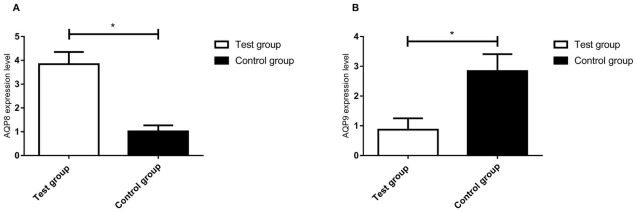
AQP8 and AQP9 mRNA expression in the
two groups of patients
AQP8 and AQP9 mRNA expression in the ovarian tissues
of the two groups of patients was detected by RT-qPCR. The levels
of AQP8 mRNA in the control group were significantly lower than
those in the test group and there was a significant difference
between the two groups (t=37.75, P<0.01; Fig. 1A). The AQP9 mRNA expression levels in
the control group were significantly higher than those in the test
group and there was a statistically significant difference between
the two groups (t=19.59, P<0.01; Fig.
1B).
Embryonic quality and number of
oocytes retrieved from the test group
Based on the median expression levels of AQP8 and
AQP9 mRNA, the patients were divided into high expression and low
expression groups. The quality of embryos and the number of oocytes
obtained were compared between these groups, and it was indicated
that the number of oocytes obtained in the AQP8 mRNA high
expression group was significantly lower than that obtained in the
low expression group (t=2.64, P<0.01); however, there was no
significant difference in the number of high-quality embryos
between the AQP8 mRNA high and low expression groups (t=1.02,
P>0.05). There was no significant difference in the number of
oocytes obtained from patients with high vs. low AQP9 mRNA
expression (t=0.71, P>0.05), and there was no significant
difference in the number of high-quality embryos between the high
and low expression groups of AQP9 mRNA (t=1.01, P>0.05; Table III).
| Table III.Embryo quality and egg count in the
test group. |
Table III.
Embryo quality and egg count in the
test group.
|
| AQP8 |
|
| AQP9 |
|
|
|---|
|
|
|
|
|
|
|
|
|---|
| Group | High level
(n=17) | Low level
(n=28) | t | P-value | High level
(n=22) | Low level
(n=23) | t | P-value |
|---|
| Oocytes retrieved
(n) | 11.84±3.54 | 14.36±2.81 | 2.64 | 0.01 | 13.58±2.52 | 13.08±2.17 | 0.71 | 0.47 |
| High-quality
embryos (n) | 10.84±2.76 | 9.86±3.29 | 1.02 | 0.31 | 12.28±2.28 | 11.48±2.94 | 1.01 | 0.31 |
Observation of pregnancy outcomes of
patients
Patients were grouped based on the median expression
levels of AQP8 and AQP9 mRNA. The pregnancy rate in the AQP8 mRNA
high expression group was not significantly different from that in
the AQP8 mRNA low expression group (P>0.05). There was no
significant difference in the rate of abortions between the two
groups (P>0.05). The pregnancy rate among patients in the high
AQP9 mRNA expression group was significantly higher than that in
the AQP9 mRNA low expression group (P<0.05). The high AQP9 mRNA
expression group also had a lower miscarriage rate than the AQP9
mRNA low expression group (P<0.05; Table IV).
| Table IV.Pregnancy-associated outcomes in the
test group. |
Table IV.
Pregnancy-associated outcomes in the
test group.
|
| AQP8 |
| AQP9 |
|
|---|
|
|
|
|
|
|
|---|
| Group | High level
(n=17) | Low level
(n=28) | P-value | High level
(n=22) | Low level
(n=23) | P-value |
|---|
| Conception |
|
| 0.76 |
|
| 0.02 |
| No | 10 (58.82) | 18 (64.29) |
| 4 (18.18) | 11 (47.83) |
|
|
Yes | 7 (41.18) | 10 (35.71) |
| 18 (81.82) | 12 (52.17) |
|
| Spontaneous
abortion |
|
| 0.49 |
|
| 0.04 |
| No | 14 (82.35) | 20 (71.43) |
| 16 (72.73) | 9 (39.13) |
|
|
Yes | 3 (17.65) | 8 (28.57) |
| 6 (27.27) | 14 (60.87) |
|
Discussion
As a gynecological endocrine disease, PCOS has a
complex range of causes, which include the metabolic system,
endocrine system and ovarian regulation system (16). Patients with PCOS encountered in the
clinic may also have varying types and degrees of metabolic
disorders (e.g., insulin resistance, high blood lipids and high
blood pressure); however, what impairs the reproductive health of
females the most is ovulation dysfunction. The mechanisms of
ovulation abnormalities are highly complex and may be induced by
factors including hypothalamic-pituitary-ovarian function disorders
and abnormal hormone feedback signals in the body (17).
As one of the gene families that have been receiving
an increased amount of attention from researchers in recent years,
AQPs constitute an important route of water transportation that is
separate from simple diffusion (18). Previous studies have indicated that
AQPs have a regulatory role in functions associated with the
mammalian reproductive system and affect the formation of the
embryo sac; they may cause changes in the uterine cavity fluid at
the implantation stage and the formation of the oviduct (19). A difference in the transportation of
substances via AQP8 has been noted among different species, and it
has been indicated that AQP8 facilitates water and urea
transportation in mice, but fails to transport glycerol, whereas it
transports only water in rats but not urea and glycerol (20). However, in recent years, the known
roles of AQP8 have expanded, i.e., in addition to the expression on
the cell membrane, expression of AQP8 has also been observed on the
mitochondrial membrane (21). The
extensive expression of AQP8 in various tissue types in the body
has been assessed numerous times and it has been indicated that
AQP8 mRNA expression is present in ovarian tissues. As one of the
members of the AQP family, AQP9 is highly expressed in the liver,
and varying degrees of differential expression among different
individuals have been reported in the testis, nervous system and
thyroid system (22). In a study by
Sales et al (23), AQP9 was
indicated to have an important role in the development of
follicular cells. However, there is a dispute on its expression in
patients with PCOS. In a previous study on PCOS, no difference in
AQP9 expression in ovarian tissues was identified between the test
group and the control group and yet, low expression was detected in
the granulosa cells of the PCOS patients (24). In the present study, a significant
reduction of AQP9 mRNA was detected in the ovarian tissue of PCOS
patients compared to the control group. The discrepancy of these
results may be due to the recruitment of more patients in the
current study, using different tissues.
In the present study, AQP8 and AQP9 mRNA expression
in the ovarian tissue of the two groups of patients was detected by
RT-qPCR. AQP8 mRNA expression in the PCOS group was significantly
higher than that in the control group, while AQP9 mRNA expression
was significantly lower. In previous studies (25–27),
ovarian tissue and follicular fluid were examined by PCR and it was
revealed that AQP9 mRNA expression was significantly lower in the
test group compared with that in the control group, suggesting that
patients with high expression of AQP8 and low expression of AQP9
may have PCOS. AQP8 and AQP9 are expected to become biological
indicators for the condition. A study by Su et al (28), indicated that in female AQP8-knockout
mice, the ovulation numbers were higher than those in wild-type
mice. In the present study, the PCOS patients were divided into
high and low expression groups based on the median value of AQP8
mRNA expression, and the number of ovulations in the low expression
group was significantly higher than that in the high expression
group in patients undergoing IVF-ET. This may be due to the water
transportation in granulosa cells being affected by the reduced
expression of AQP8, which may result in a reduction of apoptosis
among granulosa cells, as well as inhibition of follicular
disappearance and atresia. This indicates that AQP8 expression is
closely associated with the generation and development of egg
cells. At the time of completion of the present study, no
difference in the number of non-pregnant patients and the rate of
abortions was noted between the AQP8 mRNA high and low expression
groups. By contrast, the number of non-pregnant patients and
patients with miscarriage in the AQP9 mRNA high group was
significantly lower than that in the AQP9 low expression group.
This proved the association between the expression of AQP9 and
pregnancy-associated processes in PCOS patients.
Of note, the present study has several limitations.
First, only a small number of patient samples were analyzed, which
may have affected the results. Furthermore, only the ovarian tissue
of PCOS patients was examined, while the follicle fluid was not
analyzed; therefore, it remains elusive whether any possible
difference in the follicle fluid levels may have had an influence.
In addition, the underlying mechanism for the difference in AQP8
and AQP9 levels were not explored with regard to patients with
PCOS. A future study will endeavor to increase the number of
subjects and the type of samples analyzed, and further explore the
association between AQP8 and AQP9 in PCOS to determine the accuracy
of the experimental results and further confirm the validity of the
conclusions.
In conclusion, a differential expression of AQP8 and
AQP9 in ovarian tissues was identified between PCOS patients and
normal patients. AQP8 expression was closely associated with the
development of oocytes and AQP9 expression was associated with the
success of pregnancy in PCOS patients.
Acknowledgements
Not applicable.
Funding
The current study was supported by Guangdong
Provincial Science and Technology Hall (grant no.
2016A020218002).
Availability of data and materials
The complete data are available from the
corresponding author on reasonable request. The data are not
publicly available due to their containing information that could
compromise the privacy of research participants.
Authors' contributions
ZX and HL conceived the study and designed the
experiments, BL, LW and XZ contributed to data collection, and BYL
and XS performed the data analysis and interpreted the results. ZX
wrote the manuscript and HL contributed to the critical revision of
the article. All authors read and approved the final
manuscript.
Ethics approval and consent to
participate
This study was approved by the ethics committee of
Guangzhou Women and Children's Medical Center (Guangzhou Medical
University, Guangzhou, China). Written informed consent was
obtained from the patients and their families.
Patient consent for publication
Not applicable.
Competing interests
The authors declare that they have no competing
interests.
References
|
1
|
Rosenblum J and Ekhlaspour L: Polycystic
Ovary Syndrome. In: The MassGeneral Hospital for Children
Adolescent Medicine Handbook. Goldstein MA: Springer: pp. 187–193.
2017
|
|
2
|
Goetsch AL, Kimelman D and Woodruff TK:
Polycystic Ovary Syndrome. Fertility Preservation and Restoration
for Patients with Complex Medical Conditions Springer: pp. 231–248.
2017, View Article : Google Scholar
|
|
3
|
Connolly A and Beckett VA: Women's Health
in Primary Care. Cambridge University Press. (Cambridge). i–ii.
2017.
|
|
4
|
Johnston-MacAnanny EB and Berga SL:
Polycystic Ovary Syndrome. Clinical Reproductive Medicine and
Surgery Springer: pp. 123–137. 2017, View Article : Google Scholar
|
|
5
|
Franks S: Polycystic ovary syndrome. N
Engl J Med. 333:853–861. 1995. View Article : Google Scholar : PubMed/NCBI
|
|
6
|
Zaki M, Hassan N, El-Bassyouni HT, Kamal
S, Basha W, Azmy O and Amr K: Association of the Pro12Ala
polymorphism with the metabolic parameters in women with polycystic
ovary syndrome. Open Access Maced J Med Sci. 5:275–280.
2017.PubMed/NCBI
|
|
7
|
Chen WY, Du YQ, Guan X, Zhang HY and Liu
T: Effect of GnRHR polymorphisms on in vitro fertilization and
embryo transfer in patients with polycystic ovary syndrome. J Hum
Genet. 62:1065–1071. 2017. View Article : Google Scholar : PubMed/NCBI
|
|
8
|
Bienert GP and Chaumont F:
Aquaporin-facilitated transmembrane diffusion of hydrogen peroxide.
Biochim Biophys Acta. 1840:1596–1604. 2014. View Article : Google Scholar : PubMed/NCBI
|
|
9
|
Ishibashi K, Kondo S, Hara S and Morishita
Y: The evolutionary aspects of aquaporin family. Am J Physiol Regul
Integr Comp Physiol. 300:R566–R576. 2011. View Article : Google Scholar : PubMed/NCBI
|
|
10
|
Zhu C, Jiang Z, Bazer FW, Johnson GA,
Burghardt RC and Wu G: Aquaporins in the female reproductive system
of mammals. Front Biosci (Landmark Ed). 20:838–871. 2015.
View Article : Google Scholar : PubMed/NCBI
|
|
11
|
Mobasheri A, Marples D, Young IS, Floyd
RV, Moskaluk CA and Frigeri A: Distribution of the AQP4 water
channel in normal human tissues: Protein and tissue microarrays
reveal expression in several new anatomical locations, including
the prostate gland and seminal vesicles. Channels (Austin).
1:29–38. 2007. View Article : Google Scholar : PubMed/NCBI
|
|
12
|
McConnell NA, Yunus RS, Gross SA, Bost KL,
Clemens MG and Hughes FM Jr: Water permeability of an ovarian
antral follicle is predominantly transcellular and mediated by
aquaporins. Endocrinology. 143:2905–2912. 2002. View Article : Google Scholar : PubMed/NCBI
|
|
13
|
Society for Assisted Reproductive
Technology; American Society for Reproductive Medicine, . Assisted
reproductive technology in the United States: 2000 results
generated from the American Society for Reproductive
Medicine/Society for Assisted Reproductive Technology Registry.
Fertil Steril. 81:1207–1220. 2004. View Article : Google Scholar : PubMed/NCBI
|
|
14
|
European Society of Human Genetics;
European Society of Human Reproduction and Embryology, . The need
for interaction between assisted reproduction technology and
genetics: Recommendations of the European Societies of Human
Genetics and Human Reproduction and Embryology. Hum Reprod.
21:1971–1973. 2006. View Article : Google Scholar : PubMed/NCBI
|
|
15
|
Livak KJ and Schmittgen TD: Analysis of
relative gene expression data using real-time quantitative PCR and
the 2(-Delta Delta C(T)) method. Methods. 25:402–408. 2001.
View Article : Google Scholar : PubMed/NCBI
|
|
16
|
Azziz R, Carmina E, Chen Z, Dunaif A,
Laven JS, Legro RS, Lizneva D, Natterson-Horowtiz B, Teede HJ and
Yildiz BO: Polycystic ovary syndrome. Nat Rev Dis Primers.
2:160572016. View Article : Google Scholar : PubMed/NCBI
|
|
17
|
Gong JG, Campbell BK, Bramley TA,
Gutierrez CG, Peters AR and Webb R: Suppression in the secretion of
follicle-stimulating hormone and luteinizing hormone, and ovarian
follicle development in heifers continuously infused with a
gonadotropin-releasing hormone agonist. Biol Reprod. 55:68–74.
1996. View Article : Google Scholar : PubMed/NCBI
|
|
18
|
Jozefkowicz C, Scochera F and Alleva K:
Two aquaporins, multiple ways of assembly. Channels (Austin).
10:438–439. 2016. View Article : Google Scholar : PubMed/NCBI
|
|
19
|
Yeste M, Morató R, Rodríguez-Gil J, Bonet
S and Prieto-Martínez N: Aquaporins in the male reproductive tract
and sperm: Functional implications and cryobiology. Reprod Domest
Anim. 52 (Suppl 4):S12–S27. 2017. View Article : Google Scholar
|
|
20
|
Ferri D, Mazzone A, Liquori GE, Cassano G,
Svelto M and Calamita G: Ontogeny, distribution, and possible
functional implications of an unusual aquaporin, AQP8, in mouse
liver. Hepatology. 38:947–957. 2003. View Article : Google Scholar : PubMed/NCBI
|
|
21
|
Saparov SM, Liu K, Agre P and Pohl P: Fast
and selective ammonia transport by aquaporin-8. J Biol Chem.
282:5296–5301. 2007. View Article : Google Scholar : PubMed/NCBI
|
|
22
|
Elkjær M, Vajda Z, Nejsum LN, Kwon T,
Jensen UB, Amiry-Moghaddam M, Frøkiaer J and Nielsen S:
Immunolocalization of AQP9 in liver, epididymis, testis,
spleen, and brain. Biochem Biophys Res Commun. 276:1118–1128. 2000.
View Article : Google Scholar : PubMed/NCBI
|
|
23
|
Sales A, Lobo C, Carvalho A, Moura A and
Rodrigues A: Structure, function, and localization of aquaporins:
Their possible implications on gamete cryopreservation. Genet Mol
Res. 12:6718–6732. 2013. View Article : Google Scholar : PubMed/NCBI
|
|
24
|
Qu F, Wang FF, Lu XE, Dong MY, Sheng JZ,
Lv PP, Ding GL, Shi BW, Zhang D and Huang HF: Altered aquaporin
expression in women with polycystic ovary syndrome:
Hyperandrogenism in follicular fluid inhibits aquaporin-9 in
granulosa cells through the phosphatidylinositol 3-kinase pathway.
Hum Reprod. 25:1441–1450. 2010. View Article : Google Scholar : PubMed/NCBI
|
|
25
|
Maeda N, Funahashi T and Shimomura I:
Metabolic impact of adipose and hepatic glycerol channels aquaporin
7 and aquaporin 9. Nat Clin Pract Endocrinol Metab. 4:627–634.
2008. View Article : Google Scholar : PubMed/NCBI
|
|
26
|
Suzuki-Toyota F, Ishibashi K and Yuasa S:
Immunohistochemical localization of a water channel, aquaporin 7
(AQP7), in the rat testis. Cell Tissue Res. 295:279–285. 1999.
View Article : Google Scholar : PubMed/NCBI
|
|
27
|
Richard C, Gao J, Brown N and Reese J:
Aquaporin water channel genes are differentially expressed and
regulated by ovarian steroids during the periimplantation period in
the mouse. Endocrinology. 144:1533–1541. 2003. View Article : Google Scholar : PubMed/NCBI
|
|
28
|
Su W, Guan X, Zhang D, Sun M, Yang L, Yi
F, Hao F, Feng X and Ma T: Occurrence of multi-oocyte follicles in
aquaporin 8-deficient mice. Reprod Biol Endocrinol. 11:882013.
View Article : Google Scholar : PubMed/NCBI
|