Introduction
Rotavirus (RV) belongs to the reovirus subfamily and
is the most important causative agents of viral diarrhea in infants
and young children, as well as many young animals worldwide
(1). It is also the major causative
agent of acute diarrhea in children under 5 years of age, which may
lead to mortality in severe cases (2). There is currently no efficient drug for
the treatment of RV infections and vaccines remains the only
effective and economical means to prevent and control RV (3). RV-encoded structural proteins primarily
consist of VP2, VP6, VP4 and VP7, of which VP4 and VP7 are the
major neutralizing antigens (4). VP7
accounts for ~30% of the total viral proteins and mediates the
viral shedding and host invasion, which is required for RV
maturation (5). A previous study
demonstrated that the RV VP7 protein exhibits higher antigenicity
and immunogenicity than the VP4 protein (6). Therefore a large number of studies have
utilized VP7 as the primary target for engineering RV viral gene
subunit vaccines, RV nucleic acid vaccines and therapeutic
monoclonal antibodies (7,8). However, the ability to produce a large
quantity of mouse anti-human RV VP7 monoclonal antibodies is a key
factor that determines its success in clinical applications
(9). Currently, in vitro
culture methods and in vivo induction methods are used to
generate a large quantity of monoclonal antibodies. However, during
the process of monoclonal antibody production using in vitro
culture methods, the culture medium needs to be replaced several
times (once per day) and the antibody yield is low. Proliferation
of hybridoma cells in animals can overcome these weaknesses. There
are currently two methods of in vivo induction for the mass
production of monoclonal antibodies; subcutaneous and
intraperitoneal injection into the back of mice. However, in the
preparation of experimental antibodies, the most commonly used
method involves the use of ascites (10,11). To
the best of the author's knowledge, no studies published thus far
have investigated the mass preparation of mouse anti-human RV VP7
monoclonal antibodies using the ascites method. Therefore, in the
present study, the feasibility of preparing mouse anti-human RV VP7
monoclonal antibodies using the ascites method was assessed and the
protective effect of mouse anti-human RV VP7 monoclonal antibodies
on RV infection were verified. These results may enable the
screening antiviral drugs for RV and allow investigation of their
mechanisms of action.
Materials and methods
Materials
Liquid paraffin was purchased from Chengdu Kelong
Chemical Co., Ltd. (Chengdu, China). Hybridoma cells were kindly
provided by Department of Microbiology, West China School of Basic
Medical Sciences and Forensic Medicine, Sichuan University
(Chengdu, China). An ultraviolet (UV)-visible spectrophotometer was
purchased from Beijing Kaiao Technology Development Co., Ltd.
(Beijing, China). Horseradish peroxidase (HRP)-labeled goat
anti-mouse IgG was purchased from Jackson ImmunoResearch
Laboratories, Inc. (West Grove, PA, USA). Trypsin was purchased
from Gibco (Thermo Fisher Scientific, Inc.). The CO2
incubator was purchased from SANYO Electric Co., Ltd. (Osaka,
Japan). Dimethylsulfoxide (DMSO) was purchased from Sigma-Aldrich;
Merck KGaA (Darmstadt, Germany). MA-104 cells were purchased from
China Center For Type Culture (Wuhan, China). Maintenance solution
(cat. no. YP02476) was purchased from Shanghai Yuan Mu
Biotechnology Co., Ltd. (Shanghai, China).
Animals
A total of 50 female Kunming mice (age, 7 days;
weight, 6±1 g) were provided by Chengdu Dashuo Biotechnology Co.,
Ltd., (Chengdu, China). All mice were housed 20–26°C, humidity
40–70%, 12 h light-12 h dark cycle conditions, feeding full-price
nutritious pellet feed, access to safe, clean water. The animal
experiments performed in the present study were approved by the
Committee on Animal Research and Ethics of West China School of
Basic Medical Sciences and Forensic Medicine, Sichuan University.
Mice in the current study were euthanized via cervical
dislocation.
Preparation of mouse anti-human RV Wa
strain VP7 monoclonal antibodies
Each mouse received an intraperitoneal inoculation
of 0.5 ml liquid paraffin. Following 7 days, hybridoma cells were
diluted in serum-free Eagle's minimum Essential medium (EMEM; cat.
no. 77203; Beijing Bitab Biotechnology Co., Ltd., Beijing, China).
The diluted hybridoma cells were then injected intraperitoneally at
a dosage of 2×10⁶ cells/0.2 ml/cell. Following a further
7 days, mouse ascites were monitored daily. Ascites were considered
to have developed when significant abdominal swelling was observed
and skin tension was palpable.
Purification of VP7 monoclonal
antibodies
The collected ascites was subjected to agitation and
dialysis with loading buffer (20 mM phosphate buffer; 0.15 M NaCl,
pH 7.0), and run at a flow rate of 0.5 ml/min. Following dialysis,
protein content was detected by the bicinchoninic acid method. The
protein purity was detected using BandScan 5.0 software (Glyko
Inc., Novato, CA, USA). VP7 monoclonal antibodies were purified by
the ammonium sulfate precipitation method (12). Antibodies were purified using a 1 ml
HiTrap™ Protein A (GE Healthcare, Chicago, IL, USA)-prepacked
column. The column was equilibrated with 20 times the column volume
of PBS loading buffer. A 20-mg sample was selected and re-loaded
manually into the column. Loading buffer (20 times the column
volume) was then used to wash any unbound protein remaining on the
column. The column was then rinsed with elution buffer (30 mM
imidazole; flow rate, 0.2 ml/min; detection wavelength, 214 nm) and
the elution peak was collected (sample volume, 9 ml). A total of 1
M Tris-HCl buffer (pH=7.0) was immediately added to the elution
peak. The eluted protein was mixed with and dialyzed against 24X 20
mM phosphate buffer column buffer. Protein samples were collected
and the protein content was measured using a UV-visible
spectrophotometer. Samples were stored at −20°C until required.
Identification of VP7 monoclonal
antibodies
VP7 monoclonal antibodies were identified via
western blotting. RV viral proteins were purchased from American
Type Culture Collection (cat. no. ATCCVR-2018). Following the
separation of the RV (20 µg protein/lane) by SDS-PAGE using a 10%
separating gel, sufficient transfer buffer was prepared to fill the
transfer tank and polyvinylidene fluoride membranes were prepared.
Following transfer of the separated proteins, the membrane was
washed in Tris-buffered saline five times in a shaker at 4°C for 5
min. The membrane was subsequently blocked with 10 ml blocking
solution (0.1 M TBST containing 5% dried skimmed milk) and either
transferred to a shaker for 2 h at room temperature or incubated
overnight at room temperature. The membrane was then incubated with
the laboratory-produced VP7 primary monoclonal antibodies (1:1,000
in blocking solution) overnight in a shaker at 4°C. The following
day, the membrane was washed in Tris-Tween-buffer-saline (TTBS)
three times for 5 min each. The membrane was subsequently incubated
with HRP-labeled goat anti-mouse IgG secondary antibodies (cat. no.
115-035-003; 1:8,000) on a shaker at 4°C for 1 h. The secondary
antibodies were recovered and the membrane was washed with TTBS
three times for 5 min each time. The membrane was then stained with
tetramethylbenzidine (cat. no. 54827-17-7; China Chengdu Huaxia
Chemical Reagent Co., Ltd.) and the results were recorded by direct
photography.
In vitro detection of VP7 monoclonal
antibody neutralization
MA-104 cells were digested with 0.25% trypsin and
centrifuged at 447.2 × g for 3 min at 4°C. The supernatant was
discarded and the precipitate was resuspended in 1 ml serum-free
EMEM. Cells were counted using an optical microscope at a
magnification of ×100 and the concentration was adjusted to
2.5×10⁵ cells/ml. Cells (200 µl) were transferred to
each well and incubated in a 5% CO2 incubator at 37°C
for 24 h. The following day, RV viral solution was diluted 10 times
and the laboratory-produced VP7 monoclonal antibodies were added to
the diluted RV solution at a ratio of 1:10. A negative control with
maintenance solution (1:10 in RV) was prepared simultaneously.
Following mixing, samples were incubated at 37°C for 60 min. The
supernatant was discarded before the samples were added to a
96-well plate containing the MA-104 cells. The cells were washed
with serum-free EMEM three times. A total of 200 µl/well of the
solution was added to the experimental and the negative control
groups. A normal control without virus solution was also included.
Each group consisted of 6 parallel wells. The plates were incubated
in a 5% CO2 incubator for 2 h at 37°C. The medium was
then discarded and 200 µl maintenance solution was added to each
well. Following incubation at 37°C for 3 days, the medium was
discarded. To each well, 100 µl serum-free EMEM and 20 µl MTT was
added and the plates were incubated at 37°C for a further 4 h. The
culture medium was carefully removed and 150 µl DMSO was added to
each well. Samples were mixed thoroughly in order to completely
dissolve the purple precipitate. The optical density (OD) value of
each well was detected at 578 nm. Cell survival was calculated
using the following formula: Cell survival rate (%)=(OD value of
test wells/OD value of control wells) ×100%.
Determination of the neutralization
titer of the mouse anti-human RV Wa strain VP7 antibody
MA-104 cells were digested with 0.25% trypsin and
centrifuged at 447.2 × g for 3 min at 4°C. The precipitate was then
resuspended in 1 ml serum-free EMEM and the cells were counted with
an optical microscope at a magnification of ×100. The cell
concentration was adjusted to 2.5×10⁵ cells/ml. A total
of 200 µl cell sample was transferred into each well of a culture
plate and the cells were cultured in a 5% CO2 incubator
at 37°C. The following day, RV was diluted 10 times and combined
with mouse anti-VP7 monoclonal antibodies at ratios of 1:10, 1:20,
1:40, 1:80, 1:160, 1:320, 1:640 and 1:1,280. A negative control
(maintenance solution; RV=1:10) was prepared simultaneously.
Following mixing, samples were incubated at 37°C for 60 min before
the medium was discarded and the cells were washed with serum-free
EMEM three times. A total of 200 µl/well of samples in the
experimental and the negative control groups were transferred to a
fresh 96-well plate. A normal control consisting of maintenance
solution without virus and the antibodies were prepared
simultaneously. Each group consisted of six parallel wells. Cells
were incubated in a 5% CO2 incubator for 3 days at 37°C.
The cytopathic effect was monitored daily and the number of lesions
were recorded in order to calculate the antibody neutralization
titer using the Reed-Muench formula, namely, rotavirus
TCID50=10−6.59/0.2 ml. Briefly,
TCID50/0.2 ml=[log of virus dilution with lesion rate
>50% + log of the dilution coefficient × (lesion percentage
>50-50%)]/(lesion percentage >50-<50%).
Analysis of the protective effects of
VP7 monoclonal antibodies in vivo
A RV challenge test was carried out using 500 µl
10−6.59 TCID50/ml RV. Briefly, all
experimental mice were first divided into 3 groups (n=6/group).
Mice in two experimental groups were subjected to intragastric
administration of 500 µl RV solution and RV solution diluted 10
times, respectively. Mice in the control group were administered
500 µl normal saline. Alterations in body weight were recorded
daily after the virus challenge test and this was used as a basis
for establishing a model of RV infection in young mice.
In order to establish this model, the 32 Kunming
mice were randomly divided into 4 groups (n=8/group): Two
experimental groups, one positive control group and one negative
control group. Intragastric administration at
TCID50=10−6.59 (twice daily, every other day)
was performed using 200 µl RV solution for each mouse. The mental
state, including response to the external environment, activity
sensitivity, respiratory status and somnolence state in mice was
observed by perusal. The body weights of mice were measured by
using scales. The RV antigen in mouse feces was detected by the
colloidal gold method (13).
Following the establishment of the RV infection model (at 6 days
following the final infection), the four groups were administered
with different treatments three times/day for a period of 6 days,
as presented in Table I (2). Mice in the negative control group were
treated with 100 µl normal saline, 60 µg ribavirin (approval no.
Chinese Medicine Standard H19999411; China Meheco Topfond Pharma
Co., Ltd., Zhumadian, China) was administered to mice in the
positive control group and mice in the monoclonal antibody groups
were treated with 100 or 50 µl monoclonal antibody solutions.
 | Table I.Dosages for different
preparation-treated groups. |
Table I.
Dosages for different
preparation-treated groups.
| Group | Preparation and
dose |
|---|
| Negative
control | Normal saline, 100
µl/mouse |
| Positive
control | Ribavirin, 60
µg/mouse |
| High dose
monoclonal antibody | Mouse anti-RV–VP7
monoclonal antibody, 100 µl/mouse |
| Low dose monoclonal
antibody | Mouse anti-RV–VP7
monoclonal antibody, 50 µl/mouse |
At 6 days following final treatment, mice were
sacrificed, dissected and the caecum feces were used to detect RV
using immune colloid gold (cat. no. IM4101053; Sichuan Mike
Biotechnology Co., Ltd., Chengdu, China) following the
manufacturing protocol. Finally, the results of immune colloid gold
RV detection of each group were statistically analyzed and the
protective effect of mouse-anti VP7 monoclonal antibodies on RV
infection was measured. The following formulae were used: Positive
rate of colloidal gold RV detection (%)=mice with positive
colloidal gold RV detection in each group/number of surviving mice
in each group ×100% and; protection rate (%)=(positive rate of
colloidal gold RV detection in control group-positive rate of
colloidal gold RV detection in treatment group)/positive rate of
colloidal gold RV detection in control group ×100%.
Statistical analysis
SPSS 21.0 statistical software (IBM Corp., Armonk,
NY USA) was used for statistical analysis. A Fisher's exact test
was used to compare mice rotavirus antigen test results following
intragastric administration of different preparations. One-way
analysis of variance was used to compare the neutralization test
results in mice administered with anti-human rotavirus Wa strain
VP7 antibodies, with a Dunn-Bonferroni test for post-hoc
comparisons. P<0.05 was considered to indicate a statistically
significant difference.
Results
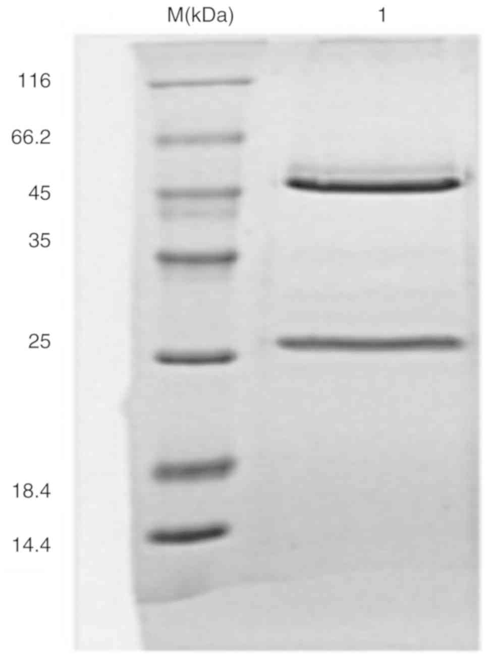
Purification of VP7 monoclonal
antibodies
The dialysis and affinity chromatography results
were obtained prior to antibody purity detection by SDS-PAGE
electrophoresis. The results showed that the protein purity was
>90% (Fig. 1).
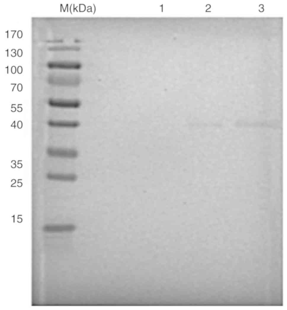
VP7 monoclonal antibody
identification
The results of western blot analysis demonstrated
that the VP7 monoclonal antibody specifically bound to the purified
human RV Wa strain and formed a specific reaction band at a
molecular weight of ~40 kD (Fig.
2).
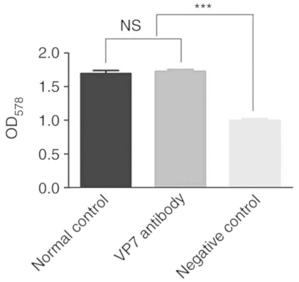
Mouse anti-human RV VP7 monoclonal
antibody in vitro neutralization test results
The in vitro neutralization effect of mouse
anti-human RV VP7 monoclonal antibodies was detected using an MTT
assay. The survival rate of cells in the virus plus monoclonal
antibody group was 102.1% (data not shown). By contrast, the
survival rate of cells in the virus plus maintenance fluid group
was 58.8% (data not shown). Therefore, the mouse anti-human RV VP7
monoclonal antibody secreted by the hybridoma cells exhibited
neutralizing activity (data not shown). The survival rate of cells
in the VP7 antibody group was higher than that of cells in the
negative control group (P<0.05), but was similar to that of the
normal control group (Fig. 3). By
subtracting the survival rate of cells in the virus plus monoclonal
antibody group from that of the virus plus maintenance fluid group,
it demonstrated that the cell protection rate of monoclonal
antibody reached 43.3%.
VP7 monoclonal antibody neutralization
titer test results
The MA-104 cell line was infected with mouse
anti-human RV VP7 monoclonal antibodies following gradient dilution
and its action against human RV was measured. A control group of
cells treated with maintenance liquid plus human RV and a normal
control group of cells treated with maintenance fluid without virus
or antibodies were also included. The number of lesions were
observed and recorded each day (7 days were observed). Further
lesions did not develop following 4 days. The TCID50 was
calculated according to the Reed-Muench formula. An antibody
neutralization titer of 1:446 was obtained, namely, 1:446 antibody
protect 50% of cells against lesions (data not shown).
RV immune colloidal gold detection in
mouse feces at 6 days following intragastric administration of
different preparations
In the control group, 8 mice infected with RV were
treated with sterile normal saline. Of these, 7 mice exhibited
positive immune colloidal gold detection in their feces, with a
positive rate of 87.5%. In mice treated with ribavirin, 3 were
infected with RV, equating to a positive rate of 37.5%. Of the mice
treated with 100 and 50 µl monoclonal antibodies, 2 and 4 mice were
infected with RV, with positive rates of 25 and 50%, respectively
(Table II).
| Table II.Mice rotavirus antigen test results
following the intragastric administration of different
preparations. |
Table II.
Mice rotavirus antigen test results
following the intragastric administration of different
preparations.
| Group | Number of positive
mice | Number of negative
mice |
|---|
| Monoclonal antibody
(100 µl) groupa | 2 | 6 |
| Monoclonal antibody
(50 µl) group | 4 | 4 |
| Ribavirin
groupa | 3 | 5 |
| Negative control
group | 7 | 1 |
Further statistical analysis was performed to
compare RV infection in different groups treated with different
preparations. The results revealed that the positive rate of
colloidal gold RV detection in the group treated with 100 µl
monoclonal antibody was significantly lower than that of the
control group (P<0.05). The remaining experimental groups
demonstrated no significant differences when compared with the
control group.
Using the aforementioned formula, the protection
rate of RV-infected mice treated with different preparations were
as follows: Ribavirin positive control group, 57.1%; 100 µl
monoclonal antibody group, 71.4% and 50 µl monoclonal antibody
group, 42.9%.
Discussion
Kunming mice are the most productive and abundant
outbred mice in China, which originate from Swiss mice (14). Kunming mice also exhibit strong
disease resistance, adaptability, a high reproductive rate, a high
survival rate and are relatively cheap to purchase (15). These mice also represent ~70% of
total mice used in biomedical experiments, involving pharmacology,
toxicology and the production and verification of drugs and
biological products in China (16).
In the present study, a large number of monoclonal antibodies were
prepared using the ascites method in Kunming mice. When using
ascites to prepare monoclonal antibodies in large quantities,
paraffin oil must be injected into the abdominal cavity (17). This is to promote immunosuppression,
preventing immune rejection reactions and accelerating tumor growth
(18). This step can also induce
mice to concentrate monocytes and lymphocytes in the peritoneal
cavity, avoiding the development of solid tumors and increasing the
production of ascites in mice (19,20). The
number of injected hybridoma cells may also affect the preparation
of monoclonal antibodies using ascites and may lead to cell death
if it is excessive, or a lack of ascites production if the number
is too low (21). Generally,
105−106 hybridoma cells are appropriate
(22). In addition, fewer injections
of hybridoma cells may lead to increased tolerance and survival
rates, delayed pathological alterations and reduced pain in mice
(23). By inoculating
2×10⁶ cells/0.2 ml into the peritoneal cavity of mice, a
large number of ascites were successfully harvested in the present
study.
Antibodies prepared using the ascites method are
mixed with a large number of contaminating proteins, including
lipid proteins, transferrin, macroglobulin, albumin and serum,
which do not meet the requirements for structural and functional
studies (24,25). Therefore, antibody purification is
required (26). The principle of
antibody purification is based on unique charge characteristics,
hydrophobicity, chelation with metal ions, specificity, affinity,
solubility and molecular size (27,28). In
the present study, the ammonium sulfate precipitation method was
employed. Due to differences in the hydrophobicity of different
proteins, altering the salt concentration allows for protein
precipitation. The RV VP7 monoclonal antibody precipitates in
ammonium sulfate with a concentration range of 30–50%, which allows
for the removal of contaminating proteins (29,30).
This purification method stabilizes the antibody, reduces the risk
of antibody activity loss, removes the majority of contaminating
proteins and concentrates the sample (31). However, a disadvantage of this method
is that some antibody activity is lost following precipitation or
co-precipitation with other contaminating proteins, which may
affect the antibody purity (32).
Therefore, following purification, antibody purity must be
determined (33). In the present
study, the results revealed that the antibody purity was 90%.
Therefore, the experimental requirements were met. Further
detection of antibody specificity by western blot analysis revealed
that the monoclonal antibody exhibited a specific reaction band at
~40 kDa, indicating that it bound to the purified human RV Wa
strain.
The MTT colorimetric method is used to detect the
activity and growth rate of cells. The OD value was measured in the
current study using a spectrophotometer at 578 nm to determine the
number of surviving cells following an MTT assay (34,35). The
results demonstrated that the mouse anti-human RV VP7 monoclonal
antibody exhibited good neutralization activity, with a cell
protection rate of 43.3% and a neutralization titer of 1:446.
In the present study, a model of RV infection in
young mice was established. Mice were treated with ribavirin, 100
or 50 µl monoclonal antibody or a negative control. The results
demonstrated that the positive rate of colloidal gold RV detection
in mice treated with 100 µl monoclonal antibody was significantly
lower than that in the negative control group. However, the
positive rate of colloidal gold RV detection in ribavirin-treated
group was not statistically lower than that of the negative control
group, which is inconsistent with a previous report on ribavirin
resistance to RV treatment (36). It
has been reported that treatment with ribavirin, a broad-spectrum
antiviral agent, phosphorylates ribavirin upon entry into
virus-infected cells (37). The
products then inhibit or reduce viral synthetases, mRNA guanosine
transferase, inosine monophosphate dehydrogenase and guanosine
triphosphate in cells (38). This
affects the synthesis of viral proteins and formation of viral RNA,
thereby inhibiting virus replication and proliferation and
effectively treating RV-induced intestinal inflammation,
alleviating clinical symptoms (39).
In the current study, the sample size was small and the selected
outcome indicator only included a feces RV antigen test without
comparisons of certain clinical symptoms, including stool
frequency. The protective effect of different preparations was
therefore evaluated by further calculations. The protection rate of
mice following the intragastric administration of 100 µl monoclonal
antibodies reached 71.4%, which confirmed that the mouse anti-human
RV VP7 monoclonal antibody produced in this study was able to
neutralize the virus in mice, thus preventing viral proliferation.
In addition, the protection rate of the 100 µl monoclonal antibody
group was 14.3% higher than that of the ribavirin group.
The present study had several limitations. For
instance, the number of mice selected was small and an increased
sample size should thus be utilized to confirm the results
obtained. In addition, future studies should assess whether the VP7
monoclonal antibody can be directly inoculated into an individual
to provide immuno-protection. Furthermore, it is unclear whether
the VP7 monoclonal antibody should only be used for the in
vitro screening of anti-viral drugs. The efficacy of the VP7
monoclonal antibody has also not been compared with other vaccines
that are currently in use, which will be investigated further in
future studies.
In conclusion, mouse anti-human RV VP7 monoclonal
antibodies can be successfully generated using the ascites method.
These monoclonal antibodies demonstrate a good neutralization
effect on the Wa strain human RV in vitro and in
vivo. A higher dose was associated with a greater protective
effect and the protective effects of high doses were superior to
that of ribavirin.
Acknowledgements
Not applicable.
Funding
No funding was received.
Availability of data and materials
The datasets used and/or analyzed during the present
study are available from the corresponding author on reasonable
request.
Authors' contributions
MZ, JY, LZ, HW, XP, ZD, YY and WL collected and
interpreted the data. MZ and YY drafted the manuscript. MZ and WL
revised it critically for important intellectual content. BW and ML
were responsible for the conception and design of the study. All
authors read and approved the final manuscript.
Ethics approval and consent to
participate
The study was approved by the Committee on Animal
Research and Ethics of West China School of Basic Medical Sciences
and Forensic Medicine, Sichuan University (Chengdu, China).
Patient consent for publication
Not applicable.
Competing interests
The authors declare that they have no competing
interests.
References
|
1
|
Yin N, Yang FM, Qiao HT, Zhou Y, Duan SQ,
Lin XC, Wu JY, Xie YP, He ZL, Sun MS and Li HJ: Neonatal rhesus
monkeys as an animal model for rotavirus infection. World J
Gastroenterol. 24:5109–5119. 2018. View Article : Google Scholar : PubMed/NCBI
|
|
2
|
Layton JB, Butler AM, Panozzo CA and
Brookhart MA: Rotavirus vaccination and short-term risk of adverse
events in US infants. Paediatr Perinat Epidemiol. 32:448–457. 2018.
View Article : Google Scholar : PubMed/NCBI
|
|
3
|
Vannie P, Capua I, Le Potier MF, Mackay
DK, Muylkens B, Parida S, Paton DJ and Thiry E: Marker vaccines and
the impact of their use on diagnosis and prophylactic measures. Rev
Sci Tech. 26:351–372. 2007.PubMed/NCBI
|
|
4
|
Teng Y, Zhao B, Pan X, Wen Y and Chen Y: A
new rotavirus VP6-based foreign epitope presenting vector and
immunoreactivity of VP4 epitope chimeric proteins. Viral Immunol.
27:96–104. 2014. View Article : Google Scholar : PubMed/NCBI
|
|
5
|
Perez CA, Eichwald C, Burrone O and
Mendoza D: Rotavirus vp7 antigen produced by Lactococcus lactis
induces neutralizing antibodies in mice. J Appl Microbiol.
99:1158–1164. 2005. View Article : Google Scholar : PubMed/NCBI
|
|
6
|
Morozova OV, Sashina TA, Fomina SG and
Novikova NA: Comparative characteristics of the VP7 and VP4
antigenic epitopes of the rotaviruses circulating in Russia (Nizhny
Novgorod) and the Rotarix and RotaTeq vaccines. Arch Virol.
160:1693–1703. 2015. View Article : Google Scholar : PubMed/NCBI
|
|
7
|
Yang SH: Research on genetic engineering
subunit vaccine of A group bovine rotavirus. Chin J Zoonoses.
27:506–510. 2011.(In Chinese).
|
|
8
|
Jalilvand S, Afchangi A, Mohajel N,
Roohvand F and Shoja Z: Diversity of VP7 genes of G1 rotaviruses
isolated in Iran, 2009–2013. Infect Genet Evol. 37:275–279. 2016.
View Article : Google Scholar : PubMed/NCBI
|
|
9
|
Macek C: Monoclonal antibodies: Key to a
revolution in clinical medicine. JAMA. 247:2463–2470. 1982.
View Article : Google Scholar : PubMed/NCBI
|
|
10
|
Peterson NC and Peavey JE: Comparison of
in vitro monoclonal antibody production methods with an in vivo
ascites production technique. Contemp Top Lab Anim Sci. 37:61–66.
1998.PubMed/NCBI
|
|
11
|
Kairemo KJ, Lappalainen AK, Kääpä E,
Laitinen OM, Hyytinen T, Karonen SL and Grönblad M: In vivo
detection of intervertebral disk injury using a radiolabeled
monoclonal antibody against keratan sulfate. J Nucl Med.
42:476–482. 2001.PubMed/NCBI
|
|
12
|
Ogawa M, Regino CA, Choyke PL and
Kobayashi H: In vivo target-specific activatable near-infrared
optical labeling of humanized monoclonal antibodies. Mol Cancer
Ther. 8:232–239. 2009. View Article : Google Scholar : PubMed/NCBI
|
|
13
|
Fernández D, Valle I, Llamos R, Guerra M,
Sorell L and Gavilondo J: Rapid detection of rotavirus in faeces
using a dipstick system with monoclonal antibodies and colloidal
gold as marker. J Virol Methods. 48:315–323. 1994. View Article : Google Scholar : PubMed/NCBI
|
|
14
|
Zhang GM and Yao GH: Investigation on
genetic background of Kunming mice (KM mice) in China. Chin J Lab
Anim Sci. 7:246–251, (In Chinese).
|
|
15
|
Shang H, Wei H, Yue B, Xu P and Huang H:
Microsatellite analysis in two populations of Kunming mice. Lab
Anim. 43:34–40. 2009. View Article : Google Scholar : PubMed/NCBI
|
|
16
|
Ma P, Wu Y, Zeng Q, Gan Y, Chen J, Ye X
and Yang X: Oxidative damage induced by chlorpyrifos in the hepatic
and renal tissue of Kunming mice and the antioxidant role of
vitamin E. Food Chem Toxicol. 58:177–183. 2013. View Article : Google Scholar : PubMed/NCBI
|
|
17
|
Weng J, Yang L, Lei C, Li T, Peng G, Fu C,
Han X, Li H, Jiang Z, Zhang Z, et al: Elimination of mycoplasma
contamination from infected human hepatocyte C3A cells by
intraperitoneal injection in BALB/c mice. Front Cell Infect
Microbiol. 7:4402017. View Article : Google Scholar : PubMed/NCBI
|
|
18
|
Tammer R, Evensen O, Lutz H and Reinacher
M: Immunohistological demonstration of feline infectious
peritonitis virus antigen in paraffin-embedded tissues using feline
ascites or murine monoclonal antibodies. Vet Immunol Immunopathol.
49:177–82. 1995. View Article : Google Scholar : PubMed/NCBI
|
|
19
|
Zheng J, Zhang H, Bao K, Gao W, Xu C and
Xia C: Preparation of monoclonal antibodies against bovine
progesterone. Monoclon Antib Immunodiagn Immunother. 34:275–277.
2015. View Article : Google Scholar : PubMed/NCBI
|
|
20
|
Ezzatifar F, Majidi J, Baradaran B,
Aghebati Maleki L, Abdolalizadeh J and Yousefi M: Large scale
generation and characterization of anti-human IgA monoclonal
antibody in ascitic fluid of BALB/c mice. Adv Pharm Bull. 5:97–102.
2015.PubMed/NCBI
|
|
21
|
Velez D, Miller L and Macmillan JD: Use of
tangential flow filtration in perfusion propagation of hybridoma
cells for production of monoclonal antibodies. Biotechnol Bioeng.
33:938–940. 1989. View Article : Google Scholar : PubMed/NCBI
|
|
22
|
Rokni M, Razavi AR, Shokri F, Ahmadi Kia
K, Solaymani-Mohammadi F, Chahardoli R and Saboor-Yaraghi AA:
Enhancement of monoclonal antibody production after single and
combination treatment of the hybridoma cells with all-trans
retinoic acid and docosahexaenoic acid: An in vitro and in vivo
study. Int Immunopharmacol,. 59:295–300. 2018. View Article : Google Scholar
|
|
23
|
Hnasko R, Lin AV, Stanker L and McGarvey
J: A bioassay for optimization of macrophage-conditioned medium as
a culture supplement to promote hybridoma cell survival and growth.
Monoclon Antib Immunodiagn Immunother. 37:126–133. 2018. View Article : Google Scholar : PubMed/NCBI
|
|
24
|
Yu YC, Huang YC and Lee TY: Purification
of antibodies from protein mixtures and mouse ascites fluid using
Zeolite X. Biotechnol Prog. 14:332–337. 1998. View Article : Google Scholar : PubMed/NCBI
|
|
25
|
Ohkawa K, Tsukada Y and Hirai H: Effect of
antibody to rat alpha-fetoprotein (AFP) on protein and DNA
synthesis of rat ascites hepatoma AH66 cells. Gan To Kagaku Ryoho.
11:227–234. 1984.(In Japanese). PubMed/NCBI
|
|
26
|
Johnson E, Miribel L, Arnaud P and Tsang
KY: Purification of IgM monoclonal antibody from murine ascitic
fluid by a two-step column chromatography procedure. Immunol Lett.
14:159–165. 1987. View Article : Google Scholar : PubMed/NCBI
|
|
27
|
Jermy A, Beeson D and Vincent A:
Pathogenic autoimmunity to affinity-purified mouse acetylcholine
receptor induced without adjuvant in BALB/c mice. Eur J Immunol.
23:973–976. 1993. View Article : Google Scholar : PubMed/NCBI
|
|
28
|
Yavuz H, Bereli N, Yılmaz F, Armutcu C and
Denizli A: Antibody purification from human plasma by
metal-chelated affinity membranes. Methods Mol Biol. 1286:43–46.
2015. View Article : Google Scholar : PubMed/NCBI
|
|
29
|
Graczyk TK and Cranfield MR: Detection of
Cryptosporidium- specific serum immunoglobulins in captive snakes
by a polyclonal antibody in the indirect ELISA. Vet Res.
28:131–142. 1997.PubMed/NCBI
|
|
30
|
Pringels L, Broeckx V, Boonen K, Landuyt B
and Schoofs L: Abundant plasma protein depletion using ammonium
sulfate precipitation and protein A affinity chromatography. J
Chromatogr B Analyt Technol Biomed Life Sci. 1089:43–59. 2018.
View Article : Google Scholar : PubMed/NCBI
|
|
31
|
Grodzki AC and Berenstein E: Antibody
purification: Ammonium sulfate fractionation or gel filtration.
Methods Mol Biol. 588:15–26. 2010. View Article : Google Scholar : PubMed/NCBI
|
|
32
|
Kent UM: Purification of antibodies using
ammonium sulfate fractionation or gel filtration. Methods Mol Biol.
34:13–21. 1994.PubMed/NCBI
|
|
33
|
Ahlstedt S, Holmgren J and Hanson LA: The
validity of the ammonium sulphate precipitation technique of
estimation of antibody amount and avidity. Immunology. 25:917–922.
1973.PubMed/NCBI
|
|
34
|
Surin AM, Sharipov RR, Krasil'nikova IA,
Boyarkin DP, Lisina OY, Gorbacheva LR, Avetisyan AV and Pinelis VG:
Disruption of functional activity of mitochondria during MTT assay
of viability of cultured neurons. Biochemistry (Mosc). 82:737–749.
2017. View Article : Google Scholar : PubMed/NCBI
|
|
35
|
Nguyen VS, Shi L, Luan FQ and Wang QA:
Synthesis of kaempferide Mannich base derivatives and their
antiproliferative activity on three human cancer cell lines. Acta
Biochim Pol. 62:547–552. 2015. View Article : Google Scholar : PubMed/NCBI
|
|
36
|
Shetty AK, Gans HA, So S, Millan MT, Arvin
AM and Gutierrez KM: Intravenous ribavirin therapy for adenovirus
pneumonia. Pediatr Pulmonol. 29:69–73. 2000. View Article : Google Scholar : PubMed/NCBI
|
|
37
|
Gross AE and Bryson ML: Oral ribavirin for
the treatment of noninfluenza respiratory viral infections: A
systematic review. Ann Pharmacother. 49:1125–1135. 2015. View Article : Google Scholar : PubMed/NCBI
|
|
38
|
Satoh S, Mori K, Onomura D, Ueda Y,
Dansako H, Honda M, Kaneko S, Ikeda M and Kato N: Ribavirin
suppresses hepatic lipogenesis through inosine monophosphate
dehydrogenase inhibition: Involvement of adenosine
monophosphate-activated protein kinase-related kinases and retinoid
X receptor α. Hepatol Commun. 1:550–563. 2017. View Article : Google Scholar : PubMed/NCBI
|
|
39
|
Musser JMB, Heatley JJ, Koinis AV,
Suchodolskin PF, Guo J, Escandon P and Tizard IR: Ribavirin
inhibits parrot bornavirus 4 replication in cell culture. PLoS One.
10:e01340802015. View Article : Google Scholar : PubMed/NCBI
|