Introduction
Osteoarthritis (OA) is a degenerative joint disease,
pathologically characterized by the progressive loss of cartilage,
osteophyte formation, subchondral sclerosis, matrix degradation and
matrix synthesis imbalance (1–3).
Symptoms of OA include inflammation, stiffness and loss of mobility
(1). Currently, OA treatment is
challenging due to its multiple etiologies and complex pathological
processes (2). New molecular targets
are therefore urgently required to prevent this disease.
Calcitonin (CT), a naturally occurring peptide that
provides benefits to articular cartilage and subchondral bone
(3). CT improves cell viability and
modulates inflammatory reactions in IL-1β- or lipopolysaccharide
(LPS)-injured rat chondrocytes (4,5).
Although CT is an ideal treatment for OA progression, the specific
mechanism by which CT exerts its effects is yet to be fully
elucidated.
Wnt/β-catenin signaling is critical for OA
development and the expression of β-catenin is necessary for the
maintenance of cartilage homeostasis (6). Furthermore, previous studies indicate
that the Wnt/β-catenin pathway controls chondrocyte proliferation
and is fundamental for differentiating stem cells into osteoblasts
rather than chondrocytes (7).
Following Wnt/β-catenin pathway activation, β-catenin is
upregulated in the cytoplasm and transfers to the nucleus, where it
binds to T-cell factor/lymphoid enhancer factor (8). This activates the transcription of
target genes associated with cell proliferation, apoptosis,
inflammation and matrix metabolism; including metallopeptidase
(MMP) 13 and metalloproteinase with thrombospondin motifs 4
(ADAMTS4) (8). A report has
demonstrated that CT inhibits β-catenin in interleukin
(IL)-1β-injured rabbit chondrocytes (9). In the present study, an n in
vitro OA model was established by isolating rat chondrocytes
and injuring these with IL-1β. Following, the current study
investigated whether the Wnt/β-catenin pathway was involved in the
protective effect of CT treatment in OA and the mechanisms
underlying this effect.
Materials and methods
Cell isolation
Male Sprague-Dawley (SD) rats (n=10; weight, 50±1.4
g; 4-week-old) were purchased from Shanghai SIPPR-Bk Lab Animal
Co., Ltd. (Shanghai, China) with the certificate number
2008001682174. The animal license was provided by Shanghai
Laboratory Animal Center (Shanghai, China) with the license number
SCXK 2013–0016. Rats were housed at 25°C with a 12:12 h light/dark
for a week, and given healthy atmosphere air, food and water ad
libitum. Rats were sacrificed by cervical dislocation and
cartilage tissue was harvested from knee joints, which was immersed
in PBS, cut into slices (2–4 mm thick) and placed in 5 ml
centrifuge tubes. The use and treatment of SD rats was approved by
the Ethics Committee of the School of Medicine, Shandong University
(Jinan, China). Chondrocytes were obtained by enzyme digestion
using collagenase II and were then maintained in high glucose DMEM
with 15% FBS (v/v; both GE Healthcare Life Sciences) at 37°C under
5% CO2 until a confluence of 50–60% was reached.
Immunohistochemical staining (IHC) was subsequently performed for
identification
Cell culture
Rat chondrocytes cultured at 37°C under 5%
CO2 in DMEM containing 10% FBS, 0.1 mg/ml streptomycin
and 100 U/ml penicillin (both P1400-100; Beijing Solarbio Science
& Technology Co., Ltd.) until achieving the logarithmic growth
phase. Cell morphology was observed by light microscopy
(magnification, ×200).
Cell treatment
To establish IL-1β-injured rat chondrocytes, cells
were exposed to various concentrations of 100 µl IL-1β (BioVision,
Inc.; 0.5, 1, 2.5, 5, 10 and 20 ng/ml). To investigate the effect
of CT (Signalway Antibody LLC, College Park, MD, USA) on
IL-1β-injured rat chondrocytes, cells were exposed to 10 ng/ml
IL-1β and treated with CT (10 and 50 nM). To assess the involvement
of the Wnt/β-catenin pathway, cells were exposed to 10 ng/ml IL-1β
and then treated with 5 µM of the β-catenin inhibitor, IWR-1-endo,
(Selleck Chemicals) or CT (50 nM) with IWR-1-endo (5 µM). After
treatment, cells were cultured as described above.
IHC
Primary cultured rat chondrocytes on cover slides
were washed with 0.1 M PBS and then fixed with 4% paraformaldehyde
(Sinopharm Chemical Reagent Co., Ltd) at 25°C for 30 min. Slides
were treated with H2O2 (Sinopharm Chemical
Reagent Co., Ltd.; 3% in methanol) to eliminate the activity of
endogenous peroxide and then blocked with 1% BSA (Beijing Solarbio
Science & Technology Co., Ltd.) at room temperature for 30 min.
Slides were then incubated with rabbit polyclonal anti-collagen II
(cat. no. ab34712) and anti-transcription factor SOX9 (cat. no.
ab185966; both dilution 1:5,000; Abcam) antibodies at 25°C
overnight at 4°C, followed by incubation with horseradish
peroxidase (HRP)-labeled secondary antibodies (cat. no. D-3004;
dilution 1:500; Shanghai Long Island Biotech Co., Ltd.) at room
temperature for 1 h. A DAB-containing substrate kit (Shanghai Long
Island Biotech. Co., Ltd.) was used to visualize collagen II and
SOX9 signals, followed by hematoxylin nuclear counterstaining at
25°C for 3 min.
Cell proliferation
A cell counting kit-8 (CCK-8) was used to measure
cell proliferation. Cells were seeded into 96-well plates (100 µl;
3.0×10³ cells/well) and cultured overnight at room
temperature prior to treatment. After treatment, cells were
cultured as described in a previous subsection. At 0, 24, 48 and 72
h, cultured cells (suspended in 90 µl of the medium in the culture)
were treated with CCK-8 solution (10 µl) for an additional 1 h.
Optical density (450 nm) was recorded to assess cell proliferation.
In the current study, proliferation of rat chondrocytes treated
with IL-1β (0.5, 1, 2.5, 5, 10 and 20 ng/ml) was assessed at 0, 24,
48 and 72 h, and the viability of rat chondrocytes treated with
IL-1β (10 ng/ml), IL-1β (10 ng/ml) + CT (10 and 50 nM), IL-1β (10
ng/ml) + IWR-1-endo and IL-1β (10 ng/ml) + CT (50 nM) + IWR-1-endo
was assessed at 24 h.
Annexin V/PI staining
A FITC-labeled recombinant Annexin V Apoptosis
Detection kit (Beyotime Institute of Biotechnology) was used to
measure apoptosis. Cells were seeded in six-well plates
(5.0×10⁴ cells/well) and cultured at 25°C overnight
prior to treatment. After treatment of IL-1β, IL-1β + CT, IL-1β +
IWR-1- endo and IL-1β + CT + IWR-1-endo, for 48 h, cells were
centrifuged at 1,000 × g for 5 min at 4°C. Supernatants were
subsequently removed. Cells were incubated with 5 µl Annexin V-FITC
at 4°C for 15 min in darkness, followed by 5 µl propidium iodide
(PI) for a further 5 min. The rate of early apoptosis was detected
using Accuri C6 FACScan flow cytometer with CFlow Plus®
version 1.0 software (BD Bioscience). Early apoptotic cells were
counted in the lower right quadrant of the display.
Reverse transcription-quantitative
(RT)-qPCR
Total RNA from rat chondrocytes was isolated using
TRIzol regent (cat. no. 1596-026; Invitrogen; Thermo Fisher
Scientific, Inc.). First-strand cDNA was obtained using a Reverse
Transcription kit (cat. no. K1622; Fermentas; Thermo Fisher
Scientific, Inc.). A SYBR Green PCR kit (cat. no. K0223; Thermo
Fisher Scientific, Inc.) and ABI Prism 7300 SDS Software (Applied
Biosystems; Thermo Fisher Scientific, Inc.) was used to perform
qPCR by the 2−ΔΔCq method (10) with the following thermocycling
conditions: 95°C for 10 min, followed by 40 cycles of 95°C for 15
sec and 60°C for 45 sec; 95°C for 15 sec; 60°C for 1 min; 95°C for
15 sec and 60°C for 15 sec. mRNA expression was normalized to that
of GAPDH. The primer sequences used in RT-qPCR are listed in
Table I.
 | Table I.Primers used in RT-qPCR analysis. |
Table I.
Primers used in RT-qPCR analysis.
| Name | GenBank | Primer (5′-3′) |
|---|
| DKK-1 | NM_001106350.1 at
164–389 position | Forward:
CGGTTCTTGGTCGTGCTTTC; |
|
|
| Reverse:
AAGGGTAGGGCTGGTAGTTG; 226 bps, 61% GC. |
| β-catenin | NM_053357.2 at
227–375 position | Forward:
TCACGCAAGAGCAAGTAG; |
|
|
| Reverse:
CTGGACATTAGTGGGATGAG; 149 bps, 55% GC. |
| MMP13 | NM_133530.1 at
2348–2572 position | Forward:
CAGACAGCAAGAATAAAGAC; |
|
|
| Reverse:
CAACATAAGCACAGTGTAAC; 225 bps; 41% GC. |
| ADAMTS4 | NM_023959.1at 714 -
955 position | Forward:
GGTGGCAGATGACAAGATG; |
|
|
| Reverse:
AGTCGTTCGGAGGGTTTAG; 242 bps; 59% GC. |
| GAPDH | NM_017008.4 at
357–593 position | Forward:
GGAGTCTACTGGCGTCTTCAC; |
|
|
| Reverse:
ATGAGCCCTTCCACGATGC; 237 bps; 55% GC. |
Western blot analysis
Total protein from rat chondrocytes was obtained
using RIPA lysis buffer (cat. no. R0010; Beijing Solarbio Science
& Technology Co., Ltd.) and supernatant levels were measured
using a BCA assay kit (cat. no. PICPI23223; Thermo Fisher
Scientific, Inc.). Total protein (30 mg) was separated using 10%
SDS-PAGE gel, followed by transfer to PVDF membranes. After
blocking with 5% non-fat milk at 4°C overnight, PVDF membranes were
subsequently incubated with the following primary antibodies at 4°C
overnight: Anti-dickkopf-1 (Dkk-1; cat. no. ab109416; 1:1,000;
Abcam), anti-β-catenin (cat. no. ab32572; 1:1,500; Abcam),
anti-MMP13 (cat. no. ab39012; 1:3,000; Abcam), anti-ADAMTS4 (cat.
no. ab185722; 1:500; Abcam), anti-Bax (cat. no. 5023; 1:1,000; Cell
Signaling Technology Inc.), anti-Bcl-2 (cat. no. 15071; 1:1,000;
Cell Signaling Technology, Inc.), anti-cleaved-caspase 3 (cat. no.
9664; 1:1,000; Cell Signaling Technology, Inc.), anti-MMP-3 (cat.
no. ab53015; 1:1,000; Abcam), anti-MMP-9 (cat. no. ab38898; 1:500;
Abcam), anti-ADAMTS5 (cat. no. ab41037; 1:1,000; Abcam) and
anti-GAPDH (cat. no. 5174; 1:2,000; Cell Signaling Technology,
Inc.). Samples were then incubated with HRP-conjugated secondary
antibodies: Goat anti-rabbit (cat. no. A0208); donkey anti-goat
(cat. no. A0181); and goat anti-mouse immunoglobulin G (cat. no.
A0216; all dilution 1:1,000; Beyotime Institute of Biotechnology)
for 1 h at room temperature. The HRP signal was assessed using ECL
Plus (GE Healthcare) and a Tanon-5200 Imaging system (Tanon Science
& Technology Co., Ltd.). ImageJ software version 1.8.0
(National Institutes of Health, Bethesda, MD, USA) was used for
densitometry analysis.
Cleaved caspase 3 activity assay
Cleaved caspase 3 activity in cell supernatants was
detected using a Caspase-3 Colorimetric Assay kit (cat. no. KGA203;
Nanjing KeyGen Biotech Co., Ltd.) according to the manufacturer's
protocol.
Statistical analyses
Graphs were generated using GraphPad Prism 7.0
software (GraphPad Software, Inc.). Data were expressed as the mean
± standard error of the mean (n=3). A student's t-test was
performed for comparisons between two groups, while a post-hoc
Tukey's test following ANOVA was used for comparisons between
multiple groups. P<0.05 was considered to indicate a
statistically significant result.
Results
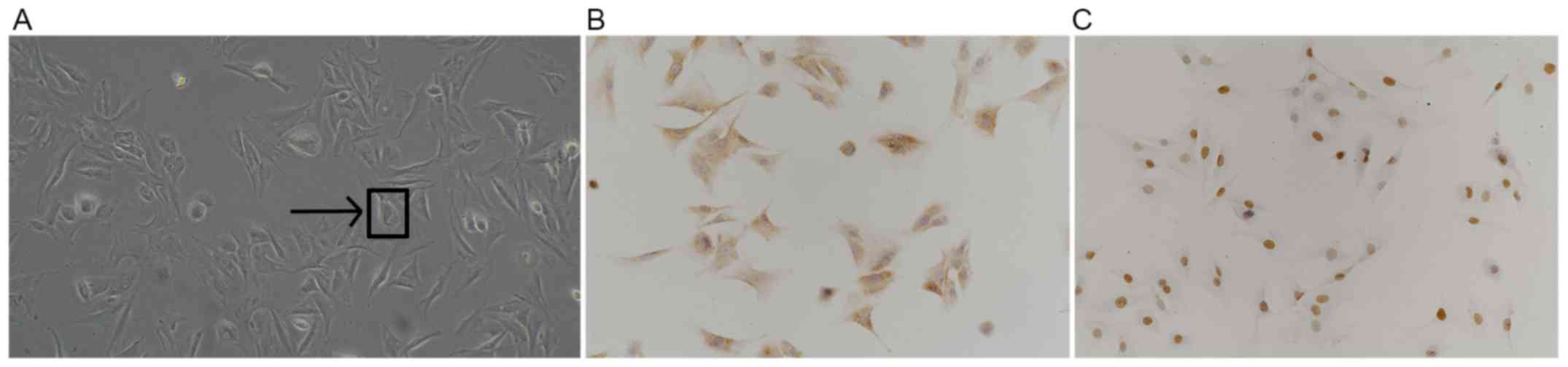
Successful isolation of rat
chondrocytes
Under light microscopy, primary isolated rat
chondrocytes were polygonal, spindle or irregularly shaped with
copious cytoplasm and a large and round nuclei. (Fig. 1A). After IHC staining for type II
collagens and transcription factor SOX9, brown-yellow granules were
observed in the cytoplasm and nucleus, the color of which stained
darker with increased cell density (Fig.
1B and C). Data collected from the current study indicates the
successful isolation of rat chondrocytes.
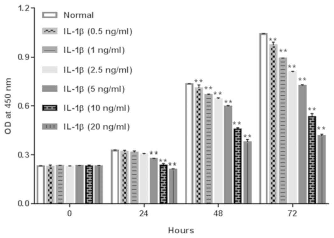
IL-1β inhibited the proliferation of
rat chondrocytes
To assess the role of IL-1β on the proliferation of
rat chondrocytes, cells were treated with IL-1β at 0.5, 1, 2.5, 5,
10 and 20 ng/ml and cell proliferation was measured using a CCK-8
assay at 0, 24, 48 and 72 h. As presented in Fig. 2, IL-1β significantly suppressed
chondrocyte viability in a dose-and time-dependent manner,
indicating the inhibitory effect of IL-1β on the proliferation of
rat chondrocytes. In the present study, 10 ng/ml IL-1β had a
relatively obvious anti-proliferation effect on chondrocytes and
did not cause >50% inhibition on cell viability, thus this
concentration was selected for the following study.
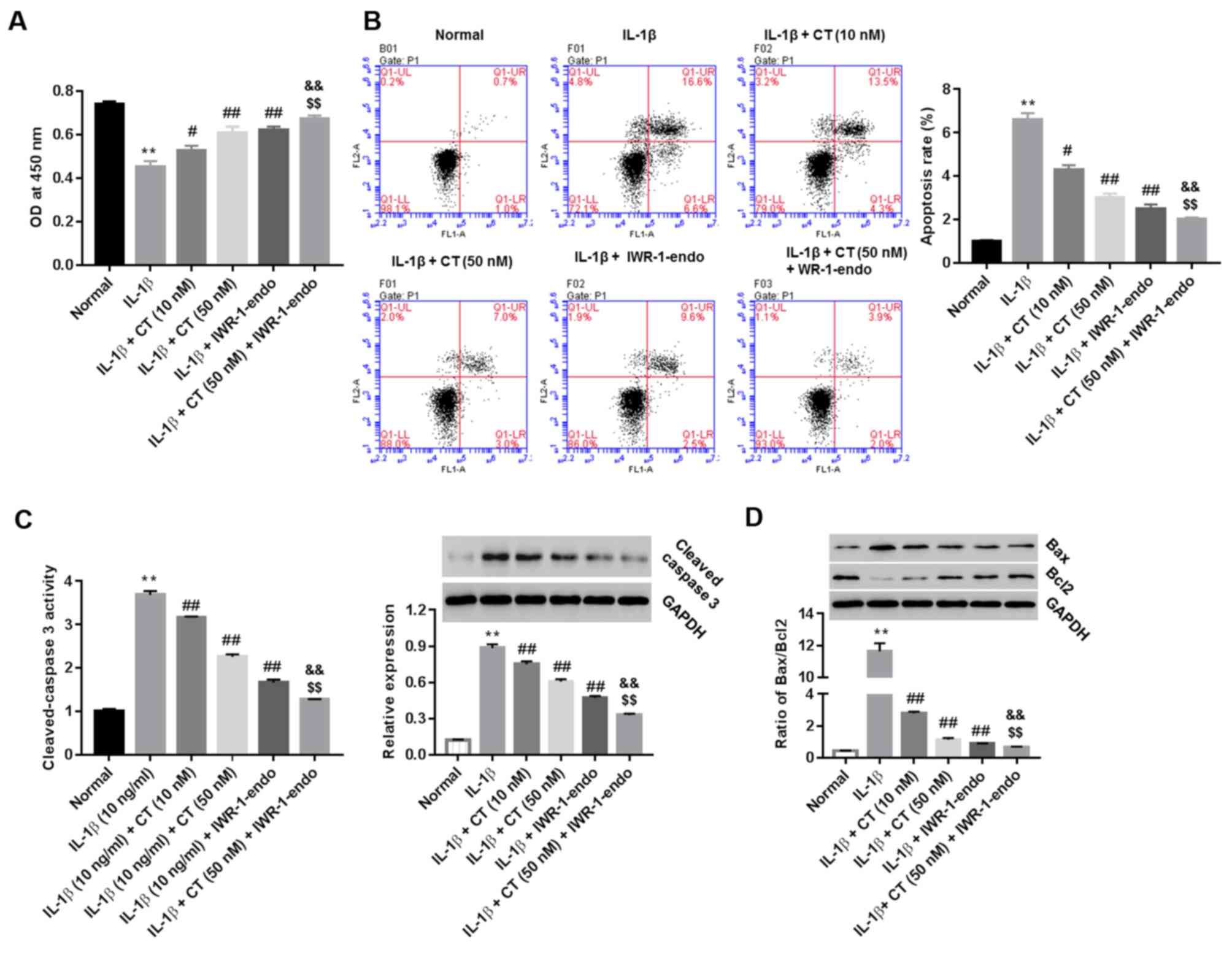
CT promotes and inhibits the viability
and apoptosis of IL-1β-injured rat chondrocytes, respectively, via
the Wnt/β-catenin pathway
IL-1β-injured rat chondrocytes were treated with CT
(10 and 50 nM), IWR-1-endo, or CT (50 nM) in addition to
IWR-1-endo. Cell viability (at 24 h) and cell apoptosis (at 48 h)
was measured using the CCK-8 assay and Annexin V/PI staining
method, respectively. Furthermore, the activity of cleaved-caspase
3 in cell supernatants, as well as the protein expression of
cleaved-caspase 3 and the ratio of pro-apoptotic Bax/anti-apoptotic
Bcl2 within cells were also measured. As indicated in Fig. 3A and B, CT treatment enhanced cell
viability and inhibited cell apoptosis compared with the IL-1β
group. CT also inhibited the activity and expression of
cleaved-caspase 3 (Fig. 3C) as well
as the ratio of Bax/Bcl2 in a dose-dependent manner (Fig. 3D), with the most significant effect
being observed at 50 nM CT when compared with the IL-1β group.
However, pro-viability and anti-apoptotic effects were augmented in
the CT (50 nM) plus IWR-1-endo group compared treated with CT (50
nM) or IWR-1-endo alone.
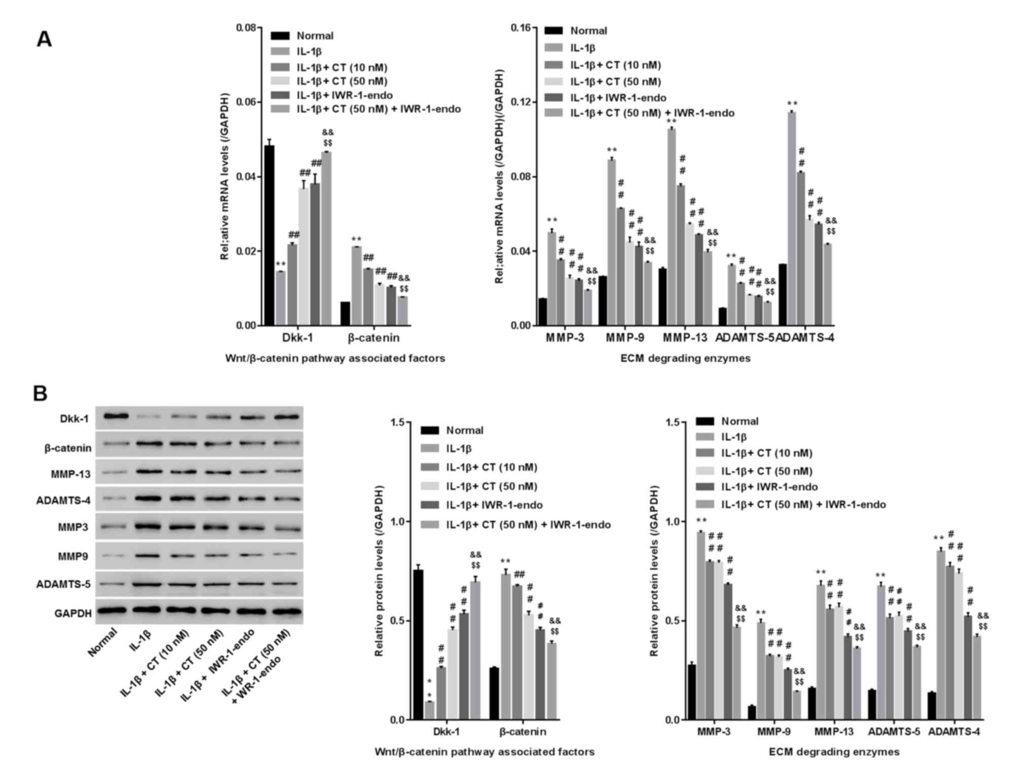
CT inhibits cartilage extracellular
matrix degradation via the Wnt/β-catenin pathway in IL-1β-injured
rat chondrocytes
IL-1β-injured rat chondrocytes were treated with CT
(10 and 50 nM), IWR-1-endo (a Wnt/β-catenin pathway inhibitor), or
CT (50 nM) in addition to IWR-1-endo. The mRNA and protein levels
of the cartilage extracellular matrix (ECM) degrading enzymes
(MMP13, MMP3, MMP9, ADAMTS4 and ADAMTS5), Dkk-1 (a specific
inhibitory protein for blocking Wnt/β-catenin) (11) and β-catenin (the core component of
Wnt/β-catenin pathway) were then measured using RT-qPCR and western
blot analysis, respectively. As presented in Fig. 4A and B, a significant increase in the
mRNA and protein levels of MMP13, MMP3, MMP9, ADAMTS4 and ADAMTS5
and β-catenin, and decrease in Dkk-1 was observed in the
IL-1β-treated group when compared with the normal group, indicating
the severe degradation of ECM and the activation of the
Wnt/β-catenin pathway in IL-1β-injured rat chondrocytes. However,
in comparison with the IL-1β group, CT treatment reduced MMP13,
MMP3, MMP9, ADAMTS4, ADAMTS5 and β-catenin while enhancing Dkk-1 in
a dose-dependent manner. These results indicated that CT inhibited
cartilage ECM degradation and during which, may have been
associated with the inactivation of Wnt/β-catenin. Furthermore,
similar to CT, IWR-1-endo consistently suppressed MMP13, MMP3,
MMP9, ADAMTS4, ADAMTS5 and β-catenin while promoting Dkk-1, and
produced an even greater effect than CT treatment. Yet, IWR-1-endo
plus CT (50 nM) was more effective than IWR-1-endo treatment alone
in inhibiting cartilage ECM degradation. However, CT treatment
inhibited β-catenin but promoted Dkk-1. The results indicate that
CT inhibits cartilage ECM degradation by blocking the activation of
Wnt/β-catenin.
Discussion
IL-1β is a major contributor to OA pathology
(12). To establish an in
vitro model of IL-1β induced OA, well-characterized rat
chondrocytes were isolated and stained using IHC for a set of
marker proteins including collagen II and SOX9 (13). A previous report has indicated that
proliferation and apoptosis are significantly inhibited in IL-1β
(10 ng/ml)-induced OA chondrocytes (14). In the present study, IL-1β (0.5, 1,
2.5, 5, 10 and 20 ng/ml) was used to injure rat chondrocytes to
assess its effect on cell proliferation. Relatively strong
responses were observed with 10 ng/ml and 20 ng/ml IL-1β. As 20
ng/ml IL-1β exposure resulted in >50% inhibition of cell
viability, IL-1β at 10 ng/ml was selected for the current
study.
CT serves an important role in improving chondrocyte
viability. Hypodermic injections of CT (5 U/kg) in OA rabbits have
been demonstrated to substantially reduce apoptotic articular
chondrocytes (15). Furthermore, in
an in vitro OA model, CT exerted pro-proliferative and
anti-apoptotic effects on LPS-injured chondrocytes (4). Moreover, CT
(10−10−10−8 M) exerted anti-apoptotic effects
in IL-1β-injured chondrocytes, (substantiated by a TUNEL assay) and
reduced the ratios of Bax/Bcl2 and cleaved caspase-3 activity
(16). In the present study, the
data revealed the proliferative and anti-apoptotic effects of CT on
IL-1β-injured rat chondrocytes. The downregulation of cleaved
caspase-3 in supernatants and cells, and the decrease of Bax/BCL2
ratios, were also revealed to be involved in the process.
MMPs and ADAMTSs serve a key role in the degradation
of ECM components, contributing to the destruction of articular
cartilage (17). CT treatment has
been previously demonstrated to inhibit MMP-13, MMP-3 and ADAMTS4
in a rat OA model (18,19), which was further confirmed in
IL-1β-injured chondrocytes. To the best of our knowledge, the
current study is the first to substantiate the inhibitory effect of
CT on MMP9 and ADAMTS5 in an IL-1β-stimulated in vitro OA
model. A previous study has revealed that growth differentiation
factor 5 reduces MMP-13 expression in human chondrocytes via
Wnt/β-catenin signaling, contributing to the homeostasis of
cartilage ECM (20). However,
whether Wnt/β-catenin signaling is involved in regulating the
expression of these matrix-degrading enzymes in IL-1β-stimulated
rat chondrocytes remains unclear.
It has been determined that CT downregulates
β-catenin while upregulating secreted frizzled related protein 1 (a
Wnt antagonist) in rat chondrocytes of the knee joint (21). To assess whether the Wnt/β-catenin
pathway was the mechanism whereby CT functioned in OA in
vitro, Wnt/β-catenin signaling was stimulated with IL-1β and
blocked by IWR-1-endo. The proliferation, apoptosis and ECM
degradation of rat chondrocytes was subsequently assessed after
treatment with CT, IWR-1-endo or CT in combination with IWR-1-endo.
The results demonstrated that CT counteracted IL-1β, as evidenced
by the promotion of cell proliferation, the inhibition of
apoptosis, the reduction in cleaved caspase-3 and Bax/Bcl2 ratios,
the upregulation of Dkk-1, the downregulation of β-catenin and the
suppression of matrix-degrading enzyme expression (MMP3, MMP9,
MMP13, ADAMTS4 and ADAMTS5). This indicated that CT exerted a
protective effect on IL-1β-injured rat chondrocytes, which was
associated with the blockade of the Wnt/β-catenin pathway. The
changes induced by IWR-1-endo in the aforementioned results were
similar to those of CT (50 nM). However, changes were strengthened
when these treatments were combined. Given the inhibitory effect of
CT on β-catenin and the promoted effect of CT on Dkk-1, the data
demonstrated that CT may protect rat chondrocytes against IL-1β
injured via the Wnt/β-catenin pathway.
The results of the current study indicate that the
Wnt/β-catenin pathway may be the mechanism by which CT exerts
chondroprotective properties in rat chondrocytes injured by IL-1β.
However, further investigation into human chondrocytes or rat OA
models is required to confirm that CT acts via the Wnt/β-catenin
pathway to improve OA pathological conditions.
Acknowledgements
Not applicable.
Funding
No funding was received.
Availability of data and materials
The datasets used and/or analyzed during the current
study are available from the corresponding author on reasonable
request.
Authors' contributions
QHJ conceived and designed the study. MXB, LG and HC
performed the experiments. MXB and QHJ wrote the manuscript. All
authors read and approved the manuscript.
Ethics approval and consent to
participate
The experimental protocols were approved by approved
by the Ethics Committee of the School of Medicine, Shandong
University (Jinan, China).
Patient consent for publication
Not applicable.
Competing interests
The authors declare that they have no competing
interests.
References
|
1
|
Yimam M, Lee YC, Wright L, Jiao P, Horm T,
Hong M, Brownell L and Jia Q: A botanical composition mitigates
cartilage degradations and pain sensitivity in osteoarthritis
disease model. J Med Food. 20:568–576. 2017. View Article : Google Scholar : PubMed/NCBI
|
|
2
|
Ickinger C and Tikly M: Current approach
to diagnosis and management of osteoarthritis. SA Fam Pract.
52:382–390. 2010.
|
|
3
|
Karsdal MA, Sondergaard BC, Arnold M and
Christiansen C: Calcitonin affects both bone and cartilage: A dual
action treatment for osteoarthritis? Ann N Y Acad Sci.
1117:181–195. 2007. View Article : Google Scholar : PubMed/NCBI
|
|
4
|
Zhang LB, Man ZT, Li W, Zhang W, Wang XQ
and Sun S: Calcitonin protects chondrocytes from
lipopolysaccharide-induced apoptosis and inflammatory response
through MAPK/Wnt/NF-κB pathways. Mol Immunol. 87:249–257. 2017.
View Article : Google Scholar : PubMed/NCBI
|
|
5
|
Yan XL, Wei LJ, Wang WY and Zhang L:
Effect of calcitonin on the inflammatory reaction in IL-1β-induced
chondrocytes in rats. Jie Fang Jun Yi Xue Za Zhi. 24:47–51.
2015.(In Chinese).
|
|
6
|
Zhou Y, Wang T, Hamilton JL and Chen D:
Wnt/β-catenin signaling in osteoarthritis and in other forms of
arthritis. Curr Rheumatol Rep. 19:532017. View Article : Google Scholar : PubMed/NCBI
|
|
7
|
Rossini M, Gatti D and Adami S:
Involvement of WNT/β-catenin signaling in the treatment of
osteoporosis. Calcif Tissue Int. 93:121–132. 2013. View Article : Google Scholar : PubMed/NCBI
|
|
8
|
Liu HY and Zheng Y: A study on
Wnt/β-catenin signal pathway in rabbit models with osteoarthritis.
Feng Shi Bing Yu Guan Jie Yan. 4:5–8. 2015.(In Chinese).
|
|
9
|
Zhang YN, Wang WY, Zhang L, Hu HY, Li B
and Wang ZY: Effect of calcitonin on beta-catenin and secreted
frizzled-related protein 1 in articular cartilages. Zhong Guo Zu
Zhi Gong Cheng Yan Jiu Lin Chuang Kang Fu. 14:7601–7604. 2010.(In
Chinese).
|
|
10
|
Livak KJ and Schmittgen TD: Analysis of
relative gene expression data using real-time quantitative PCR and
the 2(-Delta Delta C(T)) method. Methods. 25:402–408. 2001.
View Article : Google Scholar : PubMed/NCBI
|
|
11
|
Alcaraz MJ, Megías J, García-Arnandis I,
Clérigues V and Guillén MI: New molecular targets for the treatment
of osteoarthritis. Biochem Pharmacol. 80:13–21. 2010. View Article : Google Scholar : PubMed/NCBI
|
|
12
|
Kapoor M, Martel-Pelletier J, Lajeunesse
D, Pelletier JP and Fahmi H: Role of proinflammatory cytokines in
the pathophysiology of osteoarthritis. Nat Rev Rheumatol. 7:33–42.
2011. View Article : Google Scholar : PubMed/NCBI
|
|
13
|
Jacques C, Recklies AD, Levy A and
Berenbaum F: HC-gp39 contributes to chondrocyte differentiation by
inducing SOX9 and type II collagen expressions. Osteoarthritis
Cartilage. 15:138–146. 2007. View Article : Google Scholar : PubMed/NCBI
|
|
14
|
Zou J, Li XL, Shi ZM and Xue JF: Effects
of C-myc gene silencing on interleukin-1β-induced rat chondrocyte
cell proliferation, apoptosis and cytokine expression. J Bone Miner
Metab. 36:286–296. 2018. View Article : Google Scholar : PubMed/NCBI
|
|
15
|
You X: The experimental research of the
effects of calcitonin on cytokines and apoptosis of articular
chondrocytes during the development of knee osteoarthritis in
rabbits. Chin J Osteoporosis. 18:223–228. 2012.(In Chinese).
|
|
16
|
Greco KV, Nalesso G, Kaneva MK, Sherwood
J, Iqbal AJ, Moradi-Bidhendi N, Dell'Accio F and Perretti M:
Analyses on the mechanisms that underlie the chondroprotective
properties of calcitonin. Biochem Pharmacol. 91:348–358. 2014.
View Article : Google Scholar : PubMed/NCBI
|
|
17
|
Sumer EU, Qvist P and Tankó LB: Matrix
metalloproteinase and aggrecanase generated aggrecan fragments:
Implications for the diagnostics and therapeutics of destructive
joint diseases. Drug Dev Res. 68:1–13. 2007. View Article : Google Scholar
|
|
18
|
Cheng T, Zhang L, Fu X, Wang W, Xu H, Song
H and Zhang Y: The potential protective effects of calcitonin
involved in coordinating chondrocyte response, extracellular
matrix, and subchondral trabecular bone in experimental
osteoarthritis. Connect Tissue Res. 54:139–146. 2013. View Article : Google Scholar : PubMed/NCBI
|
|
19
|
Li J, Xie ZG, Xie Y and Dong QR:
Calcitonin treatment is associated with less severe osteoarthritis
and reduced toll-like receptor levels in a rat model. J Orthop Sci.
19:1019–1027. 2014. View Article : Google Scholar : PubMed/NCBI
|
|
20
|
Enochson L, Stenberg J, Brittberg M and
Lindahl A: GDF5 reduces MMP13 expression in human chondrocytes via
DKK1 mediated canonical Wnt signaling inhibition. Osteoarthritis
Cartilage. 22:566–577. 2014. View Article : Google Scholar : PubMed/NCBI
|
|
21
|
Zhang YN: Effect of calcitonin on
articular cartilage chondrocytes of osteoarthritis. Journal.
2009.
|