Introduction
Thymoma, which is derived from the epithelial cells
of the thymus, is a rare malignant tumour type. At the time of
diagnosis, patients are usually between 40 and 60 years of age
(1); certain patients present with
myasthenia graves or other symptoms, including superior vena cava
syndrome, dysphagia, cough or chest pain (2). In general, upon diagnosis with thymoma,
patients undergo surgical treatment. However, for patients with
stage III and IV disease, the 5-year overall survival rates are 74%
and <25%, respectively (3),
indicating that those patients require further treatment.
Furthermore, even after post-operative radiotherapy, the overall
survival rates of patients were not significantly improved
(4). Therefore, patients with
advanced disease are candidates for chemotherapy. According to
certain international guidelines, the first-line combination
chemotherapy regimens for thymoma include the following: i)
Cisplatin, doxorubicin and cyclophosphamide (CAP), ii) CAP with
prednisone, iii) cisplatin, doxorubicin, cyclophosphamide and
vincristine, iv) cisplatin and etoposide, v) etoposide, ifosfamide
and cisplatin and vi) carboplatin/paclitaxel, while the second-line
chemotherapy regimens include etoposide, ifosfamide, pemetrexed,
5-fluorouracil or its analogues, gemcitabine and paclitaxel,
separately (5). However, even after
chemotherapy, the 10-year disease-free survival rate is only 56%
for patients with stage III thymoma and 33% for patients with stage
IV disease (6).
Therefore, exploration of effective chemotherapeutic
agents to treat thymoma is required. However, most drug development
strategies primarily involve determining a novel therapeutic target
and then searching for compounds that fit the target. Therefore,
agent discovery is an expensive endeavor that frequently involves
issues with bioavailability first and toxicity later (7). However, drug repositioning, with the
aim of identifying novel applications for existing drugs, has been
established as a cost-effective strategy. Over the last decade,
burgeoning computer technologies have made structure-based compound
screening a prevalent tool in early drug identification (8). Among them, Connectivity Map (CMap),
which is based on RNA chip technology, is a useful data resource
for studying drug mechanisms and drug reallocation (9). Furthermore, all drugs in the database
are ranked according to a score, which is derived from conventional
measures, including the Pearson correlation coefficient. For
instance, those with a score of 1 are the most strongly positively
correlated with the query signature, and those with a score of −1
are most strongly negatively correlated. Consequently, by using the
differentially expressed genes (DEGs) between thymoma and normal
tissues, CMap is able to identify the affected pathways and
small-molecule drugs that may be considered potential therapeutic
agents for treating thymoma. Accordingly, the use of RNA chip
technology may provide novel ideas for the treatment of
thymoma.
In the present study, connectivity mapping was
performed based on DEGs identified in a large population of
patients with thymoma in The Cancer Genome Atlas (TCGA) and GTEx
databases, accompanied by the procurement of prospective candidate
drugs for the future treatment of thymoma.
Materials and methods
Screening of DEGs in thymoma
Gene expression profiling interactive analysis
(access: August 9, 2018; GEPIA; http://gepia.cancer-pku.cn/) was used to identify the
DEGs in thymoma. GEPIA uses data from TCGA and GTEx projects and is
a freely available tool to consign customizable functionalities
(10). Four-way analysis of variance
(ANOVA) was used to calculate differential expression based on
variables, including sex, age, ethnicity and disease stage. First,
on the website, all the expression profiles were transformed to the
log2 [transcripts per million (TPM)+1] scale for further
calculations. Simultaneously, the median (tumour-normal) was
defined by the log2 [fold change (FC)]. The adjusted q-value was
then obtained after multiple adjustments using the Benjamini and
Hochberg false discovery rate method for each variable. The cut-off
values for abnormally expressed mRNAs were a log2FC >3 and a
q-value of <0.05.
Gene Ontology (GO) and Kyoto
Encyclopedia of Genes and Genomes (KEGG) pathway analyses, and
protein-protein interaction (PPI) network of the DEGs
GO and KEGG analyses were used to further understand
the roles of DEGs in thymoma (11).
The Database for Annotation, Visualization and Integrated Discovery
(DAVID) 3.8 (access time: April 10, 2019, http://david.ncifcrf.gov) was applied to identify the
GO terms and KEGG pathways in which the DEGs were enriched
(12–14). A P-value of <0.05 was considered
to indicate significance. Simultaneously, the Search Tool for the
Retrieval of Interacting Genes/proteins (STRING) database (access
time: April 15, 2019, http://string-db.org), with the cut-off criterion of a
combined score of >0.9, was utilized to provide an appraisal and
integration of the PPI network (15,16).
Discovery of potential therapeutic
drugs depended on the CMap database
CMap was utilized to identify potential therapeutic
drugs for thymoma treatment (17).
First, Affymetrix gene chips, one of the most frequently used
platforms for expression profiling, were used to convert a gene
symbol to a signature in HG-U133A format (18). However, with the limitations of the
website, a log2FC >4.0 and a q-value of <0.05 were selected
as cut-off values for overexpressed genes. Cutoff values for the
downregulated expression of mRNAs were a log2FC of >3.0 and a
q-value of <0.05. After the data were inputted into CMap (access
time: April 6, 2018, http://portals.broadinstitute.org/cmap/), the
corresponding compounds with scores of <-0.75 were considered
potential drugs for the treatment of thymoma. Finally, the top 10
compounds were subjected to further analysis, and their molecular
structures were obtained from PubChem (access time: April 6, 2019,
http://www.ncbi.nlm.nih.gov/pccompound) (19).
Construction of the drug-target
network
A drug-target network was constructed to further
examine the potential mechanisms of action of the compounds. The
freely available STITCH software (access time: April 11, 2019,
http://prion.bchs.uh.edu/stitch/) was
used to construct the drug-target network and to provide genetic
identification (20). A higher score
indicated a greater likelihood that the drug targeted the tested
gene product (21).
Molecular docking analysis of the
interactions between proteins and compounds
The Surflex-Dock program in Sybyl version X-2.0 was
used to verify the interactions between drugs and targets. The
program obtained particular information on how these novel drugs
exert their anti-tumour activity. Simultaneously, the docking
scores and crash were calculated to represent binding affinities
and the degree of inappropriate penetration into the protein by the
ligand (22–24).
Functional annotation of the
targets
To further explore the functions of the compounds in
combination with the target, KEGG pathway and GO term analyses of
the targets were performed using the tools mentioned above.
Screening compounds based on
absorption, distribution, metabolism and excretion (ADME)
parameters in the Traditional Chinese Medicine Systems Pharmacology
Database and Analysis Platform (TCMSP) database
The ADME parameters were used for drug screening and
evaluation to further determine the potential clinical value of the
compounds. The TCMSP database (access time: April 6, 2018,
http://sm.nwsuaf.edu.cn/lsp/tcmsp.php) was used to
query the ADME parameters of the top 10 drugs identified above
(25). The compounds were then
screened with the following criteria: Oral bioavailability (OB)
≥30%, drug-likeness (DL) ≥0.18 and drug half-life (HL) ≥4 h.
Results
GO and pathway analyses of the DEGs
associated with thymoma
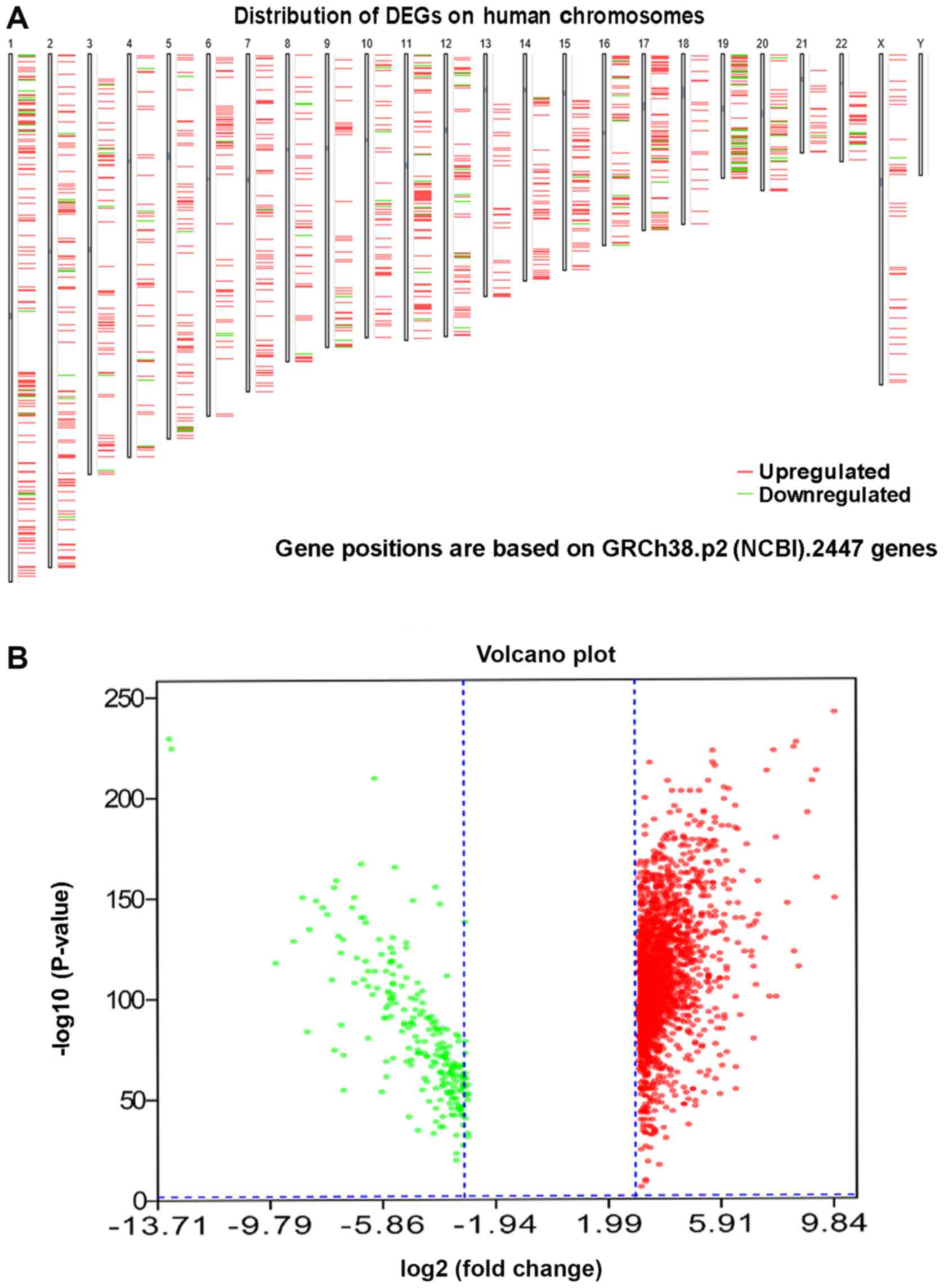
In total, 2,447 DEGs, including 2,204 upregulated
and 243 downregulated genes, were identified in 118 thymoma
patients and 339 normal samples (2 normal tissues and 337 normal
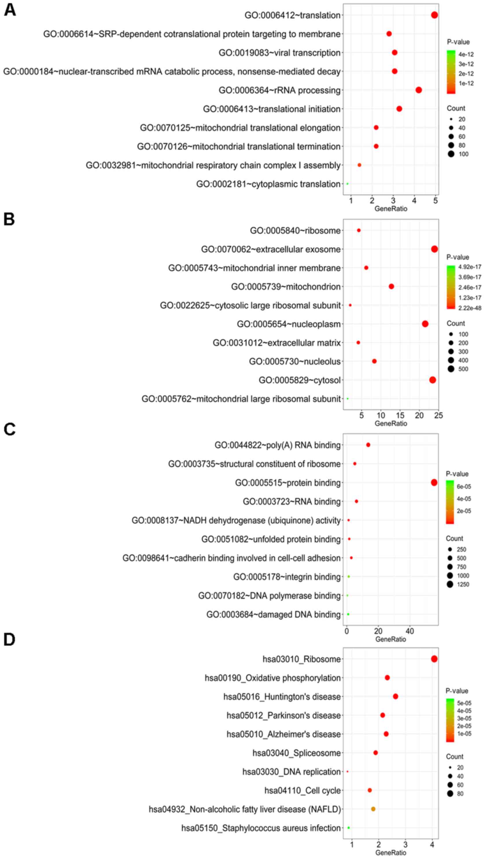
blood samples) (Fig. 1; Table I). A GO analysis was further employed
to determine the possible molecular mechanisms of the DEGs. In the
category biological process (BP), these genes were most enriched in
‘translation’, ‘SRP-dependent co-translational protein targeting to
membrane’, ‘viral transcription’, ‘nuclear-transcribed mRNA
catabolic process, nonsense-mediated decay’ and ‘rRNA processing’
(Fig. 2A). In the category cellular
component (CC), ‘ribosome’, ‘extracellular exosome’, ‘mitochondrial
inner membrane’, ‘mitochondrion’ and ‘cytosolic large ribosomal
subunit’ were the most prominent terms (Fig. 2B). In the category molecular function
(MF), these genes were significantly involved in ‘poly(A) RNA
binding’, ‘structural constituent of ribosome’, ‘protein binding’,
‘RNA binding’ and ‘NADH dehydrogenase (ubiquinone) activity’
(Fig. 2C). In the KEGG pathway
analysis, ‘ribosome’, ‘oxidative phosphorylation’, ‘Huntington's
disease’, ‘Parkinson's disease’ and ‘Alzheimer's disease’ were the
most prominent pathways (Fig. 2D).
Among these pathways, ‘ribosome’, with 93 genes included, was the
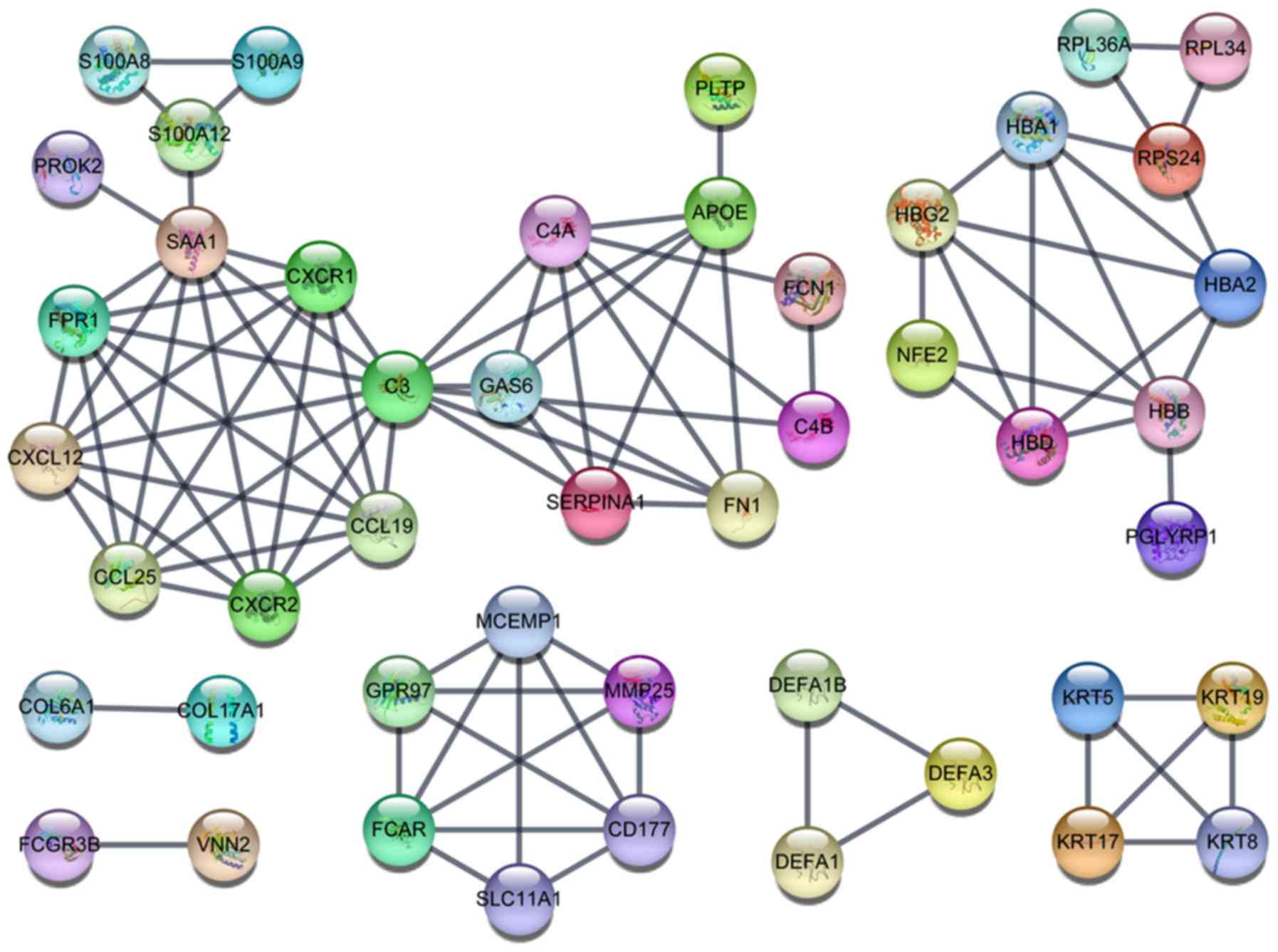
most significant pathway. The PPI networks comprising the DEGs
revealed that certain genes, including complement C3 (C3), Serum
amyloid A1 (SAA1), C-X-C motif chemokine receptor 1 (CXCR1), C-X-C
motif chemokine receptor 2 (CXCR2), C-X-C motif chemokine ligand 12
(CXCL12), C-C motif chemokine ligand 19 (CCL19), C-C motif
chemokine ligand 25 (CCL25) and Formyl peptide receptor were
closely linked with a high degree (Fig.
3).
 | Table I.Clinicopathological features of the
thymoma patients (n=118). |
Table I.
Clinicopathological features of the
thymoma patients (n=118).
| Clinicopathological
feature | Number |
|---|
| Age (years) |
|
|
<40 | 10 |
|
40–60 | 51 |
|
>60 | 57 |
| Sex |
|
|
Male | 62 |
|
Female | 56 |
| Ethnicity |
|
|
White | 98 |
|
Black | 6 |
|
Asian | 12 |
| Not
specified | 2 |
| Stage |
|
| I | 35 |
| II | 60 |
|
III | 15 |
| IV | 6 |
| Not
available | 2 |
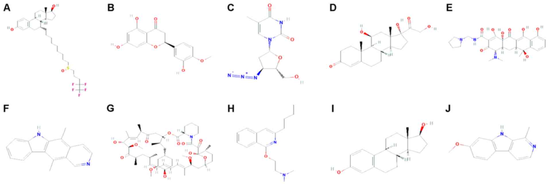
Potential therapeutic drugs identified
from the CMap database
CMap analysis identified 5,000 compounds correlated
with the DEGs. According to the score rankings, 769 drugs scored
<-0.75 and were considered potential thymoma therapeutics. The
top 10 compounds displaying the strongest negative correlation were
fulvestrant, hesperetin, zidovudine, hydrocortisone,
rolitetracycline, ellipticine, sirolimus, quinisocaine, oestradiol
and harmine (Fig. 4; Table II).
| Table II.Connectivity map results for the 10
strongest negative correlation compounds according to CMap. |
Table II.
Connectivity map results for the 10
strongest negative correlation compounds according to CMap.
| Drug name | Dose | Connectivity
score | Up score | Down score |
|---|
| Fulvestrant | 1 µM | −1.00 | −0.25 | 0.38 |
| Hesperetin | 13 µM | −0.99 | −0.25 | 0.37 |
| Zidovudine | 15 µM | −0.99 | −0.25 | 0.37 |
| Hydrocortisone | 11 µM | −0.99 | −0.24 | 0.38 |
|
Rolitetracycline | 8 µM | −0.98 | −0.23 | 0.38 |
| Ellipticine | 16 µM | −0.98 | −0.22 | 0.40 |
| Sirolimus | 100 nM | −0.98 | −0.23 | 0.39 |
| Quinisocaine | 13 µM | −0.98 | −0.23 | 0.38 |
| Estradiol | 100 nM | −0.97 | −0.24 | 0.37 |
| Harmine | 16 µM | −0.97 | −0.21 | 0.40 |
Construction of the drug-target
network
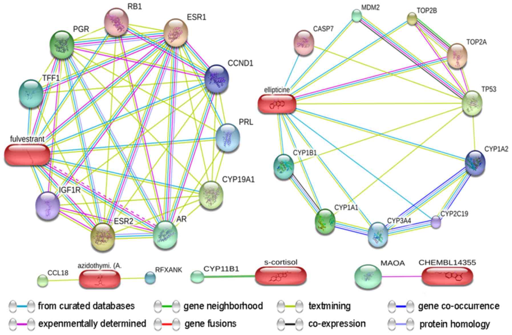
The targets of the test drugs were identified from
the STITCH database (20) (Fig. 5). The predicted targets of the top 10
drugs predicted for thymoma were confirmed, and the predicted
scores for the interactions of DNA topoisomerase IIα (TOP2A), TOP2B
and tumour protein 53 with ellipticine were 0.93, 0.86 and 0.84,
respectively. Similarly, the scores for the interactions of
estrogen receptor 1 (ESR1), ESR2 and cytochrome P450 family 19
subfamily A member 1 (CYP19A1) with fulvestrant were 0.99, 0.99 and
0.99, respectively. The score for the interaction of CYP11B1 and
s-cortisol (hydrocortisone) was 0.90. Furthermore, the scores for
the interactions between C-C motif chemokine ligand 18 and
regulatory factor X-associated ankyrin-containing protein with
azidothymidine (zidovudine) were 0.44 and 0.40, respectively. The
score for the interaction of monoamine oxidase A with CHEMBL14355
(harmine) was 0.52 (Table
III).
| Table III.Predicted targets of the drugs
identified from the STITCH database. |
Table III.
Predicted targets of the drugs
identified from the STITCH database.
| Drug/targets | Combined score |
|---|
| Ellipticine |
|
|
TOP2A | 0.93 |
|
TOP2B | 0.86 |
|
TP53 | 0.84 |
|
CYP1A1 | 0.78 |
|
MDM2 | 0.75 |
|
CYP1B1 | 0.74 |
|
CYP3A4 | 0.73 |
|
CYP1A2 | 0.73 |
|
CASP7 | 0.70 |
|
CYP2C19 | 0.70 |
| Fulvestrant |
|
|
ESR1 | 1.00 |
|
ESR2 | 0.99 |
|
CYP19A1 | 0.99 |
| AR | 0.97 |
|
PGR | 0.97 |
|
TFF1 | 0.95 |
|
PRL | 0.94 |
|
CCND1 | 0.92 |
|
IGF1R | 0.92 |
|
RB1 | 0.91 |
| S-cortisol |
|
|
CYP11B1 | 0.90 |
| Azidothymidine
(zidovudine) |
|
|
CCL18 | 0.44 |
|
RFXANK | 0.40 |
| CHEMBL14355 |
|
|
MAOA | 0.52 |
Molecular docking analysis of the
interactions between proteins and compounds
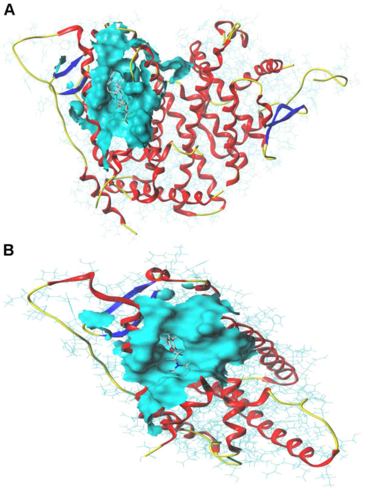
Molecular docking analysis was performed to confirm
the interactions between drugs and protein targets. The total
scores, crash and polar for the fulvestrant and ESR1 interaction
were 10.26, −3.72 and 2.08 respectively (Fig. 6A). The total scores, crash and polar
for the tamoxifen and ESR1 interaction were 6.60, −4.02 and 0,
respectively (Fig. 6B).
Functional annotation of the drug
targets
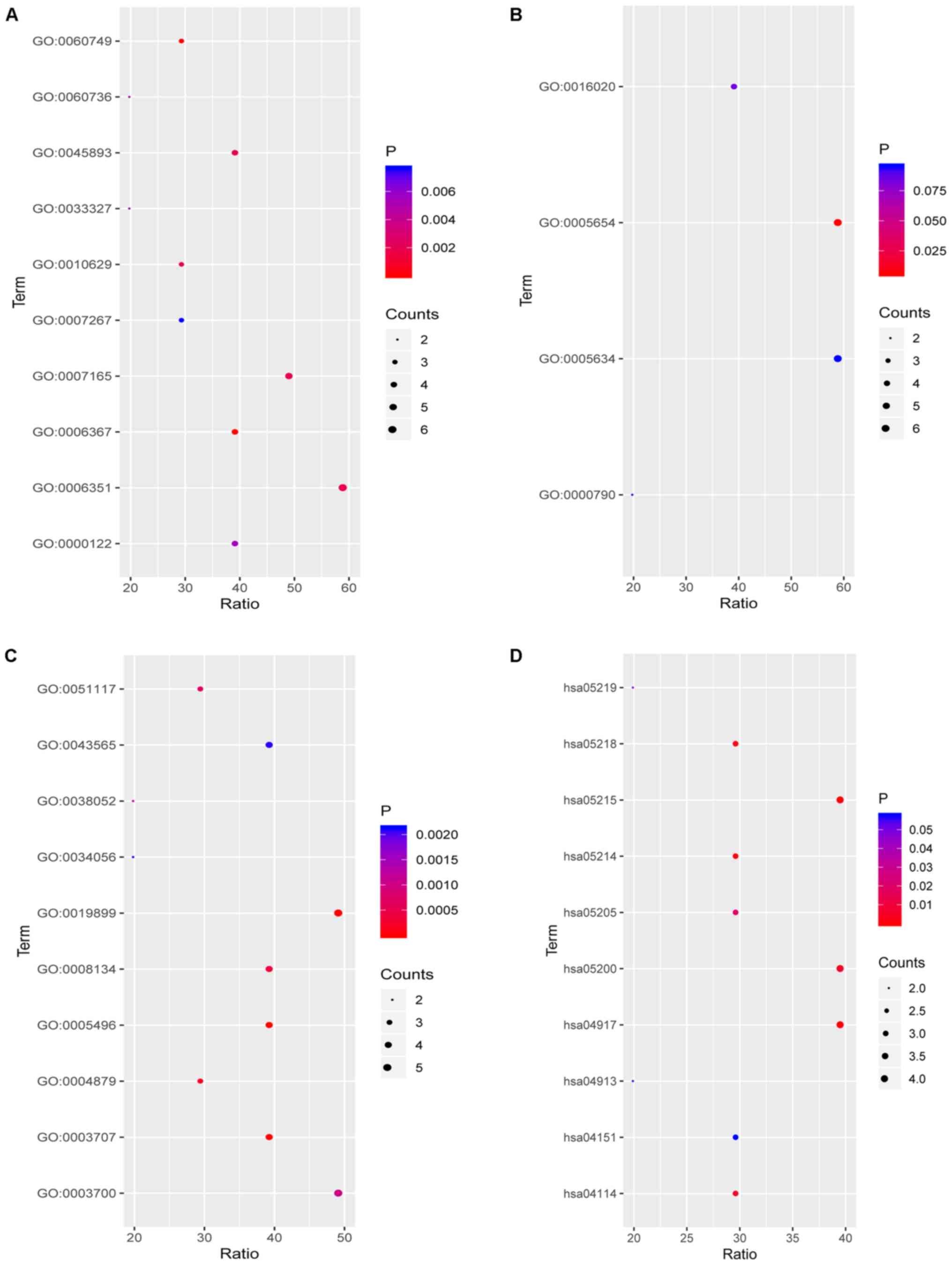
To further explore the functions of the compounds in
combination with the target, functional annotation, including GO
and KEGG pathway analyses, were performed. The results revealed
that the targets for fulvestrant in the category BP were
significantly involved in ‘mammary gland alveolus development’,
‘transcription initiation from RNA polymerase II promoter’,
‘transcription, DNA-templated’, ‘positive regulation of
transcription, DNA-templated’ and ‘signal transduction’ (Fig. 7A). However, in the CC category, the
targets were only significantly involved in ‘nucleoplasm’ (Fig. 7B). In the category MF, the targets
were mainly enriched in ‘steroid binding’, ‘steroid hormone
receptor activity’, ‘enzyme binding’, ‘RNA polymerase II
transcription factor activity, ligand’ and ‘transcription factor
binding’ (Fig. 7C). Finally, in the
KEGG pathway analysis of fulvestrant, ‘prolactin signaling
pathway’, ‘prostate cancer’, ‘glioma’, ‘melanoma’ and ‘oocyte
meiosis’ were the most prominent pathways (Fig. 7D; Table
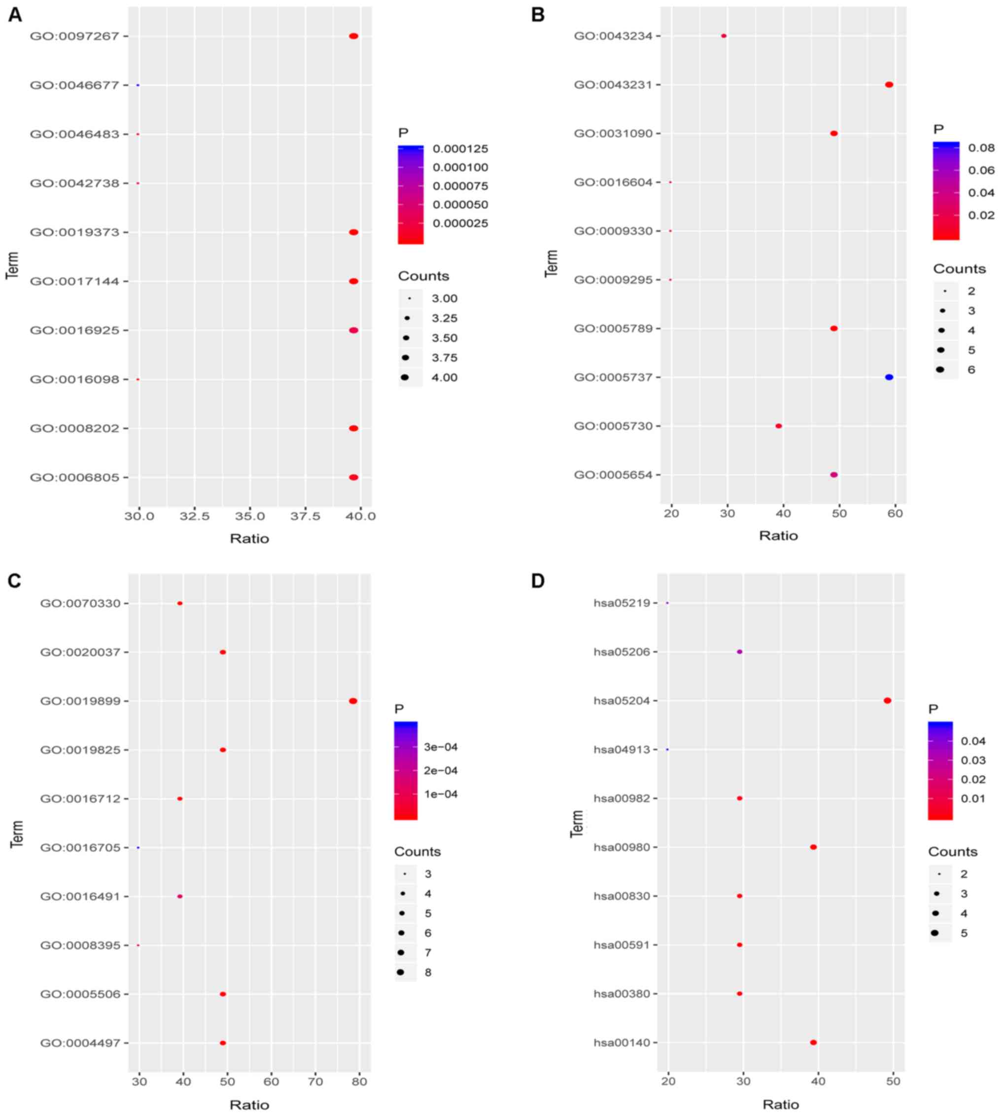
IV). Target genes of ellipticine in the category BP were mainly
enriched in ‘omega-hydroxylase P450 pathway’, ‘epoxygenase P450
pathway’, ‘drug metabolic process’, ‘steroid metabolic process’ and
‘monoterpenoid metabolic process’ (Fig.
8A). In the CC category, the targets were mainly enriched in
‘organelle membrane’, ‘intracellular membrane-bounded organelle’,
‘endoplasmic reticulum membrane’, ‘nucleoid’ and ‘DNA topoisomerase
complex (ATP-hydrolyzing)’ (Fig.
8B). Regarding the enrichment of targets of ellipticine in the
category MF, they were significantly involved in ‘enzyme binding’,
‘oxygen binding’, ‘monooxygenase activity’, ‘oxidoreductase
activity’ (acting on paired donors with incorporation or reduction
of molecular oxygen, reduced flavin or flavoprotein as one donor
and the incorporation of one atom of oxygen) and ‘aromatase
activity’ (Fig. 8C). In the KEGG
pathway analysis of ellipticine, ‘chemical carcinogenesis’,
‘steroid hormone biosynthesis’, ‘metabolism of xenobiotics by
cytochrome P450’, ‘linoleic acid metabolism’ and ‘tryptophan
metabolism’ were the most prominent pathways (Fig. 8D; Table
V).
| Table IV.Functional annotation of the targets
of fulvestrant. |
Table IV.
Functional annotation of the targets
of fulvestrant.
| ID | Description | Category/type | Gene symbol |
|---|
| GO:0060749 | Mammary gland
alveolus development |
Biological_process | AR, CCND1,
ESR1 |
| GO:0006367 | Transcription
initiation from RNA polymerase II promoter |
Biological_process | PGR, AR, ESR1,
ESR2 |
| GO:0006351 | Transcription,
DNA-templated |
Biological_process | PGR, AR, CCND1,
ESR1, RB1, ESR2 |
| GO:0045893 | Positive regulation
of transcription, DNA-template |
Biological_process | AR, ESR1, RB1,
ESR2 |
| GO:0007165 | Signal
transduction |
Biological_process | PGR, IGF1R, AR,
ESR1, ESR2 |
| GO:0010629 | Negative regulation
of gene expression |
Biological_process | PGR, ESR1, RB1 |
| GO:0060736 | Prostate gland
growth |
Biological_process | AR, CYP1A1 |
| GO:0033327 | Leydig cell
differentiation |
Biological_process | AR, CCND1 |
| GO:0000122 | Negative regulation
of transcription from RNA polymerase II promoter |
Biological_process | CCND1, ESR1, RB1,
ESR2 |
| GO:0007267 | Cell-cell
signaling |
Biological_process | PGR, AR, ESR2 |
| GO:0005654 | Nucleoplasm |
Cellular_component | PGR, AR, CCND1,
ESR1, RB1, ESR2 |
| GO:0016020 | Membrane |
Cellular_component | IGF1R, CCND1, ESR1,
CYP19A1 |
| GO:0000790 | Nuclear
chromatin |
Cellular_component | AR, ESR1 |
| GO:0005634 | Nucleus |
Cellular_component | PGR, AR, CCND1,
ESR1, RB1, ESR2 |
| GO:0005496 | Steroid
binding |
Molecular_function | PGR, AR, ESR1,
ESR2 |
| GO:0003707 | Steroid hormone
receptor activity |
Molecular_function | PGR, AR, ESR1,
ESR2 |
| GO:0019899 | Enzyme binding |
Molecular_function | PGR, AR, CCND1,
ESR1, ESR2 |
| GO:0004879 | RNA polymerase II
transcription factor activity, ligand |
Molecular_function | AR, ESR1, ESR2 |
| GO:0008134 | Transcription
factor binding |
Molecular_function | AR, CCND1, ESR1,
RB1 |
| GO:0051117 | ATPase binding |
Molecular_function | PGR, AR, ESR1 |
| GO:0003700 | Transcription
factor activity, sequence |
Molecular_function | PGR, AR, ESR1, RB1,
ESR2 |
| GO:0038052 | RNA polymerase II
transcription factor activity, estrogen |
Molecular_function | ESR1, ESR2 |
| GO:0043565 | Sequence-specific
DNA binding |
Molecular_function | PGR, AR, ESR1,
ESR2 |
| GO:0034056 | Estrogen response
element binding |
Molecular_function | ESR1, ESR2 |
| hsa04917 | Prolactin signaling
pathway | KEGG | CCND1, ESR1, ESR2,
PRL |
| hsa05215 | Prostate
cancer | KEGG | IGF1R, AR, CCND1,
RB1 |
| hsa05214 | Glioma | KEGG | IGF1R, CCND1,
RB1 |
| hsa05218 | Melanoma | KEGG | IGF1R, CCND1,
RB1 |
| hsa04114 | Oocyte meiosis | KEGG | PGR, IGF1R, AR |
| hsa05200 | Pathways in
cancer | KEGG | IGF1R, AR, CCND1,
RB1 |
| hsa05205 | Proteoglycans in
cancer | KEGG | IGF1R, CCND1,
ESR1 |
| hsa05219 | Bladder cancer | KEGG | CCND1, RB1 |
| hsa04913 | Ovarian
steroidogenesis | KEGG | IGF1R, CYP19A1 |
| hsa04151 | PI3K/Akt signaling
pathway | KEGG | IGF1R, CCND1,
PRL |
| Table V.Functional annotation of the targets
of ellipticine. |
Table V.
Functional annotation of the targets
of ellipticine.
| ID | Description | Type | Genes |
|---|
| GO:0097267 | Omega-hydroxylase
P450 pathway |
Biological_process | CYP1B1, CYP1A1,
CYP2C19, CYP1A2 |
| GO:0019373 | Epoxygenase P450
pathway |
Biological_process | CYP1B1, CYP1A1,
CYP2C19, CYP1A2 |
| GO:0017144 | Drug metabolic
process |
Biological_process | CYP3A4, CYP1A1,
CYP2C19, CYP1A2 |
| GO:0008202 | Steroid metabolic
process |
Biological_process | CYP3A4, CYP1B1,
CYP1A1, CYP2C19 |
| GO:0016098 | Monoterpenoid
metabolic process |
Biological_process | CYP3A4, CYP2C19,
CYP1A2 |
| GO:0046483 | Heterocycle
metabolic process |
Biological_process | CYP3A4, CYP2C19,
CYP1A2 |
| GO:0006805 | Xenobiotic
metabolic process |
Biological_process | CYP3A4, CYP1B1,
CYP2C19, CYP1A2 |
| GO:0042738 | Exogenous drug
catabolic process |
Biological_process | CYP3A4, CYP2C19,
CYP1A2 |
| GO:0016925 | Protein
sumoylation |
Biological_process | TP53, MDM2, TOP2B,
TOP2A |
| GO:0046677 | Response to
antibiotic |
Biological_process | CYP1A1, TP53,
MDM2 |
| GO:0031090 | Organelle
membrane |
Cellular_component | CYP3A4, CYP1B1,
CYP1A1, CYP2C19, CYP1A2 |
| GO:0043231 | Intracellular
membrane-bound organelle |
Cellular_component | CYP3A4, CYP1B1,
CYP1A1, CYP2C19, CASP7, CYP1A2 |
| GO:0005789 | Endoplasmic
reticulum membrane |
Cellular_component | CYP3A4, CYP1B1,
CYP1A1, CYP2C19, CYP1A2 |
| GO:0009295 | Nucleoid |
Cellular_component | TOP2B, TOP2A |
| GO:0009330 | DNA topoisomerase
complex (ATP-hydrolyzing) |
Cellular_component | TOP2B, TOP2A |
| GO:0005730 | Nucleolus |
Cellular_component | TP53, MDM2, TOP2B,
TOP2A |
| GO:0016604 | Nuclear body |
Cellular_component | TP53, MDM2 |
| GO:0043234 | Protein
complex |
Cellular_component | TP53, MDM2,
TOP2A |
| GO:0005654 | Nucleoplasm |
Cellular_component | CASP7, TP53, MDM2,
TOP2B, TOP2A |
| GO:0005737 | Cytoplasm |
Cellular_component | CYP3A4, CASP7,
TP53, MDM2, TOP2B, TOP2A |
| GO:0019899 | Enzyme binding |
Molecular_function | CYP3A4, CYP1A1,
CYP2C19, TP53, MDM2, CYP1A2, TOP2B, TOP2A |
| GO:0019825 | Oxygen binding |
Molecular_function | CYP3A4, CYP1B1,
CYP1A1, CYP2C19, CYP1A2 |
| GO:0004497 | Monooxygenase
activity |
Molecular_function | CYP3A4, CYP1B1,
CYP1A1, CYP2C19, CYP1A2 |
| GO:0016712 | Reduced flavin or
flavoprotein as one donor |
Molecular_function | CYP3A4, CYP1B1,
CYP1A1, CYP1A2 |
| GO:0070330 | Aromatase
activity |
Molecular_function | CYP3A4, CYP1B1,
CYP1A1, CYP1A2 |
| GO:0020037 | Heme binding |
Molecular_function | CYP3A4, CYP1B1,
CYP1A1, CYP2C19, CYP1A2 |
| GO:0005506 | Iron ion
binding |
Molecular_function | CYP3A4, CYP1B1,
CYP1A1, CYP2C19, CYP1A2 |
| GO:0008395 | Steroid hydroxylase
activity |
Molecular_function | CYP3A4, CYP1A1,
CYP2C19 |
| GO:0016491 | Oxidoreductase
activity |
Molecular_function | CYP3A4, CYP1A1,
CYP2C19, CYP1A2 |
| GO:0016705 | Incorporation or
reduction of molecular oxygen |
Molecular_function | CYP3A4, CYP1B1,
CYP2C19 |
| hsa05204 | Chemical
carcinogenesis | KEGG | CYP3A4, CYP1B1,
CYP1A1, CYP2C19, CYP1A2 |
| hsa00140 | Steroid hormone
biosynthesis | KEGG | CYP3A4, CYP1B1,
CYP1A1, CYP1A2 |
| hsa00980 | Metabolism of
xenobiotics by cytochrome P450 | KEGG | CYP3A4, CYP1B1,
CYP1A1, CYP1A2 |
| hsa00591 | Linoleic acid
metabolism | KEGG | CYP3A4, CYP2C19,
CYP1A2 |
| hsa00380 | Tryptophan
metabolism | KEGG | CYP1B1, CYP1A1,
CYP1A2 |
| hsa00830 | Retinol
metabolism | KEGG | CYP3A4, CYP1A1,
CYP1A2 |
| hsa00982 | Drug
metabolism-cytochrome P450 | KEGG | CYP3A4, CYP2C19,
CYP1A2 |
| hsa05206 | MicroRNAs in
cancer | KEGG | CYP1B1, TP53,
MDM2 |
| hsa05219 | Bladder cancer | KEGG | TP53, MDM2 |
| hsa04913 | Ovarian
steroidogenesis | KEGG | CYP1B1, CYP1A1 |
Screening of compounds based on ADME
parameters in the TCMSP database
Hesperetin, oestradiol and harmine were searched in
the TCMSP database. The OB (%), DL and HL for hesperetin were
70.31, 0.27 and 15.78, respectively; those for oestradiol were
53.56, 0.32 and 3.50, respectively; and those for harmine were
56.80, 0.13 and 5.04, respectively (Table VI).
| Table VI.Absorption, distribution, metabolism
and excretion parameters for three drugs from the Traditional
Chinese Medicine Systems Pharmacology Database and Analysis
Platform database. |
Table VI.
Absorption, distribution, metabolism
and excretion parameters for three drugs from the Traditional
Chinese Medicine Systems Pharmacology Database and Analysis
Platform database.
| Drug name | Oral
bioavailability (%) | Drug-likeness
(0–1) | Drug half-life
(h) |
|---|
| Hesperetin | 70.31 | 0.27 | 15.78 |
| Estradiol | 53.56 | 0.32 | 3.5 |
| Harmine | 56.8 | 0.13 | 5.04 |
Discussion
In the present study, it was speculated that the
potential therapeutic drugs for thymoma are compounds that are
matched with DEGs known to be associated with the occurrence and
development of tumours. First, by using the DEG data from the GEPIA
database, correlations between the genes and previously known
pharmaceutical compounds were revealed in CMap. Subsequently, the
top 10 molecules with the lowest negative correlations were
obtained and they were considered as potential therapeutic drugs
for further analysis. In addition, a drug-target network was
constructed to examine the potential mechanisms of action of the
compounds. Molecular docking analysis was then performed to confirm
the interactions between the drugs and protein targets.
Furthermore, the ADME parameters were inquired to determine the
potential clinical value of the compounds.
According to the GEPIA tool, 2,447 DEGs were
identified in 118 thymoma patients and 339 normal samples. After
functional annotation analysis, it was determined that the DEGs
were enriched in ‘ribosome’, ‘oxidative phosphorylation’,
‘spliceosome’, ‘DNA replication’ and ‘cell cycle’, which indicated
that the DEGs may affect cell growth and have an important role in
the occurrence and development of thymoma. Then, based on the DEGs,
potential therapeutic drugs were identified using the CMap
database, including fulvestrant, hesperetin, zidovudine,
hydrocortisone, rolitetracycline, ellipticine, sirolimus,
quinisocaine, oestradiol and harmine. Then, to examine the effect
of the potential drugs, a drug-target network was constructed and a
molecular docking analysis was performed. Compared with the docking
score of tamoxifen and ESR1, the score of fulvestrant and ESR1 was
high, which indicates a high interaction between fulvestrant and
ESR1. Among these compounds, tamoxifen, as an ESR1 inhibitor,
remains the first-line of medication for the treatment of
ESR1+ breast cancer (26). In addition, the ESR1 protein
localizes to the nucleus and has been proven important in the
pathological processes of several cancer types, including breast
and endometrial cancers (27,28).
Furthermore, functional enrichment analysis indicated that the
targets of fulvestrant significantly accumulated in ‘prolactin
signaling pathway’, ‘prostate cancer’, ‘glioma’ and ‘melanoma’. The
results indicated that fulvestrant may combine with ESR1 to affect
the treatment of thymoma. Finally, using the TCMSP database, it was
determined that the compounds oestrogen, oestradiol and harmine may
potentially be of high clinical value. In summary, the compounds
may have an important role in the treatment of thymoma. Among these
compounds, fulvestrant has been used to treat breast and prostate
cancers. Regardless of endocrine tolerance or the levels of hormone
receptor expression, fulvestrant significantly improves the
survival of patients with non-progressive breast cancer (29–31). In
addition, fulvestrant also represents a treatment option for
patients with recurrent hormone receptor-positive or HER2-negative
metastatic breast cancer (32).
Hesperetin is a bioflavonoid from citrus fruit.
Based on experimental evidence, hesperetin possesses anti-oxidant
and free radical-quenching activities. This compound also induces
apoptotic cell death. In addition, hesperetin reportedly exerts an
anti-cancer effect on various cancer cell lines, including breast
cancer, prostate cancer, human colon adenocarcinoma and
hepatocellular carcinoma cells (33–37).
Based on ADME values of the compound molecules from the TCMSP
database, it was revealed that hesperetin, estradiol and harmine
may have good prospects. Among them, hesperetin caught our
attention. The consequence of it for oral bioavailability was
70.31. A high OB (%) is frequently considered a key factor to judge
the drug-like properties of compounds as therapeutic agents.
Furthermore, as hesperetin is derived from citrus fruit, it may be
easy to produce on a large scale. However, due to its poor water
solubility, its clinical use is restricted. Numerous studies have
been undertaken to improve the bioavailability of flavonoids
(33). In summary, hesperetin may
not only be used as a promising compound to treat thymoma in the
future, but may also be commonly used in the treatment of other
tumour types.
Zidovudine is an inhibitor of HIV replication that
may reverse neurological dysfunction induced by HIV and ameliorate
certain clinical abnormalities (38,39).
Hydrocortisone is the major glucocorticoid and its synthetic
counterpart is used to treat inflammation, allergy, shock and
certain neoplasms (40).
Rolitetracycline is a broad-spectrum antibiotic (41). Sirolimus is a potent
immunosuppressant (42).
Quinisocaine blocks nerve conduction when applied to nervous
tissues at appropriate concentrations.
Estrogen has been consistently reported to affect
the advancement of thymoma (43–45).
However, the therapeutic value of fulvestrant and hesperetin for
thymoma has not been previously reported. Simultaneously, the other
compounds among the top 10 have also not been reported to be
suitable for the treatment of cancer.
The limitations to the present study include the
following: First, the DEGs should be further validated in
vitro to determine their specific expression in thymoma. In
addition, the exact DEGs between the different groups of patients
and at different stages of the disease should be determined in
vitro to further identify potential compounds, particularly in
patients aged 40–60 years and in stage III/IV. Finally, further
research should focus on in vitro and in vivo tests
prior to the clinical application of these compounds.
In summary, the development of compounds or
combinations of drugs remains a requirement in order to improve
chemotherapy. The present study identified compounds that may
represent novel treatments for thymoma and may reduce the range of
potential drugs for treating thymoma.
Acknowledgements
Not applicable.
Funding
No funding was received.
Availability of data and materials
The datasets used and/or analysed during this study
are available from the corresponding author on reasonable
request.
Authors' contributions
The study was designed by GC, HY, QL and RL. XW, PL,
YL, GC, HY, YH and QL were involved in the statistical analysis.
HY, YH, QL and RL were involved in drafting the manuscript and
critically revising it for important intellctual content. YH, QL
and RL gave final approval for the version of the manuscript to be
published.
Each author sufficiently participated in the work to
take public responsibility for appropriate portions of the content
and agreed to be accountable for all aspects of the work to ensure
that questions regarding the accuracy or integrity of any part of
the work are appropriately investigated and resolved.
Ethics approval and consent to
participate
Not applicable.
Patient consent for publication
Not applicable.
Competing interests
The authors declare that they have no competing
interests.
Glossary
Abbreviations
Abbreviations:
|
DEG
|
differentially expressed gene
|
|
GEPIA
|
gene expression profiling interactive
analysis
|
|
CMap
|
connectivity map
|
|
TCGA
|
The Cancer Genome Atlas
|
|
GO
|
Gene Ontology
|
|
PPI
|
protein-protein interaction
|
|
TCMSP
|
Traditional Chinese Medicine Systems
Pharmacology Database and Analysis Platform
|
|
ADME
|
absorption, distribution, metabolism
and excretion
|
References
|
1
|
Safieddine N, Liu G, Cuningham K, Ming T,
Hwang D, Brade A, Bezjak A, Fischer S, Xu W, Azad S, et al:
Prognostic factors for cure, recurrence and long-term survival
after surgical resection of thymoma. J Thorac Oncol. 9:1018–1022.
2014. View Article : Google Scholar : PubMed/NCBI
|
|
2
|
Thomas CR, Wright CD and Loehrer PJ:
Thymoma: State of the art. J Clin Oncol. 17:2280–2289. 1999.
View Article : Google Scholar : PubMed/NCBI
|
|
3
|
Kim DJ, Yang WI, Choi SS, Kim KD and Chung
KY: Prognostic and clinical relevance of the World Health
Organization schema for the classification of thymic epithelial
tumors: A clinicopathologic study of 108 patients and literature
review. Chest. 127:755–761. 2005. View Article : Google Scholar : PubMed/NCBI
|
|
4
|
Utsumi T, Shiono H, Kadota Y, Matsumura A,
Maeda H, Ohta M, Yoshioka Y, Koizumi M, Inoue T and Okumura M:
Postoperative radiation therapy after complete resection of thymoma
has little impact on survival. Cancer. 115:5413–5420. 2009.
View Article : Google Scholar : PubMed/NCBI
|
|
5
|
Ettinger DS, Riely GJ, Akerley W, Borghaei
H, Chang AC, Cheney RT, Chirieac LR, D'Amico TA, Demmy TL, Govindan
R, et al: Thymomas and thymic carcinomas: Clinical practice
guidelines in oncology. J Natl Compr Canc Netw. 11:562–576. 2013.
View Article : Google Scholar : PubMed/NCBI
|
|
6
|
Venuta F, Rendina EA, Pescarmona EO, De
Giacomo T, Vegna ML, Fazi P, Flaishman I, Guarino E and Ricci C:
Multimodality treatment of thymoma: A prospective study. Ann Thorac
Surg. 64:1585–1592. 1997. View Article : Google Scholar : PubMed/NCBI
|
|
7
|
Ma DL, Chan DS and Leung CH: Drug
repositioning by structure-based virtual screening. Chem Soc Rev.
42:2130–2141. 2013. View Article : Google Scholar : PubMed/NCBI
|
|
8
|
Kitchen DB, Decornez H, Furr JR and
Bajorath J: Docking and scoring in virtual screening for drug
discovery: Methods and applications. Nat Rev Drug Discov.
3:935–949. 2004. View
Article : Google Scholar : PubMed/NCBI
|
|
9
|
Lamb J, Crawford ED, Peck D, Modell JW,
Blat IC, Wrobel MJ, Lerner J, Brunet JP, Subramanian A, Ross KN, et
al: The Connectivity Map: Using gene-expression signatures to
connect small molecules, genes, and disease. Science.
313:1929–1935. 2006. View Article : Google Scholar : PubMed/NCBI
|
|
10
|
Tang Z, Li C, Kang B, Gao G, Li C and
Zhang Z: GEPIA: A web server for cancer and normal gene expression
profiling and interactive analyses. Nucleic Acids Res. 45:W98–W102.
2017. View Article : Google Scholar : PubMed/NCBI
|
|
11
|
He RQ, Qin MJ, Lin P, Luo YH, Ma J, Yang
H, Hu XH and Chen G: Prognostic significance of LncRNA PVT1 and its
potential target gene network in human cancers: A comprehensive
inquiry based upon 21 cancer types and 9,972 cases. Cell Physiol
Biochem. 46:591–608. 2018. View Article : Google Scholar : PubMed/NCBI
|
|
12
|
Huang da W, Sherman BT and Lempicki RA:
Bioinformatics enrichment tools: Paths toward the comprehensive
functional analysis of large gene lists. Nucleic Acids Res.
37:1–13. 2009. View Article : Google Scholar : PubMed/NCBI
|
|
13
|
Ni Z, Wang X, Zhang T, Li L and Li J:
Comprehensive analysis of differential expression profiles reveals
potential biomarkers associated with the cell cycle and regulated
by p53 in human small cell lung cancer. Exp Ther Med. 15:3273–3282.
2018.PubMed/NCBI
|
|
14
|
Huang da W, Sherman BT and Lempicki RA:
Systematic and integrative analysis of large gene lists using DAVID
bioinformatics resources. Nat Protoc. 4:44–57. 2009. View Article : Google Scholar : PubMed/NCBI
|
|
15
|
Liang L, Wei DM, Li JJ, Luo DZ, Chen G,
Dang YW and Cai XY: Prognostic microRNAs and their potential
molecular mechanism in pancreatic cancer: A study based on The
Cancer Genome Atlas and bioinformatics investigation. Mol Med Rep.
17:939–951. 2018.PubMed/NCBI
|
|
16
|
Szklarczyk D, Santos A, von Mering C,
Jensen LJ, Bork P and Kuhn M: STITCH 5: Augmenting protein-chemical
interaction networks with tissue and affinity data. Nucleic Acids
Res. 44:D380–D384. 2016. View Article : Google Scholar : PubMed/NCBI
|
|
17
|
Musa A, Ghoraie LS, Zhang SD, Glazko G,
Yli-Harja O, Dehmer M, Haibe-Kains B and Emmert-Streib F: A review
of connectivity map and computational approaches in
pharmacogenomics. Brief Bioinform. 19:506–523. 2018.PubMed/NCBI
|
|
18
|
Dalma-Weiszhausz DD, Warrington J,
Tanimoto EY and Miyada CG: The affymetrix GeneChip platform: An
overview. Methods Enzymol. 410:3–28. 2006. View Article : Google Scholar : PubMed/NCBI
|
|
19
|
Kim S, Chen J, Cheng T, Gindulyte A, He J,
He S, Li Q, Shoemaker BA, Thiessen PA, Yu B, et al: PubChem 2019
update: Improved access to chemical data. Nucleic Acids Res.
47:D1102–D1109. 2019. View Article : Google Scholar : PubMed/NCBI
|
|
20
|
Szklarczyk D, Franceschini A, Wyder S,
Forslund K, Heller D, Huerta-Cepas J, Simonovic M, Roth A, Santos
A, Tsafou KP, et al: STRING v10: Protein-protein interaction
networks, integrated over the tree of life. Nucleic Acids Res 43
(Database Issue). D447–D452. 2015. View Article : Google Scholar
|
|
21
|
Zhu D, Vaishampayan PA, Venkateswaran K
and Fox GE: STITCH: Algorithm to splice, trim, identify, track, and
capture the uniqueness of 16S rRNAs sequence pairs using public or
in-house database. Microb Ecol. 61:669–675. 2011. View Article : Google Scholar : PubMed/NCBI
|
|
22
|
Homer RW, Swanson J, Jilek RJ, Hurst T and
Clark RD: SYBYL line notation (SLN): A single notation to represent
chemical structures, queries, reactions, and virtual libraries. J
Chem Inf Model. 48:2294–2307. 2008. View Article : Google Scholar : PubMed/NCBI
|
|
23
|
Koelmel JP, Ulmer CZ, Jones CM, Yost RA
and Bowden JA: Common cases of improper lipid annotation using
high-resolution tandem mass spectrometry data and corresponding
limitations in biological interpretation. Biochim Biophys Acta Mol
Cell Biol Lipids. 1862:766–770. 2017. View Article : Google Scholar : PubMed/NCBI
|
|
24
|
Preston S, Jiao Y, Baell JB, Keiser J,
Crawford S, Koehler AV, Wang T, Simpson MM, Kaplan RM, Cowley KJ,
et al: Screening of the ‘Open Scaffolds’ collection from Compounds
Australia identifies a new chemical entity with anthelmintic
activities against different developmental stages of the barber's
pole worm and other parasitic nematodes. Int J Parasitol Drugs Drug
Resist. 7:286–294. 2017. View Article : Google Scholar : PubMed/NCBI
|
|
25
|
Ru J, Li P, Wang J, Zhou W, Li B, Huang C,
Li P, Guo Z, Tao W, Yang Y, et al: TCMSP: A database of systems
pharmacology for drug discovery from herbal medicines. J
Cheminform. 6:132014. View Article : Google Scholar : PubMed/NCBI
|
|
26
|
Wardell SE, Ellis MJ, Alley HM, Eisele K,
VanArsdale T, Dann SG, Arndt KT, Primeau T, Griffin E, Shao J, et
al: Efficacy of SERD/SERM Hybrid-CDK4/6 inhibitor combinations in
models of endocrine therapy-resistant breast cancer. Clin Cancer
Res. 21:5121–5130. 2015. View Article : Google Scholar : PubMed/NCBI
|
|
27
|
Fribbens C, O'Leary B, Kilburn L, Hrebien
S, Garcia-Murillas I, Beaney M, Cristofanilli M, Andre F, Loi S,
Loibl S, et al: Plasma ESR1 mutations and the treatment of estrogen
receptor-positive advanced breast cancer. J Clin Oncol.
34:2961–2968. 2016. View Article : Google Scholar : PubMed/NCBI
|
|
28
|
Lebeau A, Grob T, Holst F,
Seyedi-Fazlollahi N, Moch H, Terracciano L, Turzynski A, Choschzick
M, Sauter G and Simon R: Oestrogen receptor gene (ESR1)
amplification is frequent in endometrial carcinoma and its
precursor lesions. J Pathol. 216:151–157. 2008. View Article : Google Scholar : PubMed/NCBI
|
|
29
|
Dean JL, Thangavel C, McClendon AK, Reed
CA and Knudsen ES: Therapeutic CDK4/6 inhibition in breast cancer:
Key mechanisms of response and failure. Oncogene. 29:4018–4032.
2010. View Article : Google Scholar : PubMed/NCBI
|
|
30
|
Finn RS, Dering J, Conklin D, Kalous O,
Cohen DJ, Desai AJ, Ginther C, Atefi M, Chen I, Fowst C, et al: PD
0332991, a selective cyclin D kinase 4/6 inhibitor, preferentially
inhibits proliferation of luminal estrogen receptor-positive human
breast cancer cell lines in vitro. Breast Cancer Res. 11:R772009.
View Article : Google Scholar : PubMed/NCBI
|
|
31
|
VanArsdale T, Boshoff C, Arndt KT and
Abraham RT: Molecular pathways: Targeting the cyclin D-CDK4/6 axis
for cancer treatment. Clin Cancer Res. 21:2905–2910. 2015.
View Article : Google Scholar : PubMed/NCBI
|
|
32
|
Cristofanilli M, Turner NC, Bondarenko I,
Ro J, Im SA, Masuda N, Colleoni M, DeMichele A, Loi S, Verma S, et
al: Fulvestrant plus palbociclib versus fulvestrant plus placebo
for treatment of hormone-receptor-positive, HER2-negative
metastatic breast cancer that progressed on previous endocrine
therapy (PALOMA-3): Final analysis of the multicentre,
double-blind, phase 3 randomised controlled trial. Lancet Oncol.
17:425–439. 2016. View Article : Google Scholar : PubMed/NCBI
|
|
33
|
Mary Lazer L, Sadhasivam B, Palaniyandi K,
Muthuswamy T, Ramachandran I, Balakrishnan A, Pathak S, Narayan S
and Ramalingam S: Chitosan-based nano-formulation enhances the
anticancer efficacy of hesperetin. Int J Biol Macromol.
107:1988–1998. 2018. View Article : Google Scholar : PubMed/NCBI
|
|
34
|
Choi EJ: Hesperetin induced G1-phase cell
cycle arrest in human breast cancer MCF-7 cells: Involvement of
CDK4 and p21. Nutr Cancer. 59:115–119. 2007. View Article : Google Scholar : PubMed/NCBI
|
|
35
|
Sambantham S, Radha M, Paramasivam A,
Anandan B, Malathi R, Chandra SR and Jayaraman G: Molecular
mechanism underlying hesperetin-induced apoptosis by in silico
analysis and in prostate cancer PC-3 cells. Asian Pac J Cancer
Prev. 14:4347–4352. 2013. View Article : Google Scholar : PubMed/NCBI
|
|
36
|
Sivagami G, Vinothkumar R, Bernini R,
Preethy CP, Riyasdeen A, Akbarsha MA, Menon VP and Nalini N: Role
of hesperetin (a natural flavonoid) and its analogue on apoptosis
in HT-29 human colon adenocarcinoma cell line-a comparative study.
Food Chem Toxicol. 50:660–671. 2012. View Article : Google Scholar : PubMed/NCBI
|
|
37
|
Zhang J, Song J, Wu D, Wang J and Dong W:
Hesperetin induces the apoptosis of hepatocellular carcinoma cells
via mitochondrial pathway mediated by the increased intracellular
reactive oxygen species, ATP and calcium. Med Oncol. 32:1012015.
View Article : Google Scholar : PubMed/NCBI
|
|
38
|
McLeod GX and Hammer SM: Zidovudine: Five
years later. Ann Intern Med. 117:487–501. 1992. View Article : Google Scholar : PubMed/NCBI
|
|
39
|
De Clercq E: HIV resistance to reverse
transcriptase inhibitors. Biochemical pharmacology. 47:155–169.
1994. View Article : Google Scholar : PubMed/NCBI
|
|
40
|
Palacios R and Sugawara I: Hydrocortisone
abrogates proliferation of T cells in autologous mixed lymphocyte
reaction by rendering the interleukin-2 Producer T cells
unresponsive to interleukin-1 and unable to synthesize the T-cell
growth factor. Scand J Immunol. 15:25–31. 1982. View Article : Google Scholar : PubMed/NCBI
|
|
41
|
Schuttemeyer W and Reiff K: Clinical
observations on the broad spectrum antibiotic reverin. Dtsch Med J.
10:90–91. 1959.(In German). PubMed/NCBI
|
|
42
|
Sehgal SN: Sirolimus: Its discovery,
biological properties, and mechanism of action. Transplant Proc. 35
(Suppl 3):S7–S14. 2003. View Article : Google Scholar
|
|
43
|
Hengartner MO: The biochemistry of
apoptosis. Nature. 407:770–776. 2000. View Article : Google Scholar : PubMed/NCBI
|
|
44
|
Okasha SA, Ryu S, Do Y, McKallip RJ,
Nagarkatti M and Nagarkatti PS: Evidence for estradiol-induced
apoptosis and dysregulated T cell maturation in the thymus.
Toxicology. 163:49–62. 2001. View Article : Google Scholar : PubMed/NCBI
|
|
45
|
Zoller AL, Schnell FJ and Kersh GJ: Murine
pregnancy leads to reduced proliferation of maternal thymocytes and
decreased thymic emigration. Immunology. 121:207–215. 2007.
View Article : Google Scholar : PubMed/NCBI
|