Introduction
Cholangiocarcinoma (CCA), a highly aggressive type
of malignancy, is an epithelial cancer of the biliary tree
(1). According to its anatomical
location, CCA is classified into two major types, including the
extrahepatic type and the intrahepatic type, of which the
extrahepatic type is more common. In recent decades, the incidence
and mortality of CCA, particularly the intrahepatic type, has
significantly increased worldwide (2). As patients in the early stages of CCA
are frequently asymptomatic, CCA is usually diagnosed in the late
stages, resulting in a dismal survival rate and prognosis (3,4).
Furthermore, almost all types of CCA are resistant to
chemotherapies and radical surgery may be the only effective
treatment for CCA. This provides patients with only a small
benefit, given that they are diagnosed at an advanced stage
(5,6). Therefore, it is crucial to explore
novel cancer-associated genes that may serve as reliable diagnostic
or prognostic biomarkers and therapeutic targets for improving the
prognosis and therapeutic efficacy.
MicroRNAs (miRNAs/miRs) are a group of small,
endogenous non-coding RNA molecules that regulate the expression of
target genes at the post-transcriptional level. miRNAs are involved
in the regulation of various physiological and pathological
functions (7,8). They have crucial roles in various
biological processes, including inflammation, cell proliferation,
migration, invasion and differentiation (9–12).
Previous studies have indicated that miR-25 is overexpressed in a
variety of cancer types and is functionally associated with
numerous cancer-associated processes, including cancer cell
proliferation, migration and metastasis (13–16).
However, the current knowledge on the role of miR-25 in CCA is
limited.
In the present study, the expression pattern of
miR-25 in CCA tissues and cell lines was investigated. The
association between miR-25 expression and clinical characteristics
was also analyzed. The effects of miR-25 on the biological behavior
of cancer cells were investigated through in vitro cell
experiments. Furthermore, the clinical significance of miR-25 as a
potential prognostic biomarker for CCA patients was evaluated.
Materials and methods
Patients and tissue samples
The present study included 116 patients diagnosed
with CCA treated at Yidu Central Hospital (Weifang, China) between
February 2007 and October 2012. The CCA tissues and corresponding
adjacent non-tumorous tissues were snap-frozen in liquid nitrogen
after collection. None of the patients enrolled received any
treatment prior to surgery. The 5-year follow-up information was
collected for subsequent analysis. The clinicopathological
characteristics of the cohort are summarized in Table I.
 | Table I.Association between miR-25 expression
and clinicopathological parameters of patients with
cholangiocarcinoma (n=116). |
Table I.
Association between miR-25 expression
and clinicopathological parameters of patients with
cholangiocarcinoma (n=116).
|
|
| miR-25
expression |
|
|---|
|
|
|
|
|
|---|
| Characteristics | N | Low (n=51) | High (n=65) | P-value |
|---|
| Age (years) |
|
|
| 0.887 |
|
<60 | 56 | 25 | 31 |
|
| ≥60 | 60 | 26 | 34 |
|
| Sex |
|
|
| 0.733 |
| Male | 68 | 29 | 39 |
|
|
Female | 48 | 22 | 26 |
|
| Differentiation |
|
|
| 0.096 |
| Well | 65 | 33 | 32 |
|
| Moderate
+ poor | 51 | 18 | 33 |
|
| TNM stage |
|
|
| 0.026 |
|
I–II | 57 | 31 | 26 |
|
|
III–IV | 59 | 20 | 39 |
|
| Lymph node
metastasis |
|
|
| 0.032 |
|
Negative | 53 | 29 | 24 |
|
|
Positive | 63 | 22 | 41 |
|
Cell lines and transfection
The human CCA cell lines CCLP1 and HuCCT1 were
purchased from the Japan Health Science Research Resources Bank. A
normal human intrahepatic biliary epithelial cell line (HIBEC)
(17) was purchased from the
American Type Culture Collection. These cell lines were cultured in
RPMI-1640 medium (Invitrogen; Thermo Fisher Scientific, Inc.) with
10% fetal bovine serum (FBS, Invitrogen, Thermo Fisher Scientific,
Inc.) and were incubated at 37°C in a humidified incubator with 5%
CO2. miR-25 mimics (5′-AGGCGGAGACUUGGGCAAUUG-3′), mimics
negative control (mimics NC, 5′-UUCUCCGAACGUGUCACGUTT-3′), miR-25
inhibitor (5′-CAAUUGCCCAAGUCUCCGCCU-3′) or inhibitor negative
control (inhibitor NC, 5′-CAGUACUUUUGUGUAGUACAA-3′) were obtained
from Shanghai GenePharma Co., Ltd. Cell transfection was performed
using the Lipofectamine 2000 Reagent (Invitrogen; Thermo Fisher
Scientific, Inc.) following the manufacturer's protocols. A total
of 5×10⁴ cells were seeded into the wells of a 6-well
plate and after 24 h, the cells were transfected with miR-25 mimics
or mimics NC, miR-25 inhibitor or inhibitor NC for 48 h. Cells with
only transfection reagent were used as a mock group. Reverse
transcription-quantitative (RT-q)PCR analysis was used to detect
the successful knockdown or upregulation of miR-25 expression.
RNA extraction and RT-qPCR
analysis
Total RNA was extracted from the tissues and cells
using TRIzol reagent (Invitrogen; Thermo Fisher Scientific, Inc.)
according to the manufacturer's protocol. The miRNA of the purified
total RNA was then reverse-transcribed to complementary DNA (cDNA)
by using the Transcriptor First Strand cDNA Synthesis Kit (Roche).
The relative miR-25 expression was analyzed by using the qPCR
analysis, which was performed using SYBR Green (Takara Bio, Inc.)
on an ABI 7500 system (Applied Biosystems; Thermo Fisher
Scientific, Inc.). The thermocycling conditions were as follows:
Initial denaturation at 95°C for 5 min; 40 cycles of 95°C for 30
sec, 60°C for 30 sec, and 72°C for 10 sec; final extension at 72°C
for 10 min. The primers were as follows: miR-25,
5′-GTGTTGAGAGGGCGGAGACTT-3′ (forward) and
5′-TCAGACCGAGACAAGTGCAA-3′ (reverse); U6,
5′-GCTTCGGCAGCACATATACTAAAAT-3′ (forward) and
5′-CGCTTCACGAATTTGCGTGTCAT-3′ (reverse). Relative expression was
calculated using the 2−ΔΔCq method (18) with normalization to U6. Each
experiment was performed as three replicates.
MTT cell viability assay
The colorimetric MTT assay was used to evaluate the
effect of miR-25 on the viability of CCA cells. CCLP-1 and HuCCT1
cells were seeded in 96-well plates (5×10⁴ per well).
After transfection, 10 µl MTT (5 mg/ml; Sigma-Aldrich; Merck KGaA)
was added to each well and the cells were incubated at 37°C for 4
h. The medium was then removed and 150 µl DMSO (Sigma-Aldrich;
Merck KGaA) was added to each well to dissolve the formazan
crystals. The absorbance of each well at a wavelength of 490 nm was
measured with a spectrophotometer (Multiskan MK3; Thermo Fisher
Scientific, Inc.).
Colony formation assay
After transfection with miR-25 mimics, mimics NC,
miR-25 inhibitor or inhibitor NC for 48 h, CCLP-1 and HuCCT1 cells
were seeded in 6-well culture plated at a density of 300 cells/well
and incubated in complete medium at 37°C for 24 h. The medium was
removed, and the cells were washed in PBS and incubated in complete
medium for 14 days. Subsequently, the cells were washed carefully
with PBS, fixed with methanol for 15 min and stained with Giemsa
for 10 min at room temperature. The numbers of colonies (>50
cells) were counted. All experiments were performed in
triplicate.
Transwell migration and invasion
assay
The migration and invasion of CCA cells were
measured using a Transwell assay (Corning, Inc.; 8.0 µm pores).
CCLP-1 and HuCCT1 cells were transfected for 24 h, harvested and
suspended to a final concentration of 2×10⁵ cells in 100
µl serum-free RPMI-1640 medium, which were added and incubated in
the top chamber with the serum-free RPMI-1640 medium at 37°C for 24
h. RPMI 1640 with 20% FBS used as the chemotactic factor was added
to the bottom chamber. For the invasion assay, the upper chamber
was first coated with Matrigel. For the migration assay, no
Matrigel was added to the upper chamber. The cells that had
migrated or invaded to the bottom chamber were fixed in 3.7%
formaldehyde for 5 min at room temperature and stained with 0.1%
crystal violet for 15 min at room temperature and counted under a
microscope (Olympus Corp.) in five random fields of view.
Statistical analysis
Statistical analysis was performed using SPSS 21.0
software (IBM Corp.). Values are expressed as the mean ± standard
deviation. Differences between groups were analyzed by a paired
Student's t-test or one-way analysis of variance. The association
between miR-25 expression and the clinicopathological parameters of
patients was analyzed using the χ2 test. Survival curves
were drawn using the Kaplan-Meier method and statistically compared
using the log-rank test. Prognostic factors were determined using
Cox regression analysis. P<0.05 was considered to indicate
statistical significance.
Results
Expression levels of miR-25 in CCA
tissues and cell lines
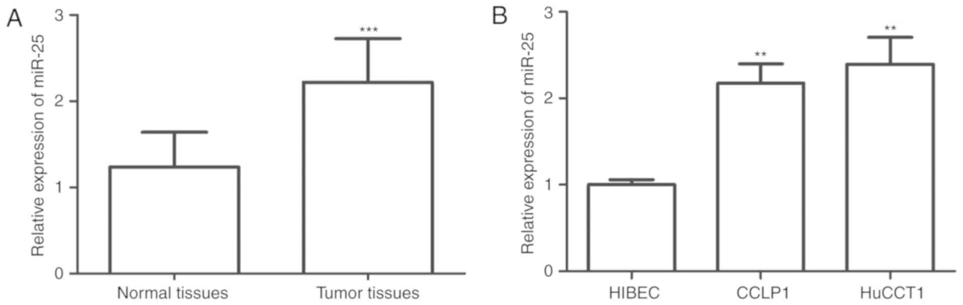
The miR-25 expression in CCA tissues and cell lines
was examined by RT-qPCR. The results suggested that the expression
levels of miR-25 in the tissues were significantly higher than
those in normal tissues (P<0.001; Fig. 1A). Furthermore, the expression levels
of miR-25 in CCLP1 cells and HuCCT1 cells were significantly higher
than those in normal HIBECs (all P<0.01; Fig. 1B).
Association between miR-25 expression
and clinicopathological features of CCA patients
It was then investigated whether miR-25 expression
is associated with the clinicopathological characteristics of the
CCA patients. For this, the CCA patients were divided into a low
miR-25 expression group (n=51) and a high expression group (n=65)
according to the mean value of relative miR-25 expression in CCA
tissues (2.219) as a cutoff. As presented in Table I, high expression of miR-25 was
closely associated with tumor-nodes-metastasis (TNM) stage
(P=0.026) and lymph node metastasis (P=0.032). Patients with a high
TNM stage had a higher expression level of miR-25 than those with a
low stage (P=0.026), and compared with CCA patients with negative
lymph node metastasis, miR-25 expression was significantly higher
in those patients with lymph node metastasis (P=0.032). High miR-25
expression was significantly associated with high TNM stage
(P=0.026) and lymph node metastasis (P=0.032). However, there was
no association of miR-25 expression with other features, including
age, gender and degree of differentiation (all P>0.05; Table I).
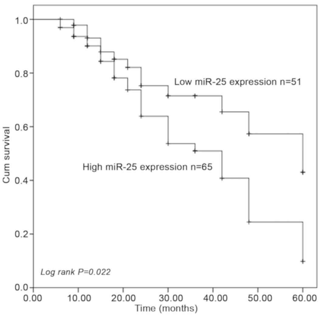
Prognostic significance of miR-25 in
CCA
To determine the prognostic value of miR-25
expression in CCA, Kaplan-Meier survival curve analysis was used.
The results indicated that the 5-year survival rate in the low
miR-25 expression group was higher than that in the high miR-25
expression group, and the difference was statistically significant
(log-rank test P=0.022; Fig. 2).
Univariate and multivariate Cox regression analysis demonstrated
that miR-25 expression was an independent prognostic predictor for
overall survival in CCA patients (hazard ratio=2.094, 95%
CI=1.051–4.171, P=0.036; Table
II).
| Table II.Univariate and multivariate Cox
logistic regression analysis of the influence of clinical
parameters on overall survival. |
Table II.
Univariate and multivariate Cox
logistic regression analysis of the influence of clinical
parameters on overall survival.
|
| Univariate
analysis | Multivariate
analysis |
|---|
|
|
|
|
|---|
|
Characteristics | HR | 95% CI | P-value | HR | 95% CI | P-value |
|---|
| miR-25 (<2.219
vs. ≥2.219) | 2.006 | 1.056–3.811 | 0.034 | 2.094 | 1.051–4.171 | 0.036 |
| Age (<60 vs.
≥60) | 1.105 | 0.597–2.046 | 0.750 | n/a | n/a | n/a |
| Sex (Male vs.
Female) | 0.966 | 0.528–1.764 | 0.909 | n/a | n/a | n/a |
| Differentiation
(Well vs. Moderate + poor) | 1.250 | 0.676–2.310 | 0.477 | n/a | n/a | n/a |
| TNM stage (I–II vs.
III–IV) | 0.895 | 0.490–1.636 | 0.719 | n/a | n/a | n/a |
| Lymph node
metastasis (negative vs. positive) | 0.996 | 0.545–1.819 | 0.989 | n/a | n/a | n/a |
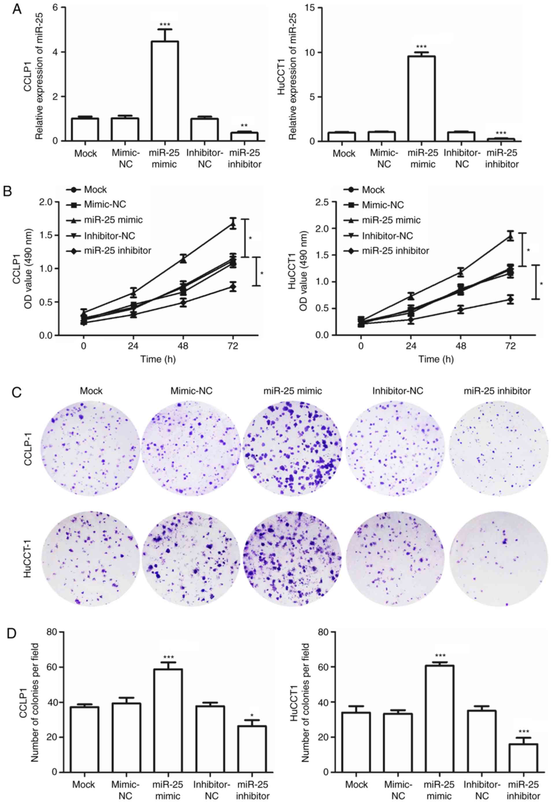
Silencing of miR-25 reduces viability,
migration and invasion of CCA cells
To determine the effects of miR-25 on cell viability
in CCA, the CCLP1 and HuCCT1 cell lines were transfected with
miR-25 mimics or miR-25 inhibitor. RT-qPCR confirmed that the
expression of miR-25 in CCA cells transfected with miR-25 mimics
was significantly elevated, while in cells transfected with the
miR-25 inhibitor, the expression was decreased, compared with that
in the mock-transfected group (P<0.05; Fig. 3A). An MTT assay was used to evaluate
the effect of transfection on cell viability, and the results
indicated that cell viability significantly increased in the cells
transfected with the miR-25 mimics but was markedly decreased
following transfection with the miR-25 inhibitor compared with that
in the mock group (P<0.05; Fig.
3B). In addition, transfection with the miR-25 mimics increased
the cologenicity of CCA cells, while miR-25 inhibitor had a
suppressive effect in the colony formation assays (P<0.05;
Fig. 3C and D). These observations
indicated that miR-25 promotes the viability of CCA cells.
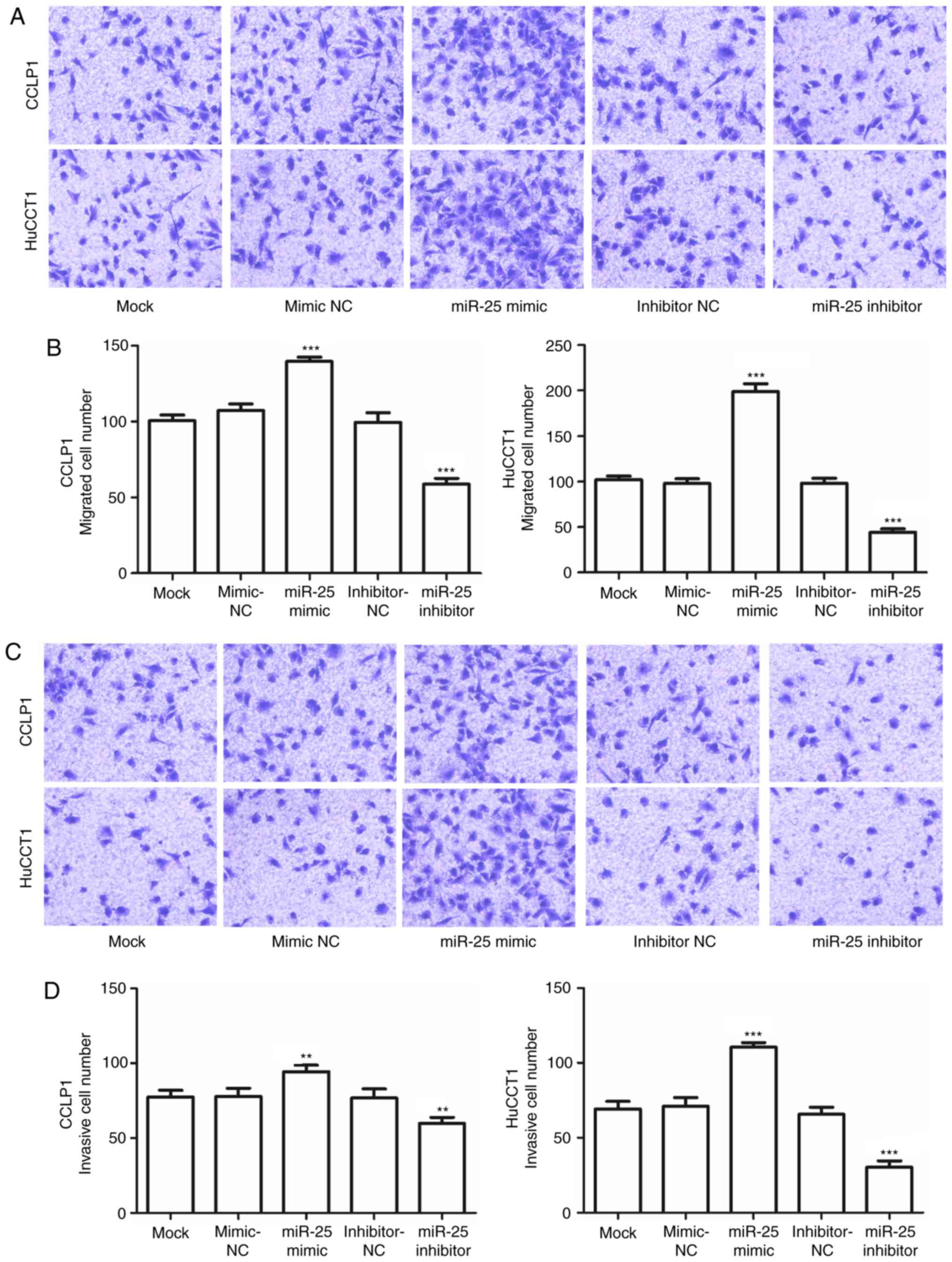
To investigate the effects of miR-25 on CCA
metastasis, the cell migration and invasive capabilities of CCLP1
and HuCCT1 cells transfected with miR-25 mimics or inhibitor were
examined. The results indicated that, compared with those in the
mock-transfected group, the migration and invasion capacities of
CCA cells transfected with the miR-25 inhibitor were significantly
decreased, but were enhanced in cells transfected with miR-25
mimics (P<0.05; Fig. 4A-D).
Discussion
Although being considered a rare tumor, owing to its
highly chemo-resistant characteristics and late diagnosis, CCA
remains a refractory disease with poor long-term overall survival.
Therefore, the prognosis of CCA patients urgently requires to be
improved. A number of studies have reported that certain molecular
biomarkers have crucial roles in tumor pathogenesis and
development, and are highly associated with the prognosis for human
malignant disease (19–21). Various prognostic biomarkers have
also been identified in CCA. For instance, Phanthaphol et al
(22) demonstrated that the
upregulation of translationally-controlled tumor protein (TCTP) was
associated with CCA progression and metastasis, while suppression
of TCTP may serve as a potential therapeutic strategy for CCA. A
member of the voltage-gated K channel superfamily, KCa3.1, was
indicated to be a promising therapeutic target in intrahepatic CCA
(23). Daya et al (24) demonstrated that progranulin has a
critical role in tumor metastasis mediated via the epithelial to
mesenchymal transition and was associated with prognosis. These
studies suggest that the development of cancer-associated genes may
help improving prognosis and explore more therapeutic targets for
identifying novel therapeutic strategies for patients.
miR-25 belongs to the miR-106b~25 cluster, which is
located within the 13th intron of minichromosome maintenance
complex component 7, an oncogene acting in cooperation with the
aforementioned cluster in promoting cancer progression (25). A number of studies demonstrated that
the miR-106b~25 cluster has numerous biological functions in
development, including viability and differentiation (26). For instance, a study by Khuu et
al (25), indicated that the
role of miRNAs encoded by the miR-106b~25 cluster is also
associated with the growth and maintenance of stem/progenitor
cells. Proper regulation of stem cell differentiation is crucial to
normal development (27). Aberrant
miRNA expression patterns have been reported to contribute to the
progression and prognosis of various cancer types (28,29). A
number of studies have indicated that miR-25 functions as an
oncogene in multiple types of malignancy, including non-small cell
lung cancer (NSCLC) and gastric cancer (13,14). In
CCA, miR-25 was also indicated to be overexpressed and negatively
regulate apoptosis signaling (30).
However, the prognostic value of miR-25 in CCA has remained
elusive.
To expand the current knowledge of the role of
miR-25 in CCA, its expression pattern was assessed in CCA tissues
and cell lines, and its prognostic significance in CCA patients was
explored. In the present study, the relative expression of miR-25
in CCA tissues and cell lines was significantly upregulated when
compared with that in adjacent normal tissues and a normal cell
line, respectively. These results were consistent with those of
Razumilava et al (30), which
demonstrated that miR-25 is upregulated in malignant CCA cell lines
as well as in patients tissue samples, compared with benign cells
and benign adjacent tissue samples, respectively. Furthermore, the
association between the expression of miR-25 and the clinical
features of CCA patients was analyzed, revealing that miR-25 was
significantly associated with TNM stage and lymph node metastasis,
i.e. that patients with a high TNM stage and positive lymph node
metastasis may have higher miR-25 expression. These above results
suggest that miR-25 may be an oncogene in CCA tumorigenesis and is
involved in the development of CCA. Considering miR-25 expression
was significantly associated with TNM stage and lymph node
metastasis, it was hypothesized that miR-25 expression may be
associated with the prognosis of CCA patients. To further
investigate the prognostic role of miR-25 in CCA patients,
Kaplan-Meier and Cox regression analyses were performed. First, it
was revealed that the expression of miR-25 was significantly
correlated with clinicopathological factors. miR-25 was also
significantly associated with the overall survival of CCA patients.
The Kaplan-Meier analysis indicated that patients in the high
miR-25 expression group had a shorter survival rate than those in
the low miR-25 expression group. Finally, miR-25 was identified as
an independent biomarker for patients with CCA by multivariate Cox
analysis. Taken together, the present results demonstrate that
miR-25 is a prognostic biomarker for CCA. A comparison between
miR-25 expression and different TNM stages of CCA patients to
determine whether miR-25 has diagnostic value in differentiating
between CCA patients in different stages will be performed in a
future study.
Recent studies have indicated the effects of miR-25
on biological behaviors during cancer progression and metastasis.
For instance, miR-25 promotes gastric cancer viability, migration
and invasion, and is associated with poor prognosis (31). To investigate the function of miR-25
in CCA cells, miR-25 mimics and inhibitor were employed and their
effects on the biological behaviour of CCA cell lines were
assessed. The results demonstrated that miR-25 mimics enhanced the
cell viability, migration and invasion, while miR-25 inhibitor had
a dampening effect, which suggested that downregulation of miR-25
expression inhibits CCA progression. Although a significant
difference in transfection efficiency between two CCA cell lines
was observed, the difference in biological functions was not
proportionally pronounced. Cancer cells exhibit different degrees
of differentiation which can produce cells with different
morphological and functional features (32). Various molecular pathways regulated
by miR-25 have been studied in a number of cancer types. For
instance, a study by Ding et al (33) demonstrated that miR-25 activates the
ERK signaling pathway by directly targeting kruppel-like factor 4,
promoting NSCLC cell migration and invasion. A recent study by
Sanchez-Mejias et al (34)
validated suppressor of cytokine signaling 5 (SOCS5) as a bona
fide target of miR-25 and identified novel molecular targets
for potential drug discovery efforts to fight hepatocellular
carcinoma. Another study by Long et al (35) demonstrated that miR-58-5-5p promotes
cancer stem cell characteristics and chemoresistance via targeting
multiple negative regulators of the STAT3 signaling pathway,
including SOCS5, leading to constitutive activation of STAT3
signaling. Therefore, it may be speculated that miR-25 may also
active the STAT3 signaling pathway by targeting SOCS5, to thereby
regulate the progression of CCA. In addition to these in
vitro results, the detailed molecular mechanisms of miR-25 in
CCA requires further elucidation.
In conclusion, the present study demonstrated that
overexpression of miR-25 was associated with poor prognosis of CCA
patients and promoted cell viability, migration, and invasion of
CCA cells in vitro. The present results provide evidence
that miR-25 may be a promising prognostic biomarker and potential
therapeutic target for the treatment of CCA patients.
Acknowledgements
Not applicable.
Funding
No funding was received.
Availability of data and materials
The datasets used and/or analyzed during the present
study are available from the corresponding author on reasonable
request.
Authors' contributions
HL and JW designed the study. HL and LM performed
the experiments, analyzed the data and prepared the manuscript.
Ethics approval and consent to
participate
All patients provided written informed consent to
participate in this study. Tissue collection and the study protocol
were approved by the Research Ethics Committee of Yidu Central
Hospital (Weifang, China).
Patient consent for publication
Not applicable.
Competing interests
The authors declare that they have no competing
interests.
References
|
1
|
Blechacz B and Gores GJ:
Cholangiocarcinoma: Advances in pathogenesis, diagnosis, and
treatment. Hepatology. 48:308–321. 2008. View Article : Google Scholar : PubMed/NCBI
|
|
2
|
Blechacz B: Cholangiocarcinoma: Current
knowledge and new developments. Gut Liver. 11:13–26. 2017.
View Article : Google Scholar : PubMed/NCBI
|
|
3
|
Fairweather M, Balachandran VP and
D'Angelica MI: Surgical management of biliary tract cancers. Chin
Clin Oncol. 5:632016. View Article : Google Scholar : PubMed/NCBI
|
|
4
|
Razumilava N and Gores GJ:
Cholangiocarcinoma. Lancet. 383:2168–2179. 2014. View Article : Google Scholar : PubMed/NCBI
|
|
5
|
Cho MS, Kim SH, Park SW, Lim JH, Choi GH,
Park JS, Chung JB and Kim KS: Surgical outcomes and predicting
factors of curative resection in patients with hilar
cholangiocarcinoma: 10-year single-institution experience. J
Gastrointest Surg. 16:1672–1679. 2012. View Article : Google Scholar : PubMed/NCBI
|
|
6
|
Jarnagin WR, Fong Y, DeMatteo RP, Gonen M,
Burke EC, Bodniewicz BS J, Youssef BA M, Klimstra D and Blumgart
LH: Staging, resectability, and outcome in 225 patients with hilar
cholangiocarcinoma. Ann Surg. 234:507–517; discussion 517–509.
2001. View Article : Google Scholar : PubMed/NCBI
|
|
7
|
Romero-Cordoba SL, Salido-Guadarrama I,
Rodriguez-Dorantes M and Hidalgo-Miranda A: miRNA biogenesis:
Biological impact in the development of cancer. Cancer Biol Ther.
15:1444–1455. 2014. View Article : Google Scholar : PubMed/NCBI
|
|
8
|
Tufekci KU, Oner MG, Meuwissen RL and Genc
S: The role of microRNAs in human diseases. Methods Mol Biol 1107.
33–50. 2014. View Article : Google Scholar
|
|
9
|
Singh RP, Massachi I, Manickavel S, Singh
S, Rao NP, Hasan S, Mc Curdy DK, Sharma S, Wong D, Hahn BH and
Rehimi H: The role of miRNA in inflammation and autoimmunity.
Autoimmun Rev. 12:1160–1165. 2013. View Article : Google Scholar : PubMed/NCBI
|
|
10
|
Chen L, Song J, Cui J, Hou J, Zheng X, Li
C and Liu L: microRNAs regulate adipocyte differentiation. Cell
Biol Int. 37:533–546. 2013. View Article : Google Scholar : PubMed/NCBI
|
|
11
|
Di Leva G, Garofalo M and Croce CM:
MicroRNAs in cancer. Annu Rev Pathol. 9:287–314. 2014. View Article : Google Scholar : PubMed/NCBI
|
|
12
|
Zhao L, Fan W, Fan Y and Gao S:
MicroRNA-214 promotes the proliferation, migration and invasion of
gastric cancer MKN28 cells by suppressing the expression of Dact2.
Exp Ther Med. 16:4909–4917. 2018.PubMed/NCBI
|
|
13
|
Xiang J, Hang JB, Che JM and Li HC: MiR-25
is up-regulated in non-small cell lung cancer and promotes cell
proliferation and motility by targeting FBXW7. Int J Clin Exp
Pathol. 8:9147–9153. 2015.PubMed/NCBI
|
|
14
|
Zhou J, Wang W, Li W, Wu L, Li G, Shi J
and Zhou S: The polymorphism in miR-25 attenuated the oncogenic
function in gastric cancer. Tumour Biol. 37:5515–5520. 2016.
View Article : Google Scholar : PubMed/NCBI
|
|
15
|
Zhang J, Gong X, Tian K, Chen D, Sun J,
Wang G and Guo M: miR-25 promotes glioma cell proliferation by
targeting CDKN1C. Biomed Pharmacother. 71:7–14. 2015. View Article : Google Scholar : PubMed/NCBI
|
|
16
|
Wang C, Wang X, Su Z, Fei H, Liu X and Pan
Q: MiR-25 promotes hepatocellular carcinoma cell growth, migration
and invasion by inhibiting RhoGDI1. Oncotarget. 6:36231–36244.
2015.PubMed/NCBI
|
|
17
|
Wang H, Li C, Jian Z, Ou Y and Ou J:
TGF-beta1 reduces mir-29a expression to promote tumorigenicity and
metastasis of cholangiocarcinoma by targeting HDAC4. PloS One.
10:e01367032015. View Article : Google Scholar : PubMed/NCBI
|
|
18
|
Livak KJ and Schmittgen TD: Analysis of
relative gene expression data using real-time quantitative PCR and
the 2(-Delta Delta C(T)) method. Methods. 25:402–408. 2001.
View Article : Google Scholar : PubMed/NCBI
|
|
19
|
Dumortier M, Ladam F, Damour I, Vacher S,
Bieche I, Marchand N, de Launoit Y, Tulasne D and Chotteau-Lelievre
A: ETV4 transcription factor and MMP13 metalloprotease are
interplaying actors of breast tumorigenesis. Breast Cancer Res.
20:732018. View Article : Google Scholar : PubMed/NCBI
|
|
20
|
Gomez-Macias GS, Garza-Rodriguez ML,
Garza-Guajardo R, Monsivais-Ovalle D, Ancer-Rodriguez J,
Barrera-Saldana HA and Barboza-Quintana O: Overexpression of the
matrix metalloproteinase 11 gene is a potential biomarker for type
1 endometrial cancer. Oncol Lett. 16:1073–1078. 2018.PubMed/NCBI
|
|
21
|
Sotgiu ML: Effects of alpha
2-adrenoceptors blockade on the inhibition of the nociceptive
jaw-opening reflex by the lateral reticular nucleus in rabbits.
Arch Ital Biol. 127:63–67. 1989.PubMed/NCBI
|
|
22
|
Phanthaphol N, Techasen A, Loilome W,
Thongchot S, Thanan R, Sungkhamanon S, Khuntikeo N, Yongvanit P and
Namwat N: Upregulation of TCTP is associated with
cholangiocarcinoma progression and metastasis. Oncol Lett.
14:5973–5979. 2017.PubMed/NCBI
|
|
23
|
Song P, Du Y, Song W, Chen H, Xuan Z, Zhao
L, Chen J, Guo D, Jin C, Zhao Y, et al: KCa3.1 as an effective
target for inhibition of growth and progression of intrahepatic
cholangiocarcinoma. J Cancer. 8:1568–1578. 2017. View Article : Google Scholar : PubMed/NCBI
|
|
24
|
Daya M, Loilome W, Techasen A, Thanee M,
Sa-Ngiamwibool P, Titapun A, Yongvanit P and Namwat N: Progranulin
modulates cholangiocarcinoma cell proliferation, apoptosis, and
motility via the PI3K/pAkt pathway. Onco Targets Ther. 11:395–408.
2018. View Article : Google Scholar : PubMed/NCBI
|
|
25
|
Khuu C, Utheim TP and Sehic A: The three
paralogous MicroRNA clusters in development and disease, miR-17-92,
miR-106a-363, and miR-106b-25. Scientifica (Cairo) 2016.
13796432016.
|
|
26
|
Brett JO, Renault VM, Rafalski VA, Webb AE
and Brunet A: The microRNA cluster miR-106b~25 regulates adult
neural stem/progenitor cell proliferation and neuronal
differentiation. Aging (Albany NY). 3:108–124. 2011. View Article : Google Scholar : PubMed/NCBI
|
|
27
|
Zwaka TP and Thomson JA: Differentiation
of human embryonic stem cells occurs through symmetric cell
division. Stem Cells. 23:146–149. 2005. View Article : Google Scholar : PubMed/NCBI
|
|
28
|
Ehrlich L, Hall C, Venter J, Dostal D,
Bernuzzi F, Invernizzi P, Meng F, Trzeciakowski JP, Zhou T,
Standeford H, et al: miR-24 Inhibition increases menin expression
and decreases cholangiocarcinoma proliferation. Am J Pathol.
187:570–580. 2017. View Article : Google Scholar : PubMed/NCBI
|
|
29
|
Wu C, Zhang J, Cao X, Yang Q and Xia D:
Effect of Mir-122 on human cholangiocarcinoma proliferation,
invasion, and apoptosis through P53 expression. Med Sci Monit.
22:2685–2690. 2016. View Article : Google Scholar : PubMed/NCBI
|
|
30
|
Razumilava N, Bronk SF, Smoot RL, Fingas
CD, Werneburg NW, Roberts LR and Mott JL: miR-25 targets
TNF-related apoptosis inducing ligand (TRAIL) death receptor-4 and
promotes apoptosis resistance in cholangiocarcinoma. Hepatology.
55:465–475. 2012. View Article : Google Scholar : PubMed/NCBI
|
|
31
|
Li BS, Zuo QF, Zhao YL, Xiao B, Zhuang Y,
Mao XH, Wu C, Yang SM, Zeng H, Zou QM and Guo G: MicroRNA-25
promotes gastric cancer migration, invasion and proliferation by
directly targeting transducer of ERBB2, 1 and correlates with poor
survival. Oncogene. 34:2556–2565. 2015. View Article : Google Scholar : PubMed/NCBI
|
|
32
|
Wang M, Yang YO, Jin Q, Shang L and Zhang
J: Function of miR-25 in the invasion and metastasis of esophageal
squamous carcinoma cells and bioinformatical analysis of the
miR-106b-25 cluster. Exp Ther Med. 15:440–446. 2018.PubMed/NCBI
|
|
33
|
Ding X, Zhong T, Jiang L, Huang J, Xia Y
and Hu R: miR-25 enhances cell migration and invasion in
non-small-cell lung cancer cells via ERK signaling pathway by
inhibiting KLF4. Mol Med Rep. 17:7005–7016. 2018.PubMed/NCBI
|
|
34
|
Sanchez-Mejias A, Kwon J, Chew XH, Siemens
A, Sohn HS, Jing G, Zhang B, Yang H and Tay Y: A novel
SOCS5/miR-18/miR-25 axis promotes tumorigenesis in liver cancer.
Int J Cancer. 144:311–321. 2019. View Article : Google Scholar : PubMed/NCBI
|
|
35
|
Long J, Jiang C, Liu B, Dai Q, Hua R, Chen
C, Zhang B and Li H: Maintenance of stemness by miR-589-5p in
hepatocellular carcinoma cells promotes chemoresistance via STAT3
signaling. Cancer Lett. 423:113–126. 2018. View Article : Google Scholar : PubMed/NCBI
|