Introduction
Intervertebral disc (IVD) degeneration is the
initiation of a series of spinal diseases, such as disc herniation,
degenerative spondylolisthesis and spinal stenosis and is
associated with a number of symptoms, such as lower back pain, neck
pain or sciatica (1). A number of
factors contribute to the pathological process of IVD degeneration,
such as age, trauma and over-load stress (1,2).
However, there is still no exact etiology to explain the process of
IVD degeneration.
The peak incidence of disc degeneration occurs among
middle-aged or older patients; however, a recent epidemiological
study demonstrated that an increasing number of young patients
(age, <45 years) also suffer from disc degeneration or disc
herniation (3). However, the reasons
for disc degeneration or herniation in young patients remain
unclear. Therefore, this poses a challenge for all spinal surgeons
and researchers worldwide.
Apart from mechanical factors or age, accumulating
evidence has indicated that low-virulence anaerobic bacteria (LVAB)
are a new pathogen involved in IVD degeneration. The study by
Stirling et al (4) first
reported that the prevalence of Propionibacterium acnes
(P. acnes) was as high as 53% in the herniated IVDs of
patients. Furthermore, a systematic review concluded that LVAB
played a critical role in disc herniation with lower back pain and
that bacterial presence was related to Modic changes associated
with disc herniation (5). In
addition, animal studies have confirmed that the inoculation of
P. acnes causes or deteriorates the degeneration of IVDs in
rats or rabbits (6,7). Therefore, the pathological role of LVAB
in IVD degeneration or herniation warrants further attention.
In this study, the prevalence of LVAB in herniated
IVDs was first analyzed. The present study hypothesized that latent
infection with LVAB may be a critical risk factor for disc
degeneration in young patients. The present study proposes a new
hypothesis regarding IVD degeneration in young patients and this
may lead to the development of novel therapeutic strategies for IVD
in the future.
Materials and methods
Patients
From January 2017 to January 2018, 176 consecutive
patients with single level IVD herniation were enrolled in this
study. The patient characteristics are shown in Table I. All of these patients suffered from
severe lower back pain and/or sciatica for several months and
conservative treatment was non-effective for all of them. IVD
samples were collected and analyzed when the patients underwent
discectomy or discectomy combined with instrumented posterior
lumbar fusion. The collection of samples was authorized by the
Committee on Ethics and the Institutional Review Board of Soochow
University and each patient signed written informed consent
forms.
 | Table I.Patient and clinical characteristics
association with bacterial presence. |
Table I.
Patient and clinical characteristics
association with bacterial presence.
| Parameters | n=170a | Positive samples
n=33a | Bacteria-free samples
n=137 | P-value |
|---|
| Age | 51.69±15.38 | 46.73±16.54 | 55.74±13.35 | 0.001 |
| Male | 92 | 21 | 71 | 0.221 |
| Female | 78 | 12 | 66 |
|
| Diabetes | 43 | 9 | 34 | 0.770 |
| Non-diabetic | 127 | 24 | 103 |
|
| Smoker | 69 | 17 | 52 | 0.154 |
| Non-smoker | 101 | 16 | 85 |
|
| Level of IVDs |
|
|
|
|
| L3/4 | 31 | 8 | 23 | 0.144 |
| L4/5 | 93 | 13 | 80 |
|
| L5/S1 | 46 | 12 | 34 |
|
Among the collected IVD samples, 33 were the level
of L3-L4, 95 were L4-L5 and 48 were L5-S1. None of the patients had
a history of antibiotics administration one month prior to the
surgery and none had any clinical or biological signs of
infection.
Preoperative imaging evaluation
To evaluate the severity of IVD degeneration,
intervertebral height was measured using the protocol reported by
Frobin et al (8) and Zhou
et al (9). In brief, lateral
X-ray films in the standing position were obtained prior to
surgery. Subsequently, a line connecting the midpoint of ventral
margin and dorsal margin of the vertebrae was drawn. The line was
defined as the midplane. An angle between two adjacent vertebrae
was measured between their midplanes. The ventral height of a
lumbar disc was then calculated as the sum of the perpendicular
distances of the corner of the cranial vertebra and the corner of
the caudal vertebra from the bisectrix between the two midplanes.
To obtain a more accurate intervertebral height which is
independent of the angle of lumbar lordosis, a correction was
applied to convert disc height measured in arbitrary angles to
height at standard angles according to the method proposed by
Frobin et al (8).
Intraoperative samples
According to the protocol of the Institutional
Review Board, all patients had to receive a single dose of 2 g
cefazolin for prophylactic antibiotic treatment during surgery. To
avoid possible contamination during tissue harvest, the incision
was disinfected at least twice with povidone iodine. Only fresh
instruments were then used for the handling of the samples. The
harvested IVD samples were rapidly stored in sterilized tubes and
transported to the laboratory of bacteriology. Finally, as
contamination markers, some muscle and ligaments surrounded the
IVDs were collected at the same time during surgery.
Microbiological culture
The collected samples were cultured under a class II
laminar flow safety cabinet at the laboratory of bacteriology in
The First Affiliated Hospital of Soochow University. Briefly, the
harvested IVD samples were dissected into 5 sections and 3 of these
were split and embedded into the plates, while the remaining 2
segments were placed into broth. The three segments were embedded
into MacConkey agar plates for aerobic culture, chocolate agar
plates in 5% CO2 and horse blood agar plates for aerobic
incubation, respectively. All plates were incubated for at least 14
days at 37°C and the results were examined on the 7th and 14th
day.
The broth used in this study was cooked meat medium
broth. One segment was first enriched for 48 h at 37°C and then
transferred to horse blood agar plates for anaerobic incubation and
then onto chocolate agar plates in 5% CO2 at 37°C for 7
days, respectively. The other segment was cultured in broth for 7
days and then anaerobically cultured on horse blood agar plates at
37°C for 7 days and with chocolate agar plates under 5%
CO2 at 37°C for 7 days. If there was any bacterial
growth in the broth or plates, the bacteria were collected and
identified with the 16S rDNA method according to the previously
described technique (9). The method
of culturing the control samples was the same as described for the
culture of IVDs.
Statistical analysis
SPSS 22.0 software (IBM Corp.) was used for
statistical analysis of data. The continuous data are expressed as
the mean ± standard deviation. When there were two groups, an
unpaired two-sided Student's t-test was used for analysis. In
addition, for the analysis of categorical variables, the Chi-Square
test was conducted. P<0.05 was considered to indciate a
statistically significant difference. Each experiment was carried
out with 3 independent repeats.
Results
Microbiological culture results of
disc samples
A total of 176 IVD samples were harvested for
microbiological culture and 39 disc samples (39/176, 22.1%) yielded
positive results. Among the 39 cases, 6 were considered as possible
contamination due to fact that the contamination markers of muscle
and ligaments were positive for bacteria at the same time and they
were excluded in the ensuing analysis (details in Supplementary
Table SI). The remaining 33 samples
(33/176, 18.7%) exhibited bacterial growth only in the discs and
they were considered to be bacteria-positive samples. Out of the 33
samples, P. acnes was detected in 31 samples (31/176, 17.6%)
and coagulase-negative Staphylococcus (CNS) was found in 2
samples (2/176, 1.1%).
With the further analysis of the epidemiological
data, a significant difference was demonstrated in the age between
the patients with or without bacteria in IVDs (P=0.001; Table I). However, no significant difference
was observed in sex, smoking status, diabetes and the levels of
discs between the positive and negative patients (Table I).
Higher proportion of bacterial
infection in younger patients
Due the statistically significant difference in age,
all patients (excluding the 6 suspicious cases) were further
divided into three subgroups based on age for analysis as follows:
Group A (age, <30 years), group B (age, 30 to 50 years) and
group C (age, >50 years). In group A, 10 patients had IVDs
infected by P. acnes and one sample infected by CNS. In
group B, 12 IVDs were infected by P. acnes and one IVD was
infected by CNS. For group C, P. acnes was found in all of
the 9 samples. The proportion of LVAB infection in each group was
34.4% (11/32), 25.5% (13/51) and 10.3% (9/87), respectively and
there was a statistically significant difference between the three
groups (P=0.005; Table II). The
rates of bacterial infection were significantly increased in the
IVDs of younger patients. The distribution of bacterial-positive
and bacterial-negative subjects by age is demonstrated in Fig. SI.
| Table II.Proportion of bacterial infection in
each group at different ages. |
Table II.
Proportion of bacterial infection in
each group at different ages.
|
| Culture on discs |
|
|
|---|
|
|
|
|
|
|---|
|
| Positive culture | Bacteria free | Total | P-value |
|---|
| Group A (Age
<30) | 11 (6.5%) | 21 (12.4%) | 32 | 0.005 |
| Group B (30≤ Age
≤50) | 13 (7.6%) | 38 (22.4%) | 51 |
|
| Group C (Age
>50) | 9 (5.3%) | 78 (45.8%) | 87 |
|
| Total | 33a | 137 | 170a |
|
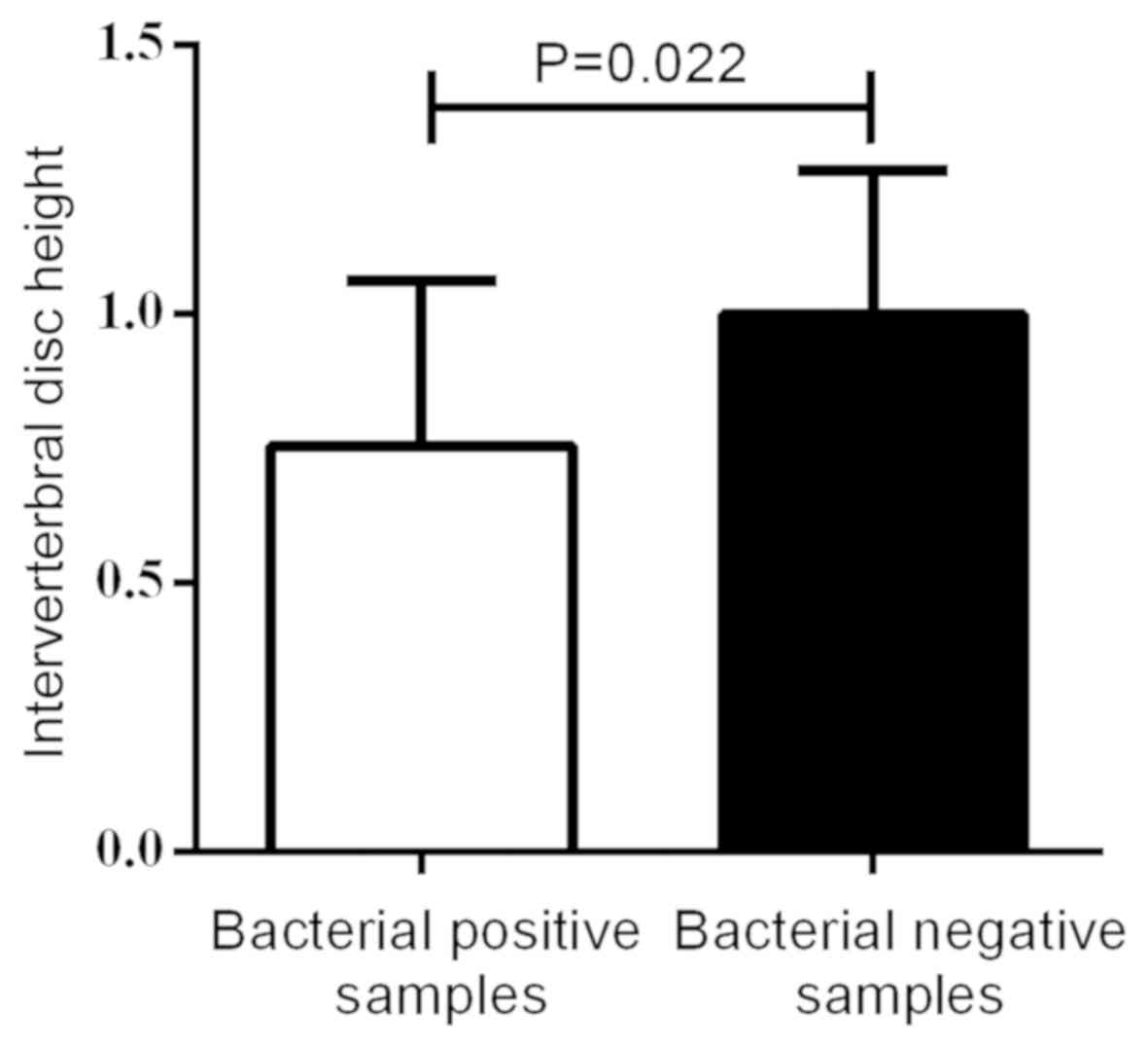
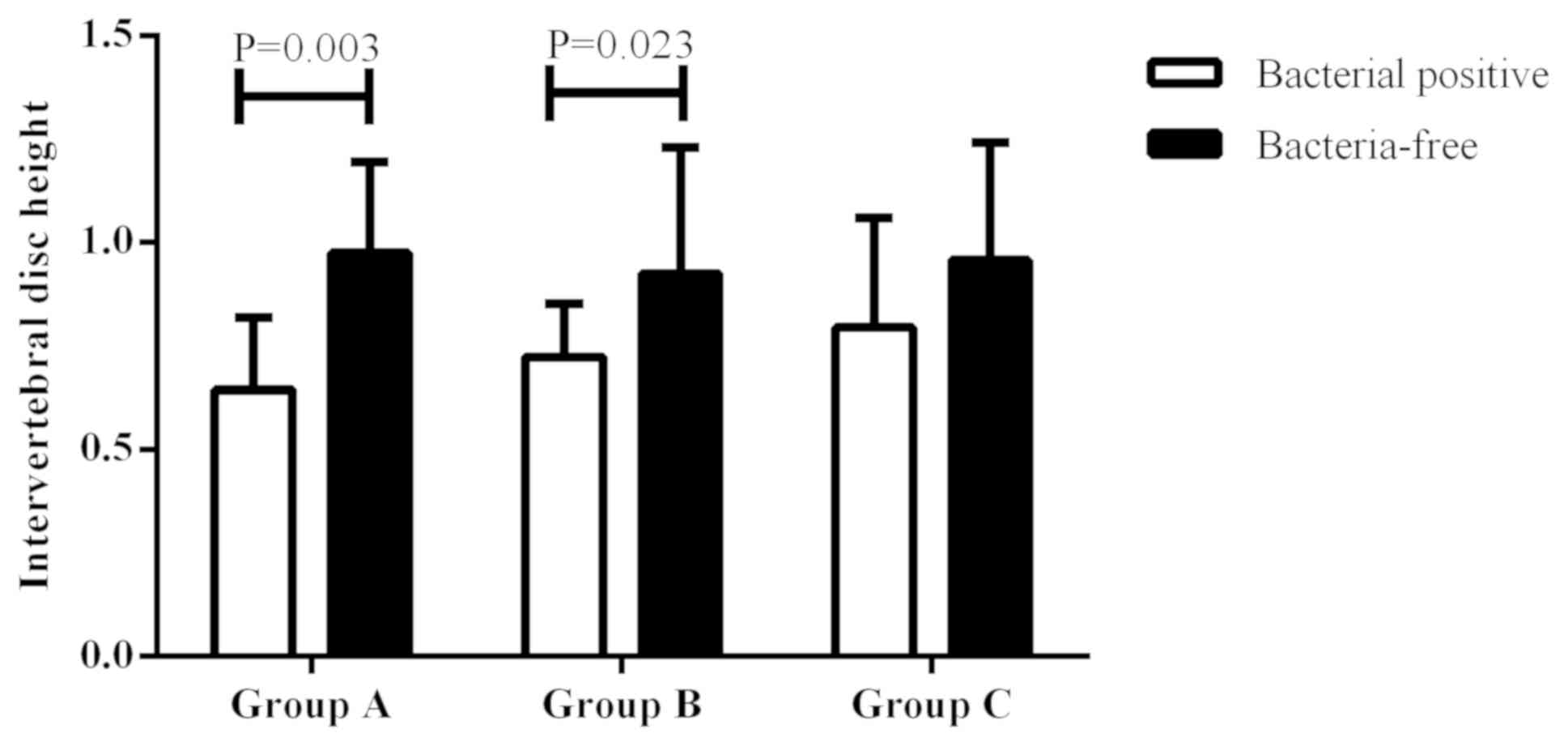
Infection of bacteria with more severe
disc degeneration
The disc height was compared between the
bacterial-positive and bacterial-negative samples. As depicted in
Fig. 1, the positive samples had a
significantly lower disc height than that of the bacterial-negative
samples (P=0.022). When comparing the disc height in each group,
the disc height was significantly lower in the bacterial-positive
samples than that of the negative samples in groups A and B, while
there was no statistically significant difference in group C
(P=0.003 and P=0.023, respectively; Fig.
2). This suggested that low virulence bacterial infection may
play a critical role in the IVD degeneration of young patients.
Discussion
There is increasing evidence to suggest that LVAB
may be a novel etiological factor for a series of spinal diseases
and contributes to plenty of symptoms, such as sciatica, neck pain
and back pain (10). In this study,
it was reported that 22.1% patients had LVAB in the IVDs, which was
in the range of 8–53% according to a previous study (10). Contamination markers were also
established during tissue harvest and culture following a
previously published protocol (9);
therefore, suspiciously contaminated cases were excluded in this
study. Therefore, the prevalence of LVAB was 18.7% in herniated
IVDs after the exclusion of six suspicious cases.
Of note, in this study, it was revealed that the
proportion of LVAB infection was much higher in younger patients.
Conventional viewpoints always considered that age and excessive
load are the key factors that induce IVD degeneration (2), however, it is hard to explain the
degeneration of IVDs in young patients with these two factors. In
this study, it was found that there was a significant difference in
that young patients had a higher rate of bacterial infection in the
IVDs, which highly suggested that the LVAB, particularly P.
acnes, may be one of the critical factors for IVD degeneration
in young patients, as previous studies have proven that P.
acnes is a definite factor responsible for the deterioration of
IVD degeneration (9,11,12). In
addition, patients with bacterial infection had a higher severity
of IVD degeneration than that of negative patients, further
demonstrating that bacteria may deteriorate IVD degeneration.
A number of studies have suggested the P.
acnes accelerates the degeneration of IVDs. For example, Lin
et al (11) and Zhou et
al (9) demonstrated that the
patients with P. acnes-positive IVDs had a lower
intervertebral disc height, which proved that infection with P.
acnes in IVDs is a novel risk factor that deteriorates disc
degeneration. Moreover, the inoculation of P. acnes into the
IVDs of rats or rabbits has been shown to cause severe disc
degeneration when compared with the control samples (6,7). Thus,
it can be reasonably concluded that P. acnes is a novel
etiological factor for IVD degeneration.
Nevertheless, it is hard to explain why young
patients had a higher prevalence of low-virulence anaerobic
bacteria, particularly P. acnes, in IVDs. To date, there is
still no theory or hypothesis to explain how P. acnes
latently infects IVDs. Albert et al (13) hypothesized that the P. acnes
enters the circulatory system via the breaking of the skin or
mucous membranes and then invades the IVDs through the tear of the
anulus fibrosus. However, the study by Li et al (12) did not support this hypothesis, as
there was no bacterial growth in the degenerated IVDs following the
injection of P. acnes into the veins of rabbits for several
days. The present study therefore hypothesized that the IVDs
harvested from younger patients may be relatively less degenerated
than that from older patients, so the younger IVDs may provide a
more suitable environment for the survival and reproduction of
LVAB. Therefore, the rates of LVAB infection in IVDs may be higher
in younger patients. In the future, further studies are required to
explore the exact mechanisms involved.
However, several limitations still exist in this
study. Firstly, the reason why young patients had a higher
prevalence of P. acnes infection remains unclear. More
evidence obtained from clinical or animal studies is required to
solve this matter. In addition, the preoperative administration of
antibiotics may reduce the bacterial positive rates in the IVDs
under the protocol of the Institutional Ethics Board. Finally, a
greater number of patients have to be analyzed in the future for a
more accurate prevalence.
In conclusion, this study reported that the
prevalence of LVAB was 18.7% in IVDs and younger patients had much
higher positive rates of bacterial infection and a greater severity
of IVD degeneration. Therefore, LVAB may play a crucial role in the
pathophysiology of intervertebral disc herniation, particularly in
younger patients.
Supplementary Material
Supporting Data
Acknowledgements
Not applicable.
Funding
The present study was supported by grants from the
Natural Science Foundation of Jiangsu Province (grant no.
BK20161274), the Natural Science Foundation of Suzhou (grant no.
kjxw2015056) and the Science and Technology Bureau of Kunshan
(grant no. KS1547).
Availability of data and materials
The datasets used and/or analyzed during the current
study are available from the corresponding author on reasonable
request.
Authors' contributions
The manuscript was approved and contributed to by
all authors. WMJ conceived and designed the study; GQT acquired the
data, interpreted the results and was involved in drafting and
revising the manuscript; YC performed the radiological analysis,
the bacterial culture and interpreted the results; JC collected and
transported the samples, acquired the epidemiological data of
patients and interpreted the results; ZW performed the statistical
analysis and interpreted the results during drafting and revising
the manuscript.
Ethics approval and consent to
participate
This study was approved by the Committee on Ethics
and the Institutional Review Board of Soochow University and each
patient signed written informed consent forms.
Patient consent for publication
Written informed consent for publication was
obtained from all individual participants included in the
study.
Competing interests
The authors declare that they have no competing
interests.
References
|
1
|
Modic MT and Ross JS: Lumbar degenerative
disk disease. Radiology. 245:43–61. 2007. View Article : Google Scholar : PubMed/NCBI
|
|
2
|
Zhang YG, Sun Z, Zhang Z, Liu J and Guo X:
Risk factors for lumbar intervertebral disc herniation in Chinese
population: A case-control study. Spine (Phila Pa 1976).
34:E918–E922. 2009. View Article : Google Scholar : PubMed/NCBI
|
|
3
|
Kim YK, Kang D, Lee I and Kim SY:
Differences in the incidence of symptomatic cervical and lumbar
disc herniation according to age, sex and national health insurance
eligibility: A pilot study on the Disease's association with work.
Int J Environ Res Public Health. 15(pii): E20942018. View Article : Google Scholar : PubMed/NCBI
|
|
4
|
Stirling A, Worthington T, Rafiq M,
Lambert PA and Elliott TS: Association between sciatica and
Propionibacterium acnes. Lancet. 357:2024–2025. 2001.
View Article : Google Scholar : PubMed/NCBI
|
|
5
|
Urquhart DM, Zheng Y, Cheng AC, Rosenfeld
JV, Chan P, Liew S, Hussain SM and Cicuttini FM: Could low grade
bacterial infection contribute to low back pain? A systematic
review. BMC Med. 13:132015. View Article : Google Scholar : PubMed/NCBI
|
|
6
|
Chen Z, Zheng Y, Yuan Y, Jiao Y, Xiao J,
Zhou Z and Cao P: Modic changes and disc degeneration caused by
inoculation of Propionibacterium acnes inside intervertebral
discs of rabbits: A pilot study. Biomed Res Int.
2016:96124372016.PubMed/NCBI
|
|
7
|
Dudli S, Liebenberg E, Magnitsky S, Miller
S, Demir-Deviren S and Lotz JC: Propionibacterium acnes
infected intervertebral discs cause vertebral bone marrow lesions
consistent with Modic changes. J Orthop Res. 34:1447–1455. 2016.
View Article : Google Scholar : PubMed/NCBI
|
|
8
|
Frobin W, Brinckmann P, Kramer M and
Hartwig E: Height of lumbar discs measured from radiographs
compared with degeneration and height classified from MR images.
Eur Radiol. 11:263–269. 2001. View Article : Google Scholar : PubMed/NCBI
|
|
9
|
Zhou Z, Chen Z, Zheng Y, Cao P, Liang Y,
Zhang X, Wu W, Xiao J and Qiu S: Relationship between annular tear
and presence of Propionibacterium acnes in lumbar
intervertebral disc. Eur Spine J. 24:2496–2502. 2015. View Article : Google Scholar : PubMed/NCBI
|
|
10
|
Chen Z, Cao P, Zhou Z, Yuan Y, Jiao Y and
Zheng Y: Overview: The role of Propionibacterium acnes in
nonpyogenic intervertebral discs. Int Orthop. 40:1291–1298. 2016.
View Article : Google Scholar : PubMed/NCBI
|
|
11
|
Lin Y, Jiao Y, Yuan Y, Zhou Z, Zheng Y,
Xiao J, Li C, Chen Z and Cao P: Propionibacterium acnes
induces intervertebral disc degeneration by promoting nucleus
pulposus cell apoptosis via the TLR2/JNK/mitochondrial-mediated
pathway. Emerg Microbes Infect. 7:12018. View Article : Google Scholar : PubMed/NCBI
|
|
12
|
Li B, Dong Z, Wu Y, Zeng J, Zheng Q, Xiao
B, Cai X and Xiao Z: Association between lumbar disc degeneration
and Propionibacterium acnes infection: Clinical research and
preliminary exploration of animal experiment. Spine (Phila Pa
1976). 14:E764–E769. 2016. View Article : Google Scholar
|
|
13
|
Albert HB, Kjaer P, Jensen TS, Sorensen
JS, Bendix T and Manniche C: Modic changes, possible causes and
relation to low back pain. Med Hypotheses. 70:361–368. 2008.
View Article : Google Scholar : PubMed/NCBI
|