Introduction
Rheumatoid arthritis (RA) is an autoimmune disease
caused by systemic inflammation affecting bones and joints. The
pathogenesis of RA involves a number of different mechanistic
pathways in both the innate and adaptive immune systems (1).
Macrophages are a class of heterogeneous, plastic
immune cells (2). To effectively
adapt to the microenvironment, macrophages form sub-populations
with different morphological functions by altering their phenotype,
namely classically activated (also known as M1 macrophages) and
alternatively activated macrophages (also known as M2 macrophages).
M1 macrophages mainly express and secrete proinflammatory
cytokines, including interleukin (IL)-6 and tumor necrosis factor-α
(TNF-α), whereas M2 macrophages are characterized by the production
of anti-inflammatory cytokines such as IL-10, which serve a role in
promoting angiogenesis and tissue repair (3,4). It has
been previously reported that macrophages serve a key role in the
development of inflammation and joint damage in patients with RA
(5), where the number of macrophages
in the synovial layer and substratum has been reported to associate
with disease progression (6).
Additionally, proinflammatory factors secreted by M1 macrophages
and/or anti-inflammatory cytokines produced by M2 macrophages have
been reported to serve important roles in RA development (7).
Lipopolysaccharide (LPS) is a major constituent of
the gram-negative bacterial cell wall. LPS is also one of the
ligands of toll-like receptor 4 (TLR4), the triggering of which can
lead to downstream signal transduction (8). Activation of this pathway results in
NF-κB activation, in turn activating the extensive transcription of
genes associated with inflammation and finally promoting the
production of factors associated with the inflammatory response
(9,10). LPS has been widely implicated in the
development of various inflammatory diseases, such that its
application to cell cultures in vitro has often been used as
a model for simulating inflammatory responses.
MicroRNAs (miRNAs) are endogenous, single-stranded
RNA molecules that are ~22 nucleotides in length and regulate the
expression of target genes by binding to the 3′-untranslated
regions (3′-UTR) of target mRNAs (11). Previous studies have demonstrated
that miRNAs are involved in the regulation of a large number of
physiological and biochemical processes, including cell
proliferation, differentiation and apoptosis (12,13). In
particular, miR-144-5p has been reported to serve important roles
in cancer development and chronic periodontitis (14–16).
However, the role of miR-144-5p in RA has not been previously
studied. Therefore, in the present study, LPS-treated THP-1
macrophages was used as the model cell line to investigate the role
of miR-144-5p in the pathophysiology of RA and associated
mechanism.
Materials and methods
Induction of THP-1 cells into
macrophages
The THP-1 cells used for the present study were
obtained from the Type Culture Collection of the Chinese Academy of
Sciences. THP-1 cells were cultured in RPMI 1640 medium (Gibco;
Thermo Fisher Scientific, Inc.) supplemented with 10% fetal bovine
serum (Gibco; Thermo Fisher Scientific, Inc.), 100 U/ml penicillin
and 100 mg/ml streptomycin (Gibco; Thermo Fisher Scientific, Inc.)
at 37°C incubator under 5% CO2.
For the differentiation of THP-1 cells into adherent
growing macrophages, THP-1 cells were first incubated for 72 h in
100 ng/ml phorbol ester dissolved in serum-free RPMI-1640 medium.
Following confirmation of successful differentiation by measuring
the expression of markers CD14 and CD11 using flow cytometry assay
(17), fresh medium was replaced 6 h
before each experiment and the cell density was then adjusted to
1×10⁶ cells/ml before subsequent experiments were
performed.
LPS-induced inflammation model
Macrophages were seeded in 24-well plates at
1×10⁶ cells/well. The cells were then divided into two
groups, an LPS (1 µg/ml, Sigma-Aldrich; Merck KGaA) treatment and
control group, following which they were either treated with LPS (1
µg/ml) or an equal amount of ultrapure water for 24 h. Each
experiment was performed in triplicate and repeated three
times.
Cell transfection
In total, 100 nM miR-144-5p mimics or the negative
control of miR-144-5p mimics (NC) (Guangzhou RiboBio Co., Ltd.), 50
nM control-siRNA (sense, 5′-UUCUCCGAACGUGUCACGUTT-3′ and antisense,
5′-ACGUGACACGUUCGGAGAATT-3′) or TLR2-siRNA (sense,
5′-GGAACAGAGUGGCAACAGUTT-3′ and antisense,
5′-ACUGUUGCCACUCUGUUCCTT-3′) (Shanghai GenePharma Co., Ltd.). were
transfected into THP-1 macrophages using Lipofectamine®
2000 (Invitrogen; Thermo Fisher Scientific, Inc.), according to the
manufacturer's protocol. Transfection efficiency was subsequently
evaluated using reverse transcription-quantitative PCR (RT-qPCR) 48
h after transfection.
Cell Counting Kit-8 (CCK-8) assay
THP-1 macrophages were first transfected with
miR-144-5p mimics, NC, control-siRNA or TLR2-siRNA for 48 h,
following which they were treated with LPS (1 µg/ml) for a further
24 h before cell viability was measured using the CCK-8 kit. The
cell concentration was adjusted to 1×10⁴/ml, and 100 µl
cell suspension were added to each well of the 96-well plates then
cultured for 12, 24 or 48 h. Subsequently, 10 µl CCK-8 reagent
(Sigma-Aldrich; Merck KGaA) was added to each well followed by a
further 2 h incubation. Absorbance values at the wavelength of 450
nm was then measured for each well using an ELISA plate reader
(Thermo Fisher Scientific, Inc.). Each experiment was repeated
three times.
ELISA
THP-1 macrophages (1×10⁶ cells/well) were
cultured in a 12-well culture plates and transfected with
miR-144-5p mimics, NC, control-siRNA or TLR2-siRNA for 48 h before
the cells were treated with LPS (1 µg/ml) for 24 h. Subsequently,
the levels of TNF-α, IL-6 and IL-8 in the culture supernatant was
analyzed using single ELISA kits (Qiagen GmbH), according to the
manufacturer's protocols.
Western blot analysis
THP-1 macrophages were transfected with miR-144-5p
mimics or NC for 48 h before the cells were treated with LPS (1
µg/ml) for 24 h. Cells were then washed twice with PBS and lysed in
RIPA buffer (Gibco; Thermo Fisher Scientific, Inc.). Bicinchoninic
Acid Protein Assay kit (Thermo Fisher Scientific, Inc.) was used to
quantify protein concentrations. The protein samples (20 µg per
lane) were separated using 10% SDS-PAGE and transferred to PVDF
membranes (Bio-Rad laboratories, Inc.). After blocking nonspecific
binding with TBS containing 0.1% Tween-20 supplemented with 5%
non-fat milk for 1 h at room temperature, the membranes were then
incubated with the respective primary antibodies at 4°C overnight.
The next day, the membranes were incubated with horseradish
peroxidase (HRP)-conjugated goat anti-rabbit secondary antibody for
2 h at room temperature. The protein bands were detected and
visualized using ECL Western Blotting Detection Reagent (EMD
Millipore). The bands were semi-quantitively analyzed using Image J
(version 4.0; National Institutes of Health). All primary and
secondary antibodies, including toll-like receptor 2 (TLR2; cat.
no. 12276; 1:1,000), phosphorylated (p-)p65 (cat. no. 3033;
1:1,000), p65 (cat. no. 8242; 1:1,000), GAPDH (cat. no. 5174;
1:1,000) and HRP-conjugated goat anti-rabbit secondary antibody
(cat. no. 7074; 1:1,000) were purchased from Cell Signaling
Technology, Inc.
RT-qPCR
Total RNA was isolated from THP-1 macrophages using
TRIzol® (Invitrogen; Thermo Fisher Scientific, Inc.)
according to the manufacturer's protocol. RNA was reverse
transcribed into cDNA using a Transcriptor First Strand cDNA
Synthesis kit (Roche Molecular Systems, Inc.), according to
manufacturer's protocol. The temperature protocol for reverse
transcription was 5 min at 25°C followed by 60 min at 42°C. qPCR
was performed in a LightCycler® 480 system (Roche
Molecular Systems, Inc.) using Fast SYBR® Green Master
Mix (Thermo Fisher Scientific, Inc.), according to the
manufacturer's protocols. The thermocycling conditions were as
follows: Initial denaturation at 95°C for 5 min, followed by 38
cycles of denaturation at 95°C for 15 sec and annealing/elongation
at 60°C for 30 sec. GAPDH or U6 was used as the internal control
for mRNA and miRNA expression, respectively. Relative gene
expression was quantified using the 2−ΔΔCq method
(18). Primer sequences used for
RT-qPCR were as follows: U6 forward,
5′-GCTTCGGCAGCACATATACTAAAAT-3′ and reverse,
5′-CGCTTCACGAATTTGCGTGTCAT-3′; GAPDH forward,
5′-CTTTGGTATCGTGGAAGGACTC-3′ and reverse,
5′-GTAGAGGCAGGGATGATGTTCT-3′; and TLR2 forward,
5′-AGCTTCATTGTTCCCTGTGTTAC-3′ and reverse,
5′-AGTTCACAGGAGCAGATGAAATG-3′.
Dual luciferase reporter assay
TargetScan bioinformatics software (www.targetscan.org/vert_71) (19) was used to predict the potential
target genes of miR-144-5p, using which TLR2 was found. To confirm
the prediction, the wild-type (WT-TLR2,
5′-UCAUAAGUCUAUUACUGAUAUCU-3′) and mutant (MUT-TLR2,
3′GAAUGUCAUAUACUACUAUAGG-5′) 3′UTR of TLR2, containing the
miR-144-5p-binding elements, were generated by reverse
transcription using a Transcriptor First Strand cDNA Synthesis Kit
(temperature protocol, 5 min at 25°C followed by 60 min at 42°C;
Roche Molecular Systems, Inc.) from total RNA preps extracted from
THP-1 macrophages, and were cloned via BamHI and AscI
sites of the pMIR-RB-Report™ dual luciferase reporter plasmid
vector (Guangzhou RiboBio Co., Ltd.). THP-1 macrophages were then
co-transfected with 500 ng each reporter construct (WT-TLR2 or
MUT-TLR2) and miR-144-5p mimic or NC (100 nM) using
Lipofectamine® 3000 (Invitrogen; Thermo Fisher
Scientific, Inc.), according to the manufacturer's protocol.
Luciferase activity was subsequently measured using a
Dual-Luciferase® Reporter Assay system (Promega
Corporation) 48 h after transfection, according to the
manufacturer's protocol. All luciferase activities were normalized
to that of Renilla luciferase.
Statistical analysis
Statistical analysis was performed using the SPSS
16.0 statistical package (SPSS, Inc.). Quantitative data area
expressed as the mean ± SD. Student's t-test or one-way analysis of
variance followed by Tukey's post-hoc test was performed for
comparison. P<0.05 was considered to indicate a statistically
significant difference.
Results
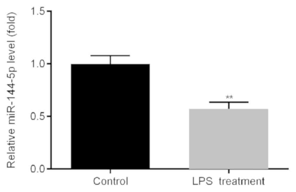
Expression of miR-144-5p in THP-1
macrophages treated with LPS is significantly lower compared with
that in the control group
THP-1 macrophages were treated with LPS or ultrapure
water for 24 h, following which RT-qPCR was used to measure the
expression of miR-144-5p. miR-144-5p expression was found to be
significantly lower in the cells treated with LPS compared with
water-treated cells (Fig. 1).
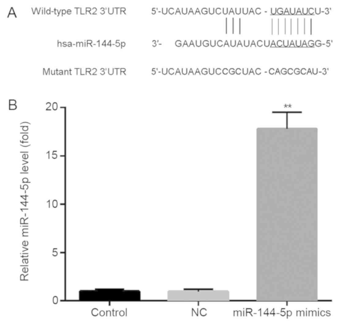
Prediction of binding sites between
TLR2 and miR-144-5p
Analysis via Targetscan revealed potential
miR-144-5p binding sites in the TLR2 3′UTR (Fig. 2A). THP-1 macrophages were then
transfected with miR-144-5p mimics or NC, followed by RT-qPCR
analysis of miR-144-5p expression. Transfection with miR-144-5p
mimics significantly increased miR-144-5p expression compared with
the NC group (Fig. 2B).
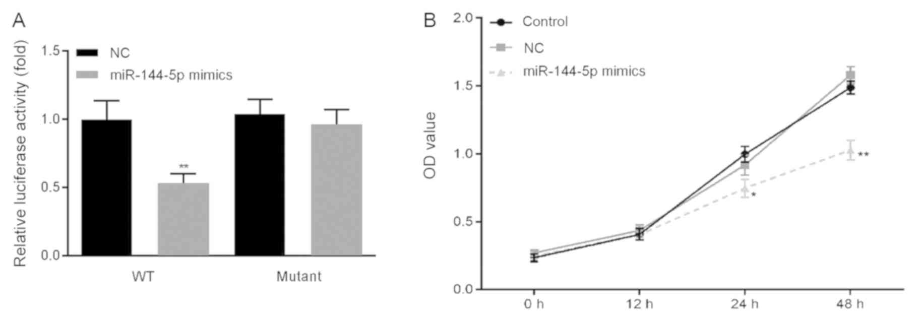
Overexpression of miR-144-5p reduces
the proliferation of macrophages
Previous studies have reported that miR-144 directly
targets TLR2 (20,21). In the present study, a dual
luciferase reporter assay was used to confirm the relationship
between TLR2 and miR-144-5p in THP-1 macrophages. In cells
transfected with the wt-TLR2 3′-UTR luciferase reporter vector,
luciferase activity in the miR-144-5p mimics group was
significantly lower compared with that in the NC group (Fig. 3A), whilst no significant difference
was observed in the miR-144-5p mimics group transfected with the
mut-TLR2 3′-UTR luciferase reporter vector.
Cell viability was then measured by CCK-8 assay.
miR-144-5p overexpression significantly reduced THP-1 macrophage
viability compared with the NC group (Fig. 3B).
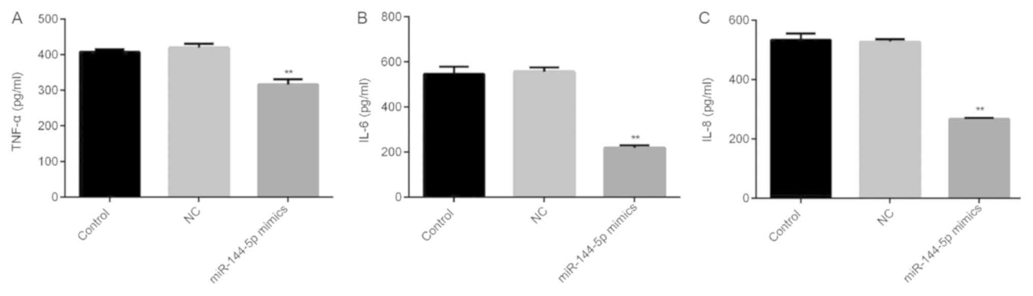
Overexpression of miR-144-5p reduces
the secretion of TNF-α, IL-6 and IL-8
THP-1 macrophages were transfected with miR-144-5p
mimics or NC for 48 h prior to treatment with LPS (1 µg/ml) for 24
h. The levels of TNF-α, IL-6 and IL-8 in the cell culture
supernatant were subsequently measured using ELISA. miR-144-5p
overexpression reduced the expression of TNF-α (Fig. 4A), IL-6 (Fig. 4B) and IL-8 (Fig. 4C) compared with the NC group.
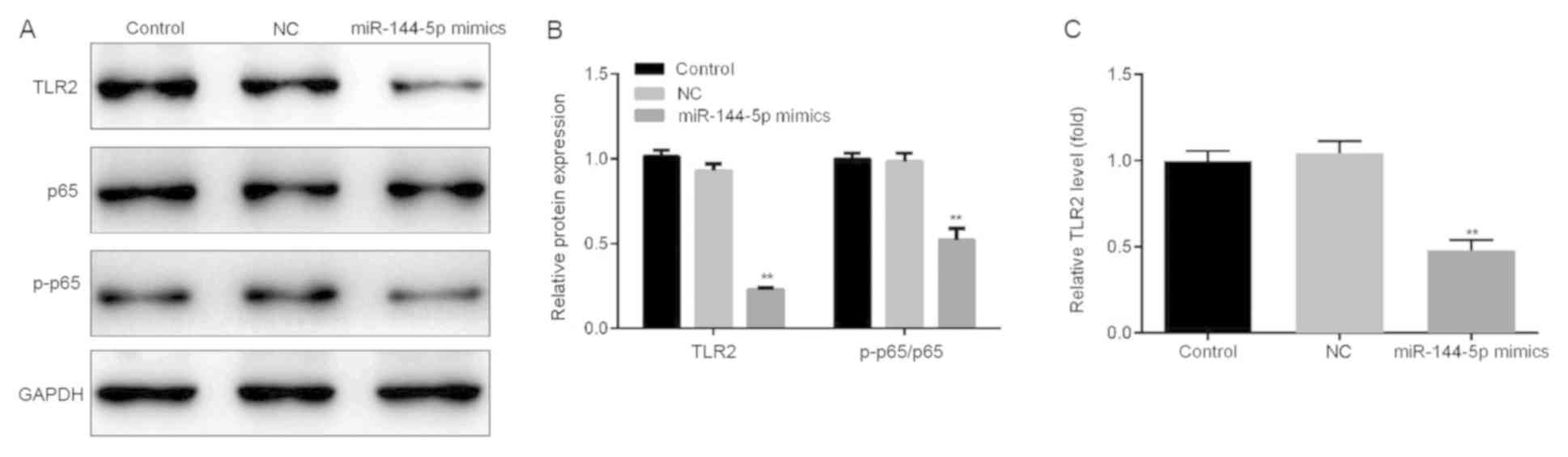
Effect of miR-144-5p overexpression on
the NF-κB signaling pathway
The effect of miR-144-5p overexpression on the NF-κB
pathway was investigated. THP-1 macrophages were first transfected
with miR-144-5p mimics or NC for 48 h before the cells were
stimulated with LPS (1 µg/ml) for a further 24 h. It was found that
the overexpression of miR-144-5p reduced the protein (Fig. 5A and B) and mRNA (Fig. 5C) expression of TLR2 in addition to
p65 phosphorylation compared with the NC group (Fig. 5A and B).
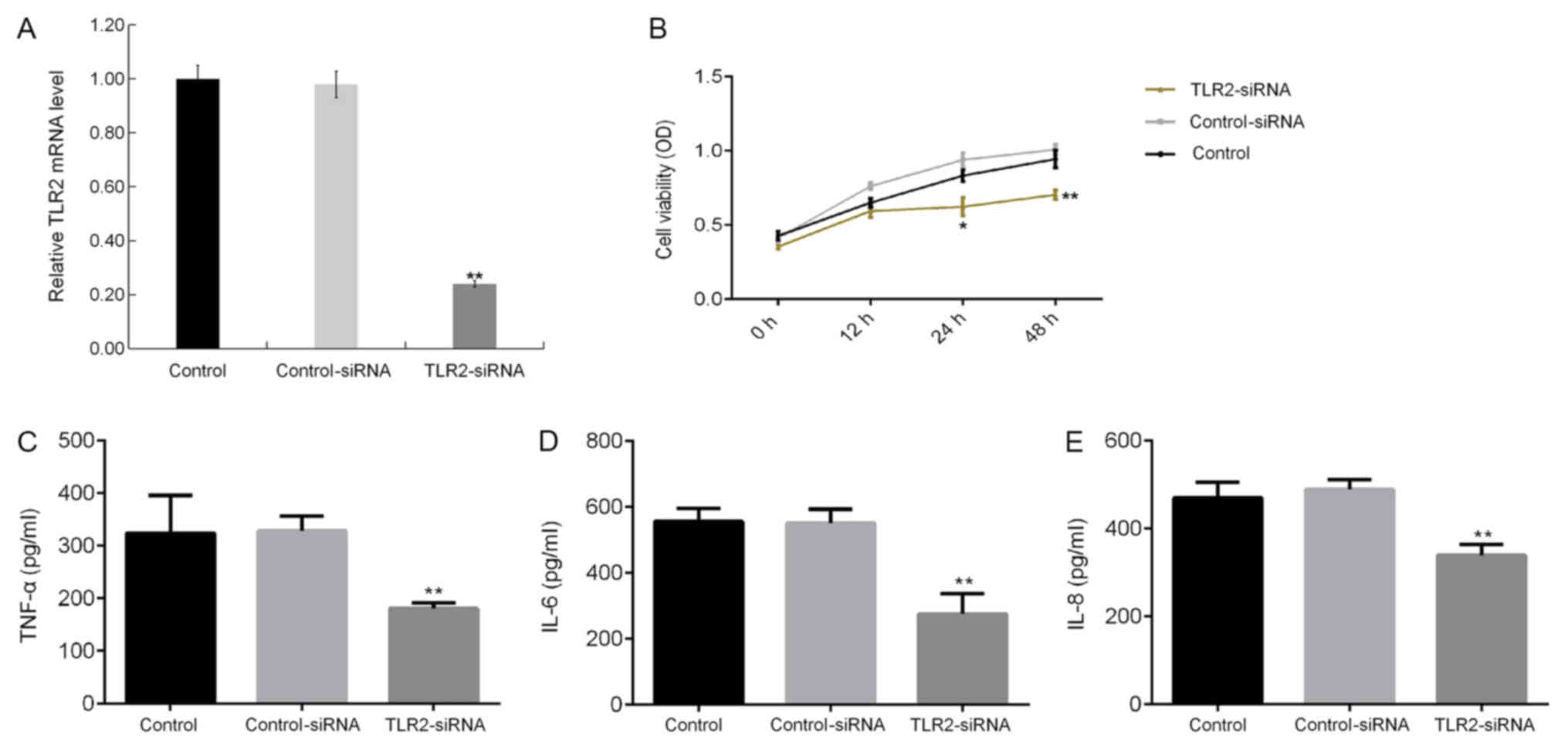
Effect of TLR2 silencing on
LPS-induced macrophages
The effect of TLR2 knockdown on LPS-treated THP-1
macrophages was next determined. TLR2-siRNA transfection
significantly reduced TLR2 mRNA expression in THP-1 macrophages
(Fig. 6A). TLR2 knockdown
significantly reduced THP-1 macrophage cell viability (Fig. 6B), in addition to significantly
reducing the levels of TNF-α (Fig.
6C), IL-6 (Fig. 6D) and IL-8
(Fig. 6E) in the culture supernatant
of THP-1 macrophages.
Discussion
Macrophages are inflammatory cells that can be
activated by a large number of cytokines or intercellular
interactions. Following activation, macrophages have the ability to
produce high levels of cytokines and chemokines, including IL-1β,
TNF-α, IL-8 and monocyte chemoattractant protein-1 (22), some of which can aggravate the
progression of chronic inflammation. In particular, TNF-α serves a
pivotal role in inflammation associated with RA (23). TNF-α is mainly produced by
macrophages, neutrophils, natural killer cells and endothelial
cells, whilst activated lymphocytes can also produce small levels
of TNF-α (24). The presence of
TNF-α can also induce the production of other proinflammatory
factors, including IL-1, IL-8, IL-6 and matrix metalloproteinases
(25). Previous studies have
demonstrated that serum TNF-α expression is elevated in patients
with RA, which is associated with inflammatory disease and joint
damage (26). Of interest, a number
of biological agents exploiting this concept have been applied for
the treatment of RA clinically (27), where the use of anti-TNF-α antibodies
or soluble pseudo receptors have been reported to successfully
reduce the degree of inflammation in RA (28).
miRNA has attracted widespread attention over the
past decade (11), due to reports of
a large quantity of aberrantly expressed miRNAs associated with the
pathogenesis of RA (29–32). Studying the abnormal expression of
these miRNAs in RA can potentially facilitate the understanding of
RA pathogenesis. In the present study, the relative expression
levels of miR-144-5p were measured in LPS-treated THP-1
macrophages, which found that miR-144-5p expression was
significantly reduced by LPS treatment, indicating the important
role of miR-144-5p in RA.
To explore the role of miR-144-5p LPS-treated THP-1
macrophages further, the potential targets of miR-144-5p were
predicted using the Targetscan software, the binding sites on the
TLR2 3′UTR were identified. This interaction between TLR2 and
miR-144-5p was subsequently confirmed using a dual-luciferase
reporter assay. Previous studies have reported that TLR2 serves
important roles in the inflammatory response during RA pathogenesis
(33–35), wherein TLR2 activation induces
migratory and invasive mechanisms (36). Therefore, it was hypothesized in the
present study that miR-144-5p may be involved in the development
and progression of Rheumatoid arthritis by regulating TLR2
expression. To test this, the effects of miR-144-5p on LPS-treated
THP-1 macrophages were then investigated. Consistent with previous
studies (37,38), LPS-treated THP-1 macrophages had high
levels of inflammatory factors, including TNF-α, IL-6, IL-1β and
IL-8. In addition, the overexpression of miR-144-5p reduced cell
viability and reduced the levels of TNF-α, IL-6 and IL-8 in the
cell culture supernatants. NF-κB is a predominant transcription
factor in amplifying the inflammatory response, such that the NF-κB
inflammatory cascade in RA highlights the crucial role of the
inhibition of the activity of NF-κB in RA development (39–41).
Therefore, in the present study, the effect of miR-144-5p
overexpression on NF-κB signaling was examined in LPS-treated THP-1
macrophages, specifically by measuring the phosphorylation status
of p65, a subunit of NF-κB. Overexpression of miR-144-5p
significantly reduced p65 phosphorylation, suggesting suppression
of the NK-κB pathway. Additionally, silencing of TLR2 expression by
siRNA transfection significantly reduced the viability of
LPS-treated THP-1 macrophages and reduced the levels of TNF-α, IL-6
and IL-8 in the cell culture supernatants.
However, it should be noted that the present study
is a preliminary study of the role of miR-144-5p and TLR2 in RA. To
verify these findings, further in-depth studies are required. For
example, the expression levels of CD14 and TLR4, which may
influence the extent of the LPS response (42), should be measured following
miR-144-5p overexpression. In addition, the role of miR-144-5p and
TLR2 in an in vivo model of RA and the correlation of
miR-144-5p expression with the clinicopathological characteristics
of patients with RA will also need to be studied in the future.
In conclusion, miR-144-5p overexpression inhibited
the secretion of TNF-α, IL-6 and IL-8 in LPS-treated THP-1
macrophages by suppressing TLR2 expression, in turn reducing NK-κB
activation whilst also reducing cell viability. This may serve as a
potential mechanism through which the development of RA can be
inhibited. Therefore, miR-144-5p may serve as an important target
for the development of novel therapeutic interventions for RA.
Acknowledgements
Not applicable.
Funding
No funding was received.
Availability of data and materials
The datasets used and/or analyzed during the current
study are available from the corresponding author on reasonable
request.
Authors' contributions
GZ wrote the manuscript, analyzed and interpreted
the data. YL designed the study and revised the manuscript. JN
analyzed and interpreted the data. PJ and ZB collected the
experimental data.
Ethics approval and consent to
participate
Not applicable.
Patient consent for publication
Not applicable.
Competing interests
The authors declare that they have no competing
interests.
References
|
1
|
Abeles AM and Pillinger MH: The role of
the synovial fibroblast in rheumatoid arthritis: Cartilage
destruction and the regulation of matrix metalloproteinases. Bull
NYU Hosp Jt Dis. 64:20–24. 2006.PubMed/NCBI
|
|
2
|
Mosser DM and Edwards JP: Exploring the
full spectrum of macrophage activation. Nat Rev Immunol. 8:958–969.
2008. View
Article : Google Scholar : PubMed/NCBI
|
|
3
|
Jeannin P, Paolini L, Adam C and Delneste
Y: The roles of CSFs on the functional polarization of
tumor-associated macrophages. FEBS J. 285:680–699. 2018. View Article : Google Scholar : PubMed/NCBI
|
|
4
|
Zhou D, Chen L, Yang K, Jiang H, Xu W and
Luan J: SOCS molecules: The growing players in macrophage
polarization and function. Oncotarget. 8:60710–60722.
2017.PubMed/NCBI
|
|
5
|
Kokkonen H, Söderström I, Rocklöv J,
Hallmans G, Lejon K and Rantapää Dahlqvist S: Up-regulation of
cytokines and chemokines predates the onset of rheumatoid
arthritis. Arthritis Rheum. 62:383–391. 2010.PubMed/NCBI
|
|
6
|
Ha M and Kim VN: Regulation of microRNA
biogenesis. Nat Rev Mol Cell Biol. 15:509–524. 2014. View Article : Google Scholar : PubMed/NCBI
|
|
7
|
Wang Y, Han CC, Cui D, Li Y, Ma Y and Wei
W: Is macrophage polarization important in rheumatoid arthritis?
Int Immunopharmacol. 50:345–352. 2017. View Article : Google Scholar : PubMed/NCBI
|
|
8
|
Takeuchi O, Hoshino K, Kawai T, Sanjo H,
Takada H, Ogawa T, Takeda K and Akira S: Differential roles of TLR2
and TLR4 in recognition of gram-negative and gram-positive
bacterial cell wall components. Immunity. 11:443–451. 1999.
View Article : Google Scholar : PubMed/NCBI
|
|
9
|
Park H, Park SG, Kim J, Ko YG and Kim S:
Signaling pathways for TNF production induced by human
aminoacyl-tRNA synthetase-associating factor, p43. Cytokine.
20:148–153. 2002. View Article : Google Scholar : PubMed/NCBI
|
|
10
|
Elias JA, Reynolds MM, Kotloff RM and Kern
JA: Fibroblast interleukin 1 beta: Synergistic stimulation by
recombinant interleukin 1 and tumor necrosis factor and
posttranscriptional regulation. Proc Natl Acad Sci USA.
86:6171–6175. 1989. View Article : Google Scholar : PubMed/NCBI
|
|
11
|
Krol J, Loedige I and Filipowicz W: The
widespread regulation of microRNA biogenesis, function and decay.
Nat Rev Genet. 11:597–610. 2010. View
Article : Google Scholar : PubMed/NCBI
|
|
12
|
Wang Y and Lee CG: MicroRNA and
cancer-focus on apoptosis. J Cell Mol Med. 13:12–23. 2009.
View Article : Google Scholar : PubMed/NCBI
|
|
13
|
Bueno MJ, Pérez de Castro I and Malumbres
M: Control of cell proliferation pathways by microRNAs. Cell Cycle.
7:3143–3148. 2008. View Article : Google Scholar : PubMed/NCBI
|
|
14
|
Yamada Y, Arai T, Kojima S, Sugawara S,
Kato M, Okato A, Yamazaki K, Naya Y, Ichikawa T and Seki N:
Regulation of antitumor miR-144-5p targets oncogenes: Direct
regulation of syndecan-3 and its clinical significance. Cancer Sci.
109:2919–2936. 2018. View Article : Google Scholar : PubMed/NCBI
|
|
15
|
Song L, Peng L, Hua S, Li X, Ma L, Jie J,
Chen D, Wang Y and Li D: miR-144-5p enhances the radiosensitivity
of non-small-cell lung cancer cells via targeting ATF2. Biomed Res
Int. 2018:51094972018. View Article : Google Scholar : PubMed/NCBI
|
|
16
|
Li J, Wang R, Ge Y, Chen D, Wu B and Fang
F: Assessment of microRNA-144-5p and its putative targets in
inflamed gingiva from chronic periodontitis patients. J Periodontal
Res. 54:266–277. 2019. View Article : Google Scholar : PubMed/NCBI
|
|
17
|
Sokol RJ, Hudson G, James NT, Frost IJ and
Wales J: Human macrophage development: A morphometric study. J
Anat. 151:27–35. 1987.PubMed/NCBI
|
|
18
|
Livak KJ and Schmittgen TD: Analysis of
relative gene expression data using real-time quantitative PCR and
the 2(-Delta Delta C(T)) method. Methods. 25:402–408. 2001.
View Article : Google Scholar : PubMed/NCBI
|
|
19
|
Agarwal V, Bell GW, Nam J and Bartel DP:
Predicting effective microRNA target sites in mammalian mRNAs.
Elife. 4:2015. View Article : Google Scholar
|
|
20
|
Li D, Wang X, Lan X, Li Y, Liu L, Yi J, Li
J, Sun Q, Wang Y, Li H, et al: Down-regulation of miR-144 elicits
proinflammatory cytokine production by targeting toll-like receptor
2 in nonalcoholic steatohepatitis of high-fat-diet-induced
metabolic syndrome E3 rats. Mol Cell Endocrinol. 402:1–12. 2015.
View Article : Google Scholar : PubMed/NCBI
|
|
21
|
Wang X, Lan X, Liu L, Yi J, Li J, Li Y,
Wang M, Li J, Song LM and Li D: MicroRNA 144 negatively regulates
Toll-like receptor 2 expression in rat macrophages. Nan Fang Yi Ke
Da Xue Xue Bao. 35:319–325. 2015.(In Chinese). PubMed/NCBI
|
|
22
|
Ma Y and Pope RM: The role of macrophages
in rheumatoid arthritis. Curr Pharm Des. 11:569–580. 2005.
View Article : Google Scholar : PubMed/NCBI
|
|
23
|
Wei ST, Sun YH, Zong SH and Xiang YB:
Serum levels of IL-6 and TNF-α may correlate with activity and
severity of rheumatoid arthritis. Med Sci Monit. 25:4030–4038.
2015. View Article : Google Scholar
|
|
24
|
Korani S, Kazemi B, Haghighi A, Nikpoor AR
and Bandehpour M: The effect of human recombinant tumor necrosis
factor receptor-2 on reducing inflammatory of collagen -induced
arthritis in Balb/c Mice. Iran J Biotechnol. 17:e21532019.
View Article : Google Scholar : PubMed/NCBI
|
|
25
|
Vasanthi P, Nalini G and Rajasekhar G:
Role of tumor necrosis factor-alpha in rheumatoid arthritis: A
review. APLAR J Rheumatol. 10:270–274. 2007. View Article : Google Scholar
|
|
26
|
Edrees AF, Misra SN and Abodou NI:
Anti-tumor necrosis factor (TNF) therapy in rheumatoid arthritis:
Correlation of TNF-alpha serum level with clinical response and
benefit from changing dose or frequency of infliximab infusions.
Clin Exp Rheumatol. 23:469–474. 2005.PubMed/NCBI
|
|
27
|
Venkatesha SH, Dudics S, Acharya B and
Moudgil KD: Cytokine-modulating strategies and newer cytokine
targets for arthritis therapy. Int J Mol Sci. 16:887–906. 2014.
View Article : Google Scholar : PubMed/NCBI
|
|
28
|
Doyle MK, Rahman MU, Frederick B, Birbara
CA, de Vries D, Toedter G, Wu X, Chen D, Ranganath VK, Westerman ME
and Furst DE: Effects of subcutaneous and intravenous golimumab on
inflammatory biomarkers in patients with rheumatoid arthritis:
Results of a phase 1, randomized, open-label trial. Rheumatology
(Oxford). 52:1214–1219. 2013. View Article : Google Scholar : PubMed/NCBI
|
|
29
|
Huang RY, Wu JQ, Liu ZH and Sun SL:
MicroRNAs in rheumatoid arthritis: What is the latest with regards
to diagnostics? Expert Rev Mol Diagn. 19:363–366. 2019. View Article : Google Scholar : PubMed/NCBI
|
|
30
|
Liu C, Pan A, Chen X, Tu J, Xia X and Sun
L: MiR-5571-3p and miR-135b-5p, derived from analyses of microRNA
profile sequencing, correlate with increased disease risk and
activity of rheumatoid arthritis. Clin Rheumatol. 38:1753–1765.
2019. View Article : Google Scholar : PubMed/NCBI
|
|
31
|
Gao J, Kong R, Zhou X, Ji L, Zhang J and
Zhao D: Correction to: MiRNA-126 expression inhibits IL-23R
mediated TNF-α or IFN-γ production in fibroblast-like synoviocytes
in a mice model of collagen-induced rheumatoid arthritis.
Apoptosis. 24:3822019. View Article : Google Scholar : PubMed/NCBI
|
|
32
|
Yu FY, Xie CQ, Jiang CL, Sun JT, Feng HC,
Li C and Huang XW: MiR-92a inhibits fibroblast-like synoviocyte
proliferation and migration in rheumatoid arthritis by targeting
AKT2. J Biosci. 43:911–919. 2018. View Article : Google Scholar : PubMed/NCBI
|
|
33
|
McGarry T, Biniecka M, Gao W, Cluxton D,
Canavan M, Wade S, Wade S, Gallagher L, Orr C, Veale DJ and Fearon
U: Resolution of TLR2-induced inflammation through manipulation of
metabolic pathways in Rheumatoid Arthritis. Sci Rep. 7:431652017.
View Article : Google Scholar : PubMed/NCBI
|
|
34
|
Arjumand S, Shahzad M, Shabbir A and
Yousaf MZ: Thymoquinone attenuates rheumatoid arthritis by
downregulating TLR2, TLR4, TNF-α, IL-1, and NFκB expression levels.
Biomed Pharmacother. 111:958–963. 2019. View Article : Google Scholar : PubMed/NCBI
|
|
35
|
Quero L, Hanser E, Manigold T, Tiaden AN
and Kyburz D: TLR2 stimulation impairs anti-inflammatory activity
of M2-like macrophages, generating a chimeric M1/M2 phenotype.
Arthritis Res Ther. 19:2452017. View Article : Google Scholar : PubMed/NCBI
|
|
36
|
McGarry T, Veale DJ, Gao W, Orr C, Fearon
U and Connolly M: Toll-like receptor 2 (TLR2) induces migration and
invasive mechanisms in rheumatoid arthritis. Arthritis Res Ther.
17:1532015. View Article : Google Scholar : PubMed/NCBI
|
|
37
|
Xie J, Li Q, Zhu XH, Gao Y and Zhao WH:
IGF2BP1 promotes LPS-induced NFκB activation and pro-inflammatory
cytokines production in human macrophages and monocytes. Biochem
Biophys Res Commun. 513:820–826. 2019. View Article : Google Scholar : PubMed/NCBI
|
|
38
|
Robertson RC, Guihéneuf F, Bahar B, Schmid
M, Stengel DB, Fitzgerald GF, Ross RP and Stanton C: The
anti-inflammatory effect of algae-derived lipid extracts on
lipopolysaccharide (LPS)-stimulated human THP-1 macrophages. Mar
Drugs. 13:5402–5424. 2015. View Article : Google Scholar : PubMed/NCBI
|
|
39
|
Roman-Blas JA and Jimenez SA: NF-kappaB as
a potential therapeutic target in osteoarthritis and rheumatoid
arthritis. Osteoarthritis Cartilage. 14:839–848. 2006. View Article : Google Scholar : PubMed/NCBI
|
|
40
|
Aravilli RK, Vikram SL and Kohila V:
Phytochemicals as potential antidotes for targeting NF-κB in
rheumatoid arthritis. 3 Biotech. 7:2532017. View Article : Google Scholar : PubMed/NCBI
|
|
41
|
Zhang HJ, Wei QF, Wang SJ, Zhang HJ, Zhang
XY, Geng Q, Cui YH and Wang XH: LncRNA HOTAIR alleviates rheumatoid
arthritis by targeting miR-138 and inactivating NF-κB pathway. Int
Immunopharmacol. 50:283–290. 2017. View Article : Google Scholar : PubMed/NCBI
|
|
42
|
Luo X, Xiao B and Xiao Z:
Anti-inflammatory activity of adenosine 5′-trisphosphate in
lipopolysaccharide-stimulated human umbilical vein endothelial
cells through negative regulation of toll-like receptor MyD88
signaling. DNA Cell Biol. 2019. View Article : Google Scholar
|