Introduction
Pneumonia is a leading cause of disease among
children in China (1). For children
with suspected pneumonia, the diagnosis is confirmed by a single
lateral view chest X-ray, to decrease radiological exposure
(2). Computed tomography (CT)
provides a three-dimensional view, which not only makes it
user-friendly, but also allows the characterization of the lung
consolidation in pneumonia (3).
Indeed, CT is the only imaging technique that can assess the whole
lung parenchyma (3). Furthermore, CT
displays the highest sensitivity and specificity for the majority
of lung diseases, except for pleural effusions (3), but has a high risk of radiation
exposure in children (4). Magnetic
resonance imaging (MRI) has several advantages over CT, including
the lack of ionizing radiation, the ability to better characterize
tissues and the provision of high soft-tissue contrast (5). However, MRI is rarely the preferred
choice for the evaluation of pulmonary parenchyma due to the
decreased likelihood of having proton content in the lungs, as well
as possible respiratory and cardiac pulsation artifacts (5). Ultrasonography is a non-radiating
imaging method (6) that can identify
subpleural lung consolidation in adults with pneumonia (7). Lung ultrasound is a promising
adjunctive technique in patients with community-acquired pneumonia,
by which radiation can be avoided (8) and secondary technical parameters (for
example, a thinner chest wall) can be successfully evaluated
(9). However, lung ultrasound is
operator-dependent and requires skilled sonographers (10), therefore, ultrasonography is not
typically used in routine clinical practice or included as a
diagnostic method for pneumonia in children (11). The chest X-ray is typically requested
by pediatricians in children with suspected pneumonia (12), however, this method has been
suggested to be insensitive and has relatively low accuracy
(13,14).
The objective of the present study was to compare
diagnostic parameters of lung ultrasound with chest X-ray in
children with suspected pneumonia, in a Chinese setting. In the
present study, chest CT was considered as a reference standard and
the pneumonia severity index was used as a level of confidence.
Materials and methods
Ethics
The protocol was approved by the Ethics Committee of
the Affiliated Hospital of Inner Mongolia Medical University.
Study population
Children that were admitted to the Department of
Pediatrics at the Affiliated Hospital of Inner Mongolia Medical
University and the Department of Pediatrics at the Maternal and
Child Health Care Hospital of Linhe District between 12 January
2018 and 28 December 2018 were evaluated for inclusion in the
present study. All patients from the Maternal and Child Health Care
Hospital of Linhe District were referred to the Department of
Pediatrics at the Affiliated Hospital of Inner Mongolia Medical
University. In total, 989 children presented with fever, dyspnea,
cough, expectoration of purulent sputum, decreased breath sounds,
pleuritic chest pain and/or the other clinical signs suggesting
pneumonia. Among them, seven patients were aged >16 years,
therefore, data from these patients were excluded from the present
analysis. According to the Institute Guidelines, patients are
considered children when aged ≤16 years. Additionally, 12 children
had not undergone all examinations for the diagnostic methods
(ultrasound and/or CT) and 21 children had confirmed pneumonia and
were on antibiotic treatment, therefore these data were also
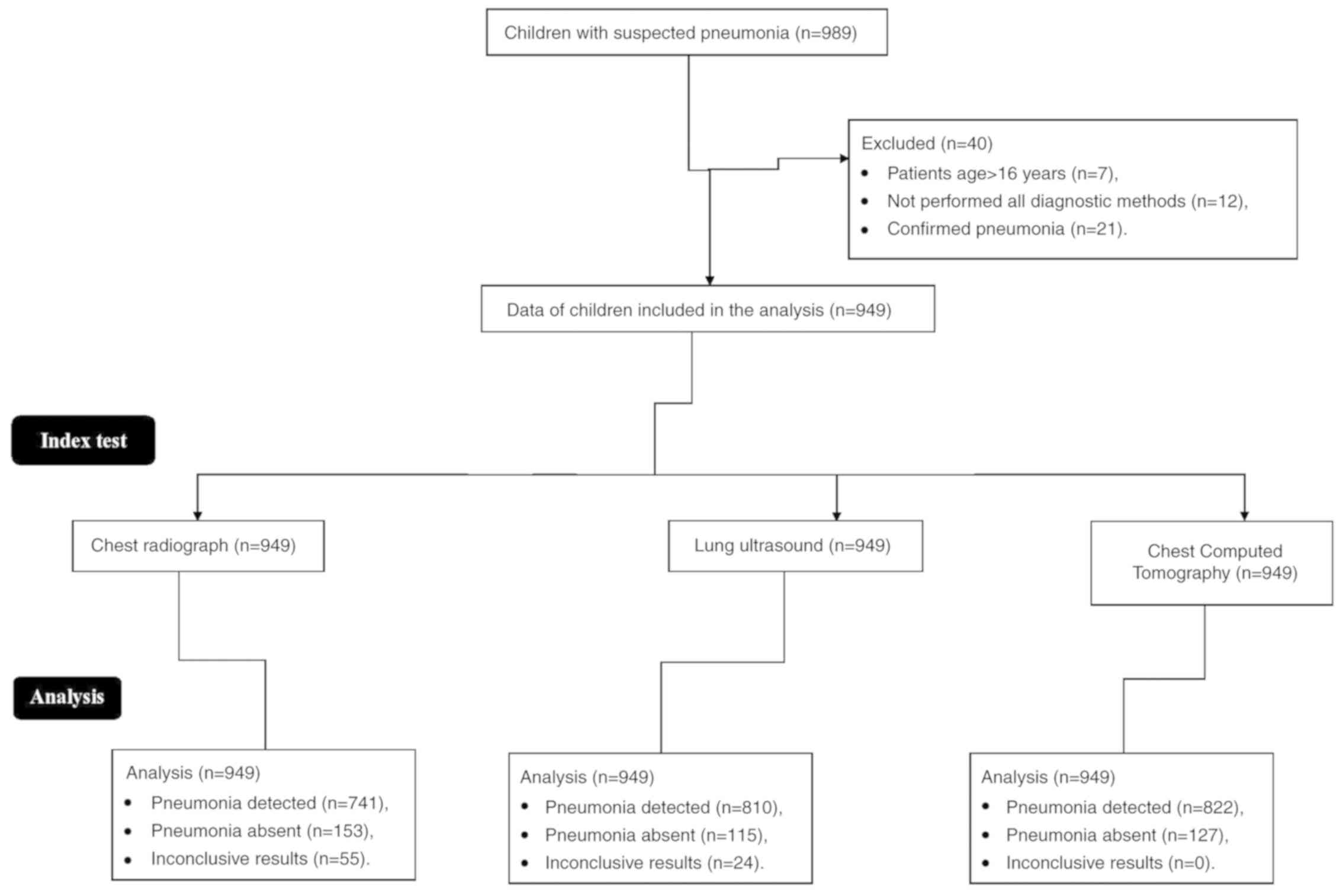
excluded from the analysis. The flowchart of analysis is presented
in Fig. 1.
Data collection
Data regarding clinicopathological conditions,
demographical characteristics, clinical examinations, the chest
X-ray, transthoracic sonography and the chest CT of 949 children
were collected from the institutional records. Data were collected
electronically and anonymized.
Clinical examinations
Within 24 h of admission, blood samples were
collected and sent to a laboratory at the Affiliated Hospital of
Inner Mongolia Medical University for pathological examination, and
data regarding blood chemistry were evaluated.
Chest radiograph
Children in a seated position received frontal and
lateral view chest X-rays using DuraDiagnost (Koninklijke Philips
N.V.). Image interpretation was performed by three pediatric
radiologists with ≥5 years' experience, who were blinded to the
results of the clinical examination, at each institute.
Lung ultrasound
Transthoracic sonography was performed by
sonographers (≥3 years of experience) at each institute, on
patients in a seated or supine position; using EPIQ Elite
(Koninklijke Philips N.V.) in B-mode connected to 7.5 MHz linear
probes (Koninklijke Philips N.V.). Scans included two rib spaces
per lung field for the anterior, mild axillary and posterior
fields, totaling to six scanning windows per hemithorax (15). Image interpretation was performed by
sonographers with ≥5 years' experience, who were blinded to the
results of clinical examinations and chest X-rays, in consultation
with pulmonologists (≥3 years' experience) at each institute.
Transthoracic ultrasound was performed in real time.
Chest CT
Chest CT scans were performed on patients from the
level of the thoracic inlet to the diaphragm using an eight-sliced
scanner (Koninklijke Philips N.V.) at 100 kV, 25 mAs, 0.14 volume,
0.5 sec rotation and 2×10 collimation. CT scans were performed by
radiologists (≥5 years' experience; blinded regarding results of
clinical examinations, chest X-rays and lung ultrasound) at each
institute. Images were analyzed by radiologists in consultation
with pulmonologists (≥3 years' experience) at each institute
(16). When images were reviewed, ≥3
independent interpretations of the results were performed. Pleural
effusion, perilesional inflammatory edema and lung consolidations
were considered as pneumonia. True, false or inconclusive results
of lung ultrasound and chest radiography were decided based on the
results obtained from chest CT.
Beneficial analysis
Beneficial score analysis was performed to analyze
an area to detect positive disease (pneumonia) by imaging methods,
or for the evaluation of imaging modality performance. Beneficial
score analysis was evaluated according to the pneumonia severity
index for each diagnostic modality, using the following equation
(17): (true positive
pneumonia/949)-[(false positive pneumonia/949) × ([5-LC]/LC)],
where true positive pneumonia means that pneumonia was detected by
imaging modalities as well as by the chest CT; false positive means
that pneumonia was detected by imaging modalities but not detected
by the chest CT; LC is the level of confidence above which children
were put on antibiotics, which was determined by the pneumonia
severity index, a 0 to 5 scale, according to Institutional
Guidelines for Pediatric Pneumonia. The scale was defined as
follows: 0, absent (no clinical signs and symptoms); 1, mild
pneumonia (clinical signs and symptoms but no hospitalization
required); 2, mild to moderate pneumonia (hospitalization but no
intensive care admission); 3, moderate pneumonia (intensive care
admission but no mechanical ventilation required); 4, moderate to
severe pneumonia (mechanical ventilation but no death); and 5,
severe pneumonia (shock or death). The ratio of true positive
pneumonia detected by each modality to true positive pneumonia
detected by chest CT was considered sensitivity. The ratio of true
negative pneumonia detected by each modality to true negative
pneumonia detected by chest CT was accuracy.
Statistical analysis
Categorical variables are presented as a number and
percentage. Continuous variables are presented as the mean ± SD.
SPSS software (version 24.0; IBM Corporation) was used for
statistical analysis. Categorical data were analyzed by the
χ2 test. A P<0.05 was considered to indicate a
significant difference.
Results
Clinical manifestations
Dyspnea and cough were the most commonly reported
symptoms among the patients, followed by expectoration of purulent
sputum, fever and pleuritic chest pain. Further demographical
characteristics of the patients are presented in Table I.
 | Table I.Clinicopathological and demographical
characteristics. |
Table I.
Clinicopathological and demographical
characteristics.
| Characteristic | Patient
population |
|---|
| Age (years) |
|
|
Minimum | 2 |
|
Maximum | 16 |
| Mean ±
SD | 12.45±3.12 |
| Sex |
|
| Male | 446 (47) |
|
Female | 503 (53) |
| Ethnicity |
|
| Han
Chinese | 871 (92) |
|
Mongolian | 62 (2) |
|
Tibetan | 16 (2) |
| Fever | 361 (38) |
| Dyspnea | 711 (75) |
| Cough | 778 (82) |
| Expectoration of
purulent sputum | 3,445 (47) |
| Decreased breath
sounds | 145 (15) |
| Pleuritic chest
pain | 345 (36) |
| Chills | 281 (30) |
| Headache | 157 (17) |
Laboratory tests
Children displayed a higher erythrocyte
sedimentation rate than normal. A total of 171 (18%) patients
displayed severe hypoxemia and were subsequently admitted into the
Emergency Department. The results of further laboratory tests are
presented in Table II.
| Table II.Biological characteristics. |
Table II.
Biological characteristics.
| Characteristic | Patient
population |
|---|
| Erythrocyte
sedimentation rate (mm/h)a | 20.12±7.22 |
| White blood
cell/dl |
12,545.00±3,415.00 |
| Procalcitonin
(μg/l) | 2.11±0.54 |
| Serum reactive
protein (mg/l) | 111.85±12.17 |
| Urine area >11
mM/l | 141 (15) |
| Blood pH
<7.35 | 10 (1) |
| Severe hypoxemia
(partial pressure of oxygen in blood <60 mmHg) | 171 (18) |
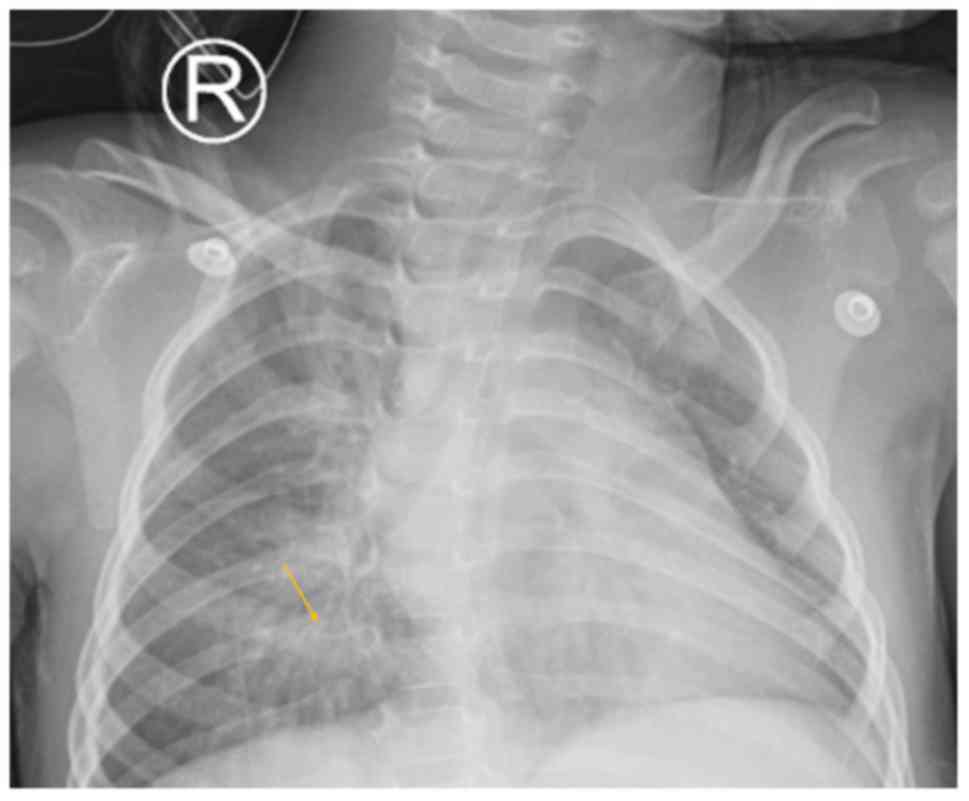
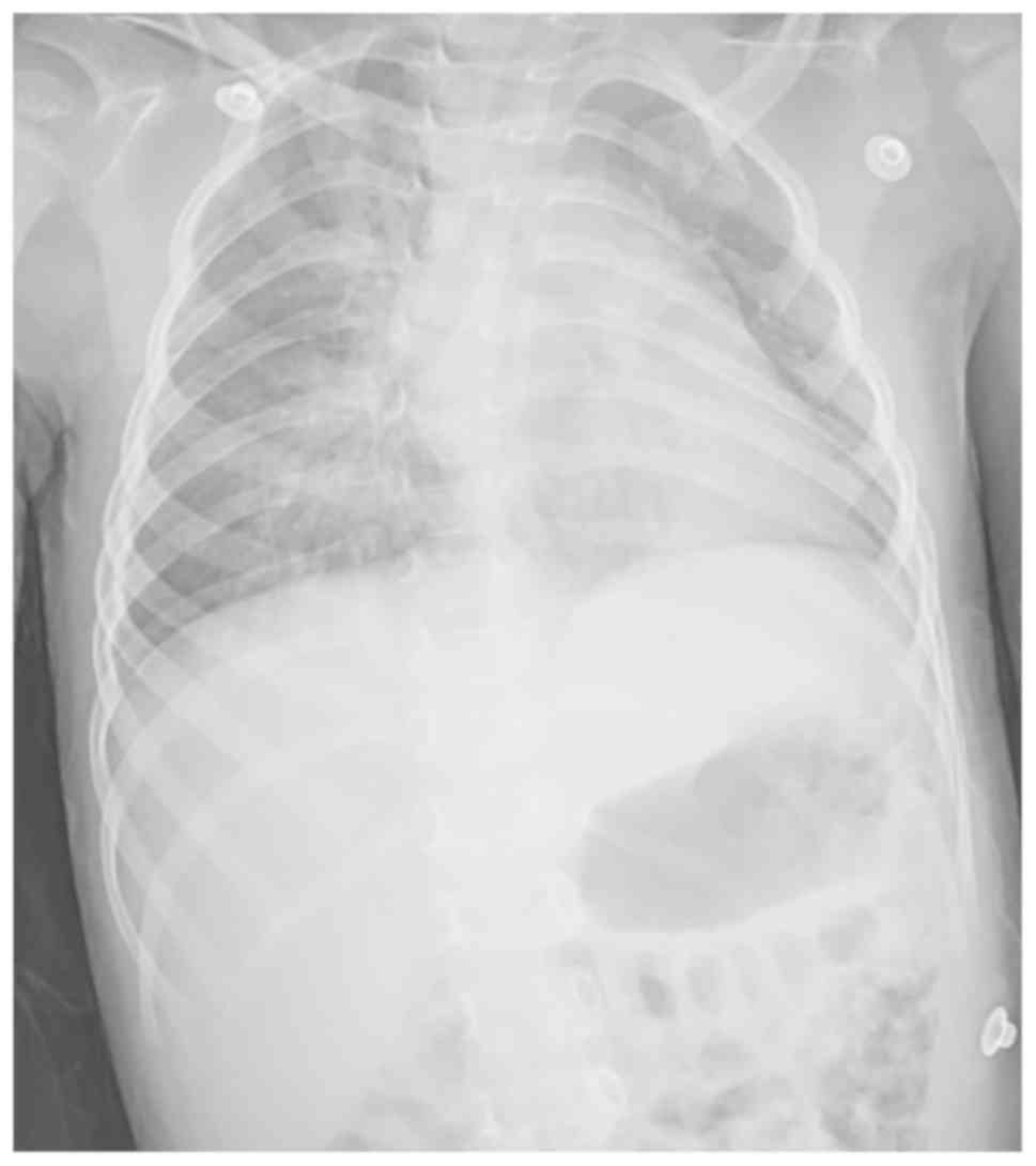
Chest radiograph
The chest X-ray successfully detected subpleural
lung consolidation (Fig. 2) and
dense opacity in the lungs, but was unable to identify whether the
lung had undergone suppurative, necrotic change or was congested
(Fig. 3).
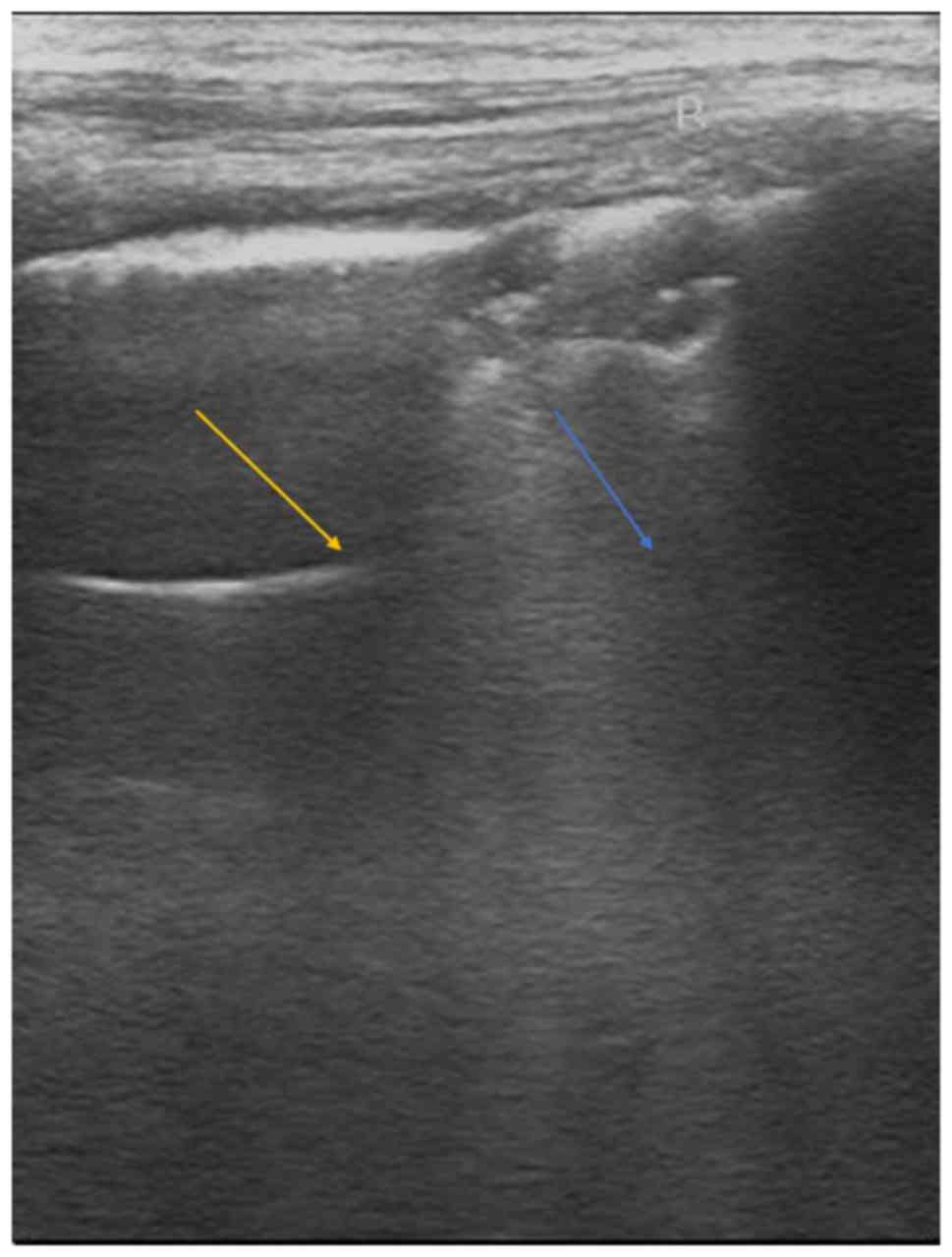
Lung ultrasound
Transthoracic sonography successfully detected a
minimal pleural effusion and perilesional inflammatory edema
(Fig. 4). However, a lung
consolidation with an air bronchogram, as well as the
consolidations that did not reach the pleura were missed. Lung
ultrasound was also not able to define the hyperechoic spots
present in the consolidations as an air bronchogram.
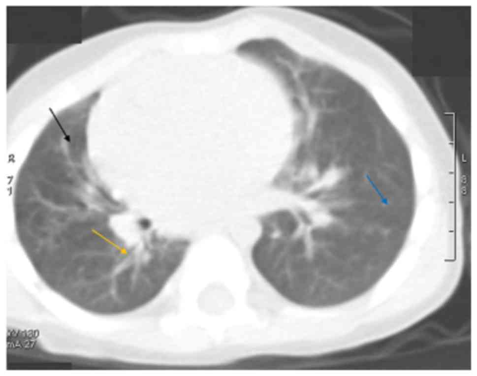
Chest CT
The chest CT successfully detected liquefied areas,
inflammation and necrosis of the lungs (Fig. 5).
Diagnostic parameters
Compared with chest CT, lung ultrasound displayed
0.906 sensitivity and 0.661 accuracy, while chest radiograph
displayed 0.793 sensitivity and 0.559 accuracy. Further diagnostic
parameters of imaging modalities are presented in Table III.
| Table III.Diagnostic parameters of imaging
modalities. |
Table III.
Diagnostic parameters of imaging
modalities.
|
| Imaging
modality |
|---|
|
|
|
|---|
|
| Chest computed
tomography | Lung
ultrasound | Chest
radiograph |
|---|
|
|
|
|
|
|---|
| Parameter | Patient
population | Patient
population | P-value | Patient
population | P-value |
|---|
| True positive
pneumonia | 822 (87) | 745 (79) | <0.0001 | 652 (69) | <0.0001 |
| True negative
pneumonia | 127 (13) | 84 (9) | 0.0020 | 71 (7) | <0.0001 |
| False positive
pneumonia | 0 (0) | 65 (7) | <0.0001 | 89 (9) | <0.0001 |
| False negative
pneumonia | 0 (0) | 31 (3) | <0.0001 | 82 (9) | <0.0001 |
| Inconclusive
results | 0 (0) | 24 (2) | <0.0001 | 55 (6) | <0.0001 |
| Sensitivity | 1 | 0.906 | <0.0001 | 0.793 | <0.0001 |
| Accuracy | 1 | 0.661 | <0.0001 | 0.559 | <0.0001 |
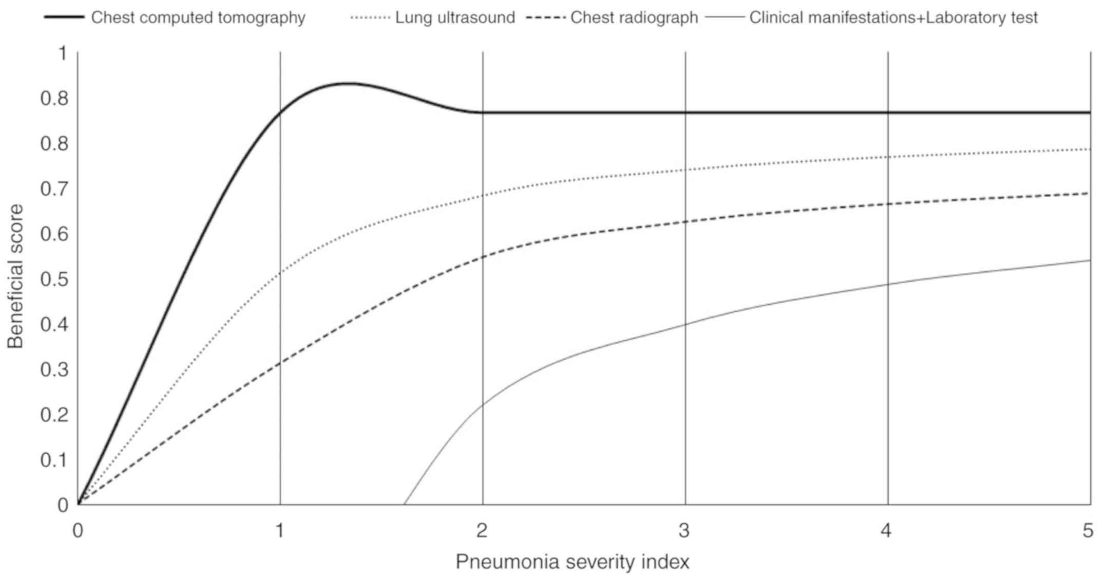
Beneficial analysis
All diagnostic modalities displayed considerable
beneficial scores when the pneumonia severity index was ≥3. For
pneumonia severity indexes <3, the chest CT provided the highest
beneficial score, followed by lung ultrasound and chest
radiography, respectively (Fig.
6).
Discussion
The present study reported that chest X-ray was
unable to identify necrosis and congestion of the lungs in
pediatric patients with suspected pneumonia. The air bronchogram
was only identified by chest CT. The results from the present study
were consistent with prospective studies (16,18) and
retrospective reviews on adult patients (13,14).
However, the results obtained were not in line with a prospective
observational cohort study on lung ultrasound in children and young
adults (19). Chest CT is considered
as ‘gold standard’ in the detection of pneumonia (20), but cannot be used routinely in
children because children are radiosensitive and display a greater
risk associated with radiation than adults (3). Therefore, there is a need for an
alternative diagnostic modality for the detection of pneumonia in
children, with a reduced risk of radiation exposure.
In the present study, for the detection of
pneumonia, lung ultrasound displayed 0.906 sensitivity and 0.661
specificity, while chest X-ray displayed 0.793 sensitivity and
0.559 specificity, compared with chest CT. These results were
consistent with results reported by a number of prospective
diagnostic accuracy studies (4,11,21). The
chest X-ray is routinely recommended as an important diagnostic
modality for pneumonia because it can predict pneumonia without
clinical signs (9). Likewise, lung
ultrasound is successful in the differential diagnosis of
atelectasis and lung consolidation (22), because fluid or solid material that
accumulates in the lung can be easily visualized by a transthoracic
sonograph (10). Additionally,
children have a smaller lung mass and thinner chest walls compared
to adults, which makes lung ultrasound ideal for the diagnosis of
pneumonia in children (23). Even a
low dose (<0.2 mGy) of chest X-ray is harmful to children
(20). Furthermore, chest X-ray does
not display 100% specificity or accuracy and also has issues of
intra- and inter-observer disagreements (23). Alternatively, the results of the
current study indicated that lung ultrasound was a safe, reliable
and superior diagnostic method compared with chest X-ray for the
diagnosis of pneumonia in children.
Lung ultrasound displayed 65 (7%) false-positive
results, whereas chest X-ray resulted in 89 (9%) false-positive
results (P=0.053) for the diagnosis of pneumonia. These results
were consistent with a prospective diagnostic accuracy study
(4). However, these results were not
in line with a prospective observational cohort study investigating
the use of ultrasound for children and young adults (19). Chest X-ray has an issue resulting in
the blending of various tissue images, whereas lung ultrasound
allows dynamic analysis of each intercostal space during breathing
(4). Ultrasonography of the left
lower chest is problematic as artifacts are caused by the
interaction between the ultrasound beam and even a small amount of
fluid and air. This implies that pleural effusion enhances artifact
generation (19). In the present
study lung ultrasound resulted in fewer false-negative results than
chest X-ray (31 vs. 82; P<0.0001). The results from the present
study were consistent with a number of prospective diagnostic
accuracy studies (7,19). X-ray has poor sensitivity for small
subpleural consolidations (4) and
can only detect lung consolidations that are ≥1.5 cm (19). The results of the current study
reported that false predictive values of lung ultrasound were lower
than those for chest X-ray and could be eliminated by integrating
cardiac and abdominal scans of the lung, through the diaphragm,
with the intercostal approach.
In the present study, for pneumonia severity indexes
<3, lung ultrasound displayed a higher beneficial score than
chest X-ray, indicating that lung ultrasound may aid in the
clinical decision-making process regarding putting children on
antibiotics in the early stages of pneumonia, when symptoms are not
severe.
There were several limitations of the present study,
for example, the study was a retrospective study and lacked a
prospective trial. The present study performed chest CT to confirm
pneumonia, but generally chest CT is not preferred as a diagnostic
module in children for safety reasons. Typically, there is less
inter-and intra-operator reproducibility for lung ultrasound than
chest X-ray (16). The amount of
experience each sonographer has can also impact the accuracy of the
ultrasound findings (9).
Furthermore, lung ultrasound has fewer accuracies in identifying
lung abscesses than chest CT (16).
Combining lung ultrasound and chest X-ray has been reported to
display results consistent with chest CT (16), however, the present study did not
include such analysis. Additionally, a large randomized trial is
required to assess the combination of lung ultrasound and chest
X-ray in the diagnosis of pneumonia in children.
In conclusion, lung ultrasound is a non-invasive and
simple method for the diagnosis of suspected pneumonia in children.
Unlike chest X-ray and CT, lung ultrasound is an easy method to
perform in children at the bedside without ionizing radiation
exposure. The present study recommended lung ultrasound for the
diagnosis of pneumonia instead of using chest radiography or
CT.
Acknowledgements
Not applicable.
Funding
No funding was received.
Availability of data and materials
The datasets used and/or analyzed during the present
study are available from the corresponding author on reasonable
request.
Authors' contributions
All authors read and approved the manuscript for
publication. CY was the project administrator and contributed to
the conceptualization, software and the literature review. RH
contributed to formal analysis, validation, resources and the
literature review. ZL contributed to the resources, data curation,
investigation and the literature review. YZ contributed to the
software, formal analysis and literature review of the study. YZ
also drafted, reviewed and edited the manuscript for intellectual
content. Authors agree to be accountable for all aspects of work
ensuring integrity and accuracy.
Ethics approval and consent to
participate
The present study was approved by the Ethics
Committee of the Affiliated Hospital of Inner Mongolia Medical
University. The present study complied with the 2008 Declaration of
Helsinki. Written informed consent was provided by the parents of
each of the pediatric patients in the present study.
Patient consent for publication
Written informed consent for the publication of the
present study was provided by the parents of each of the pediatric
patient, including the publication of personal data and images.
Competing interests
The authors declare that they have no competing
interests.
Glossary
Abbreviations
Abbreviations:
|
CT
|
computed tomography
|
|
MRI
|
magnetic resonance imaging
|
References
|
1
|
Ning G, Wang X, Wu D, Yin Z, Li Y, Wang H
and Yang W: The etiology of community-acquired pneumonia among
children under 5 years of age in mainland China, 2001–2015: A
systematic review. Hum Vaccin Immunother. 13:2742–2750. 2017.
View Article : Google Scholar : PubMed/NCBI
|
|
2
|
Harris M, Clark J, Coote N, Fletcher P,
Harnden A, McKean M and Thomson A; British Thoracic Society
Standards of Care Committee, : British Thoracic Society guidelines
for the management of community acquired pneumonia in children:
Update 2011. Thorax. 66 (Suppl 2):ii1–ii23. 2011. View Article : Google Scholar : PubMed/NCBI
|
|
3
|
Brenner DJ and Hall EJ: Computed
tomography-an increasing source of radiation exposure. N Engl J
Med. 357:2277–2284. 2007. View Article : Google Scholar : PubMed/NCBI
|
|
4
|
Reali F, Sferrazza Papa GF, Carlucci P,
Fracasso P, Di Marco F, Mandelli M, Soldi S, Riva E and Centanni S:
Can lung ultrasound replace chest radiography for the diagnosis of
pneumonia in hospitalized children? Respiration. 88:112–115. 2014.
View Article : Google Scholar : PubMed/NCBI
|
|
5
|
Ekinci A, Yucel Ucarkus T, Okur A, Öztürk
M and Doğan S: MRI of pneumonia in immunocompromised patients:
Comparison with CT. Diagn Interv Radiol. 23:22–28. 2017. View Article : Google Scholar : PubMed/NCBI
|
|
6
|
Reissig A and Kroegel C: Sonographic
diagnosis and follow-up of pneumonia: A prospective study.
Respiration. 74:537–547. 2007. View Article : Google Scholar : PubMed/NCBI
|
|
7
|
Reissig A, Copetti R, Mathis G, Mempel C,
Schuler A, Zechner P, Aliberti S, Neumann R, Kroegel C and Hoyer H:
Lung ultrasound in the diagnosis and follow-up of
community-acquired pneumonia: A prospective, multicenter,
diagnostic accuracy study. Chest. 142:965–972. 2012. View Article : Google Scholar : PubMed/NCBI
|
|
8
|
Medford ARL: Chest ultrasonography as a
replacement for chest radiography for community-acquired pneumonia.
Chest. 143:877–878. 2013. View Article : Google Scholar : PubMed/NCBI
|
|
9
|
Copetti R and Cattarossi L: Ultrasound
diagnosis of pneumonia in children. Radiol Med. 113:190–198.
2008.(In English, Italian). View Article : Google Scholar : PubMed/NCBI
|
|
10
|
Roić G: Lung ultrasound in the diagnosis
of pediatric pneumonia: Are we ready for routine use? Acta Med
Acad. 45:82–83. 2016.PubMed/NCBI
|
|
11
|
Caiulo VA, Gargani L, Caiulo S, Fisicaro
A, Moramarco F, Latini G, Picano E and Mele G: Lung ultrasound
characteristics of community-acquired pneumonia in hospitalized
children. Pediatr Pulmonol. 48:280–287. 2013. View Article : Google Scholar : PubMed/NCBI
|
|
12
|
Caiulo VA, Gargani L, Caiulo S, Fisicaro
A, Moramarco F, Latini G and Picano E: Sensitivity and feasibility
of lung ultrasound in bronchiolitis-reply to the correspondence
letter by Catalano. Eur J Pediatr. 173:407–408. 2014. View Article : Google Scholar : PubMed/NCBI
|
|
13
|
Hayden GE and Wrenn KW: Chest radiograph
vs. computed tomography scan in the evaluation for pneumonia. J
Emerg Med. 36:266–270. 2009. View Article : Google Scholar : PubMed/NCBI
|
|
14
|
Self WH, Courtney DM, McNaughton CD,
Wunderink RG and Kline JA: High discordance of chest X-ray and
computed tomography for detection of pulmonary opacities in ED
patients: Implications for diagnosing pneumonia. Am J Emerg Med.
31:401–405. 2013. View Article : Google Scholar : PubMed/NCBI
|
|
15
|
Caiulo VA, Gargani L, Caiulo S, Fisicaro
A, Moramarco F, Latini G and Picano E: Lung ultrasound in
bronchiolitis: Comparison with chest X-ray. Eur J Pediatr.
170:1427–1433. 2011. View Article : Google Scholar : PubMed/NCBI
|
|
16
|
Hajalioghli P, Nemati M, Dinparast SL and
Fouladi DF: Can chest computed tomography be replaced by lung
ultrasonography with or without plain chest radiography in
pediatric pneumonia? J Thorac Imaging. 31:247–252. 2016. View Article : Google Scholar : PubMed/NCBI
|
|
17
|
Fitzgerald M, Saville BR and Lewis RJ:
Decision curve analysis. JAMA. 13:409–410. 2015. View Article : Google Scholar
|
|
18
|
Claessens YE, Debray MP, Tubach F, Brun
AL, Rammaert B, Hausfater P, Naccache JM, Ray P, Choquet C, Carette
MF, et al: Early chest computed tomography scan to assist diagnosis
and guide treatment decision for suspected community-acquired
pneumonia. Am J Respir Crit Care Med. 192:974–982. 2015. View Article : Google Scholar : PubMed/NCBI
|
|
19
|
Shah VP, Tunik MG and Tsung JW:
Prospective evaluation of point-of-care ultrasonography for the
diagnosis of pneumonia in children and young adults. JAMA Pediatr.
167:119–125. 2013. View Article : Google Scholar : PubMed/NCBI
|
|
20
|
Caiulo VA, Gargani L, Caiulo S, Fisicaro
A, Moramarco F, Latini G, Picano E and Mele G: The role of
ultrasound in community-acquired pneumonia. Pediatr Pulmonol.
48:1043–1044. 2013. View Article : Google Scholar : PubMed/NCBI
|
|
21
|
Cortellaro F, Colombo S, Coen D and Duca
PG: Lung ultrasound is an accurate diagnostic tool for the
diagnosis of pneumonia in the emergency department. Emerg Med J.
29:19–23. 2012. View Article : Google Scholar : PubMed/NCBI
|
|
22
|
Lichtenstein D, Mezière G and Seitz J: The
dynamic air bronchogram. A lung ultrasound sign of alveolar
consolidation ruling out atelectasis. Chest. 135:1421–1425. 2009.
View Article : Google Scholar : PubMed/NCBI
|
|
23
|
Caiulo VA, Gargani L, Caiulo S, Fisicaro
A, Moramarco F, Latini G, Picano E and Mele G: Response to lung
ultrasound as an additional imaging tool for the evaluation of
pneumonia. Pediatr Pulmonol. 49:619–620. 2014. View Article : Google Scholar : PubMed/NCBI
|