Open Access

Atorvastatin inhibits endoplasmic reticulum stress through AMPK signaling pathway in atherosclerosis in mice

  • Authors:
    • Wangqiong Xiong
    • Minzhong Fei
    • Chuntao Wu
    • Wei Wang
    • Rong Luo
    • Liping Shen
    • Zheng Zhang
  • View Affiliations

  • Published online on: December 27, 2019     https://doi.org/10.3892/etm.2019.8379
  • Pages: 2266-2272
  • Copyright: © Xiong et al. This is an open access article distributed under the terms of Creative Commons Attribution License.

Metrics: Total Views: 0 (Spandidos Publications: | PMC Statistics: )
Total PDF Downloads: 0 (Spandidos Publications: | PMC Statistics: )


Abstract

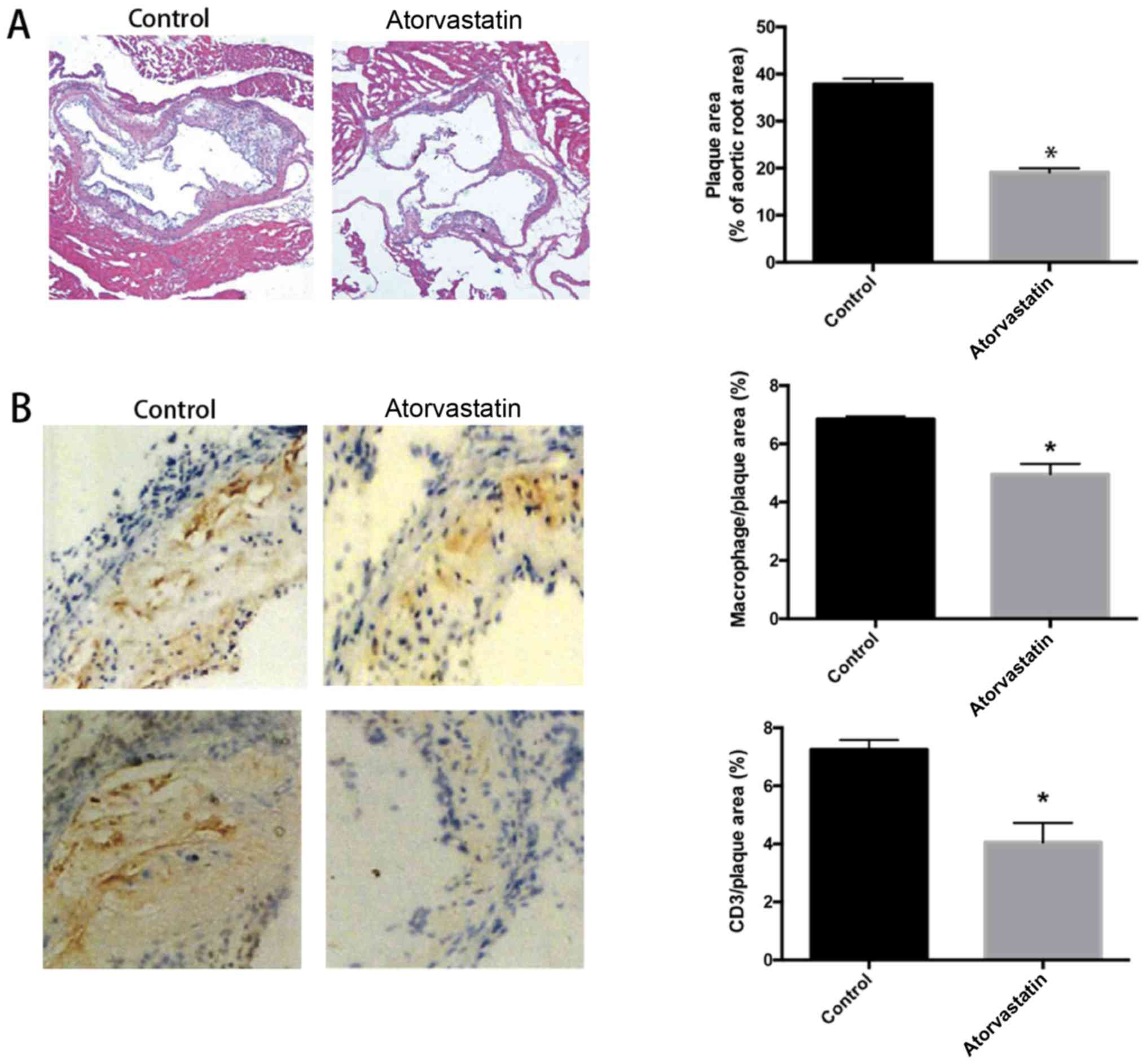
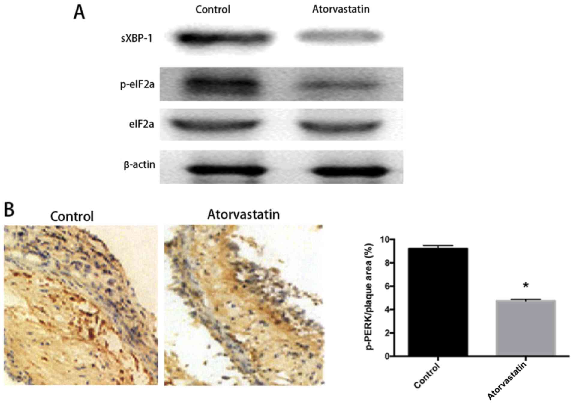
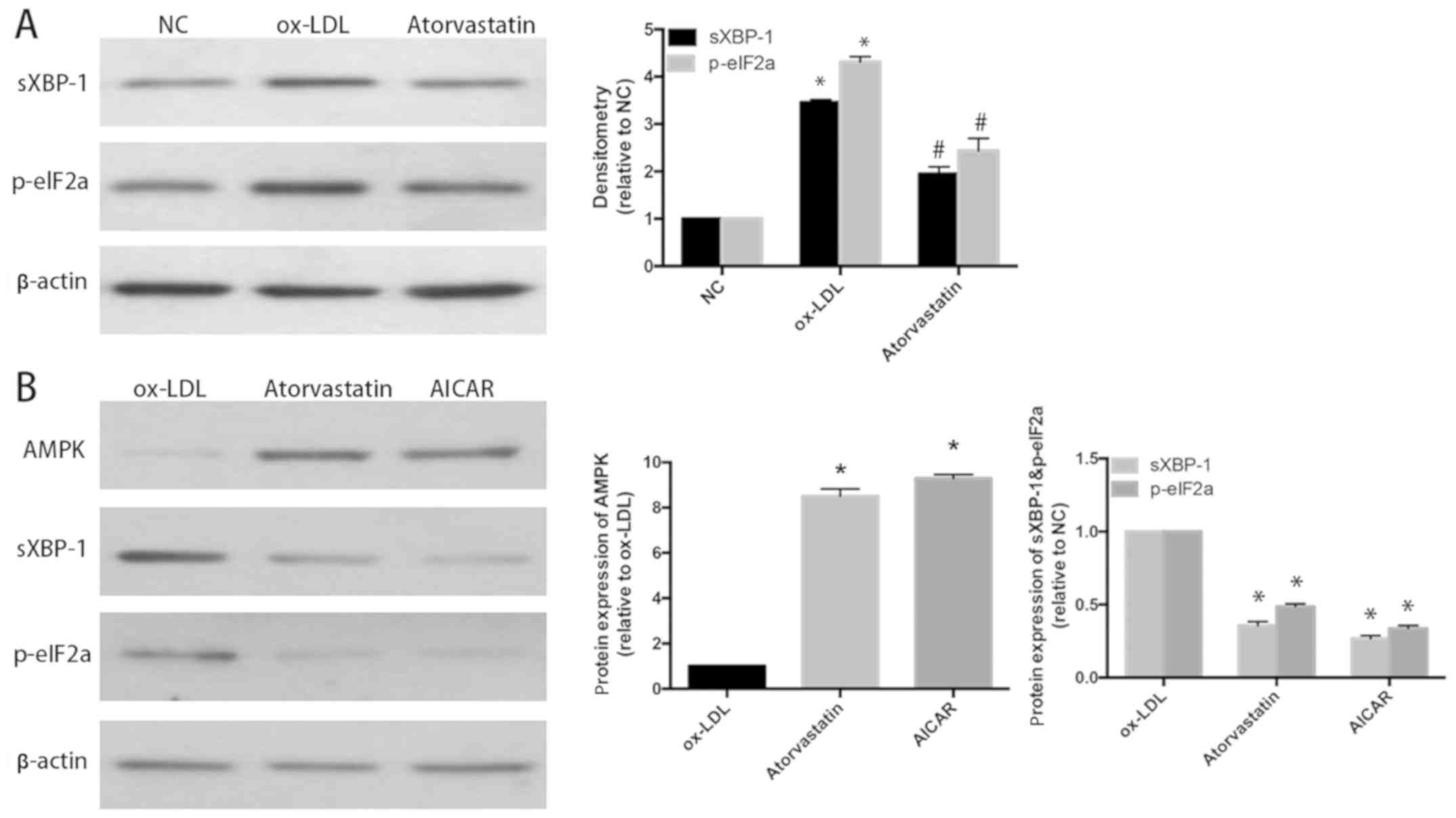
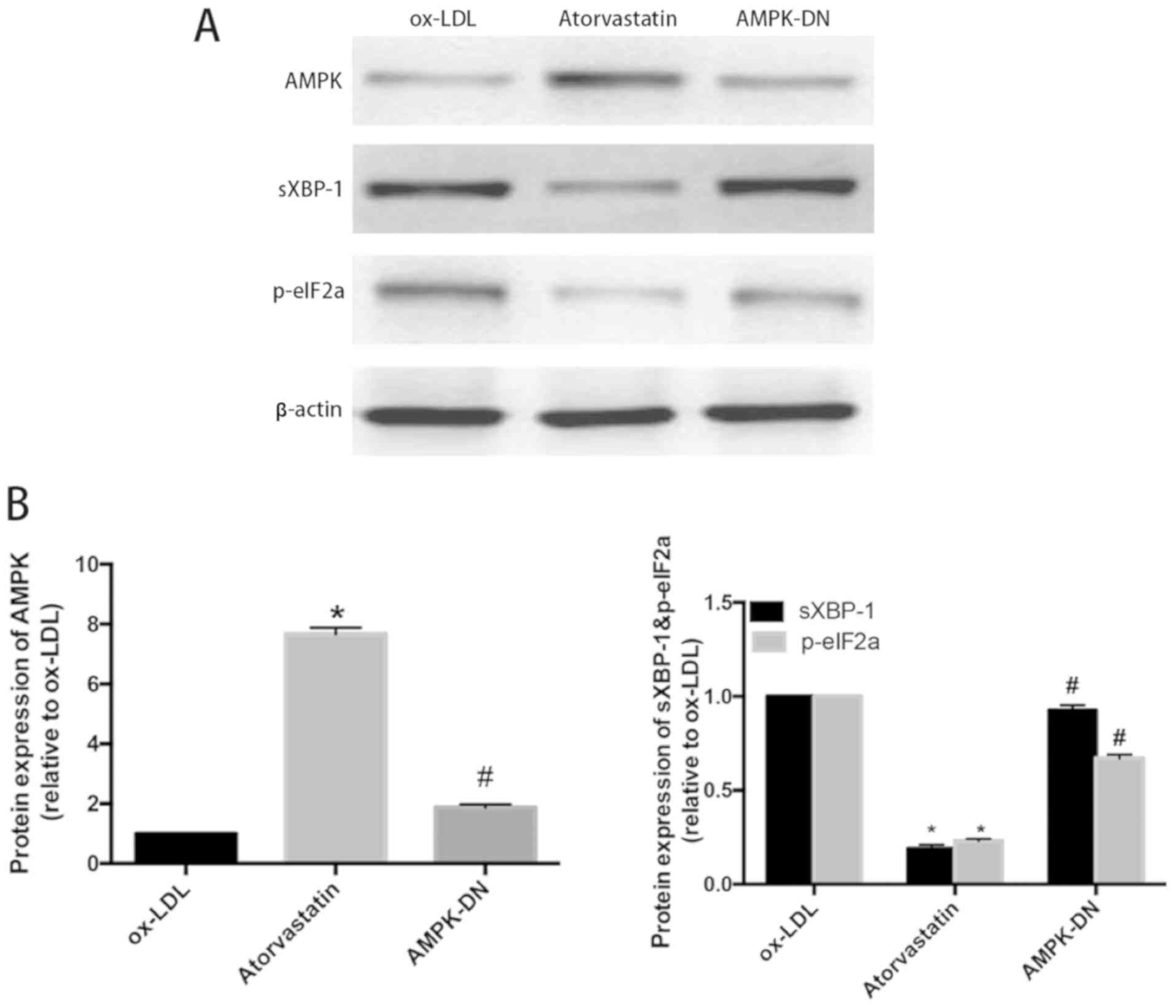
Effect of atorvastatin inhibition of endoplasmic reticulum stress and amelioration of atherosclerosis through AMPK pathway were studied. Eight‑week‑old male apolipoprotein E‑deficient (ApoE‑/‑) mice were fed with high‑fat diet for 2 weeks and randomly divided into two groups: Atorvastatin treatment group was given atorvastatin (5 mg/kg/day) injection for a total of 6 weeks; control group was given the same dose of PBS through intraperitoneal injection for a total of 6 weeks. H&E staining was used to detect plaque size; immunohistochemical staining was used to detect T cells, macrophages and phospho‑protein kinase‑like ER kinase (phospho‑PERK) in localized plaques. Proteins were extracted from mouse thoracic and abdominal aortic tissues. Western blot analysis was used to detect the protein expression levels of endoplasmic reticulum stress‑related molecules phospho‑eukaryotic initiation factor‑2α (p‑eIF2α), eukaryotic initiation factor (eIF2a), and sliced x‑box binding protein 1 (sXBP‑1). Cultured human umbilical vein endothelial cells (HUVECs), induced endoplasmic reticulum stress with human oxidized low density lipoprotein (ox‑LDL), were treated with atorvastatin, AMPK agonist 5‑amino‑4‑imidazolecarboxamide riboside‑I‑β‑D‑ribofuranoside (AICAR) and AMPK‑DN that expressed a dominant‑negative mutant of AMPK. Western blot analysis was used to test the expression levels of endoplasmic reticulum stress‑related molecules p‑elF2a and sXBP‑1. The area of aortic plaques in atorvastatin group was obviously decreased, and the infiltrations of CD3+ T cells and macrophages in the localized plaques were reduced. The endoplasmic reticulum stress‑related proteins sXBP‑1 and p‑eIF2a were significantly reduced. The results of immunohistochemistry also showed a significant decrease in the level of phospho‑PERK (p‑PERK) in atorvastatin group. The results in ox‑LDL‑induced HUVECs showed that atorvastatin inhibited ox‑LDL‑induced endoplasmic reticulum stress, and the AMPK agonist AICAR also had the same effect, which was offset by DN‑AMPK treatment. Atorvastatin inhibits ER stress both in vitro and in vivo and this protective effect is mediated by AMPK activation.
View Figures
View References

Related Articles

Journal Cover

March-2020
Volume 19 Issue 3

Print ISSN: 1792-0981
Online ISSN:1792-1015

Sign up for eToc alerts

Recommend to Library

Copy and paste a formatted citation
x
Spandidos Publications style
Xiong W, Fei M, Wu C, Wang W, Luo R, Shen L and Zhang Z: Atorvastatin inhibits endoplasmic reticulum stress through AMPK signaling pathway in atherosclerosis in mice. Exp Ther Med 19: 2266-2272, 2020
APA
Xiong, W., Fei, M., Wu, C., Wang, W., Luo, R., Shen, L., & Zhang, Z. (2020). Atorvastatin inhibits endoplasmic reticulum stress through AMPK signaling pathway in atherosclerosis in mice. Experimental and Therapeutic Medicine, 19, 2266-2272. https://doi.org/10.3892/etm.2019.8379
MLA
Xiong, W., Fei, M., Wu, C., Wang, W., Luo, R., Shen, L., Zhang, Z."Atorvastatin inhibits endoplasmic reticulum stress through AMPK signaling pathway in atherosclerosis in mice". Experimental and Therapeutic Medicine 19.3 (2020): 2266-2272.
Chicago
Xiong, W., Fei, M., Wu, C., Wang, W., Luo, R., Shen, L., Zhang, Z."Atorvastatin inhibits endoplasmic reticulum stress through AMPK signaling pathway in atherosclerosis in mice". Experimental and Therapeutic Medicine 19, no. 3 (2020): 2266-2272. https://doi.org/10.3892/etm.2019.8379