Introduction
An avulsion fracture of the dorsal articular surface
of the lunate fossa in the distal radius was initially described as
a die-punch fracture in 1962 (1).
Subsequently, fractures of the articular surface of the lunate
fossa caused by an axial force and transferred via the lunar bone
to the lunate fossa were collectively referred to as die-punch
fractures of the distal radius. The most typical manifestation was
that of a collapsing fracture of the articular surface (2,3).
Die-punch fractures of the articular surface of the distal tibia,
of the posterior articular surface of the calcaneus and of the
articular surface of the patella have also been described (4–6).
Therefore, a die-punch fracture refers only to the mechanism of the
fracture and does not reflect either the fracture site or the
affected area.
Due to differences in the extent and complexity of
force at the time of injury, wrist position and bone condition,
die-punch fractures may not only affect the ulnar demifacet of the
distal radius but also the radial demifacet of the distal radius.
These fractures include articular surface collapsed fractures and
articular surface split fractures, as well as marginal fractures on
the volar or dorsal side (2).
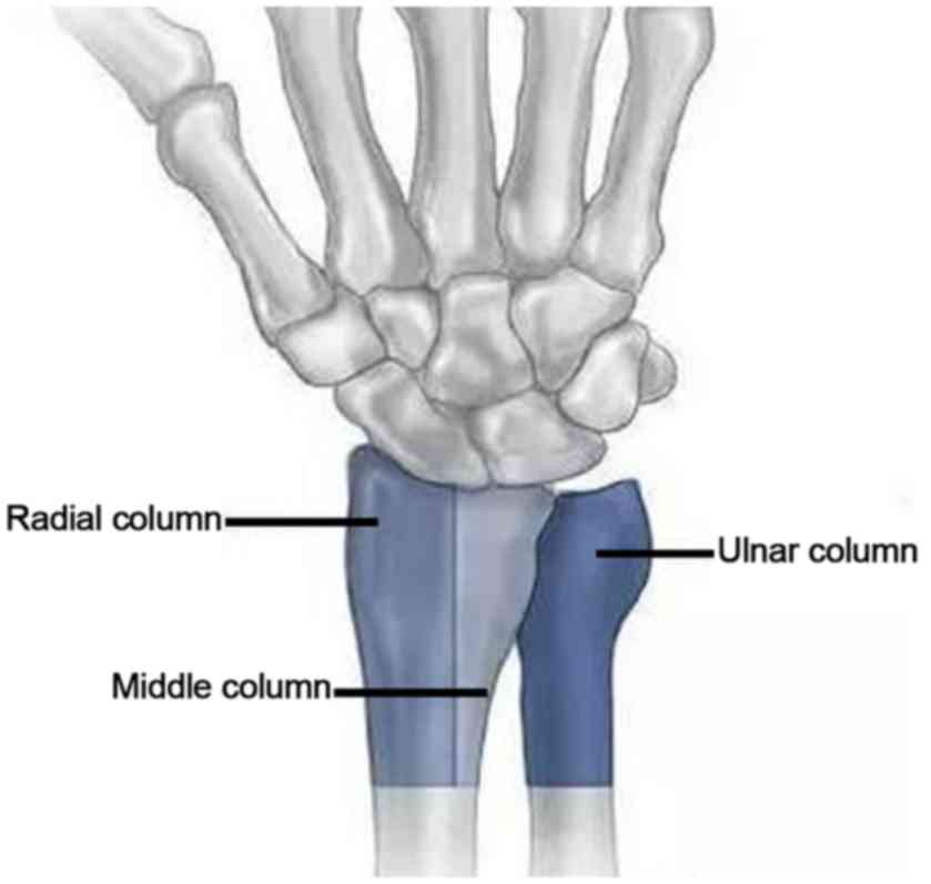
According to the ‘three-column theory’ of distal radius fractures
proposed by Rikli and Regazzoni (7)
(Fig. 1), the die-punch fractures
may be classified into the following two types: Single column and
double column (Figs. 2 and 3A). A die-punch fracture is a rare
occurrence. The Müller-AO classification of distal radius fractures
includes die-punch fractures, although no specific system currently
exists for the classification of die-punch fractures. Although
certain studies have described a classification system for
single-column die-punch fractures of the distal radius (2,8), the
classification of double-column die-punch fractures remains to be
established.
Fracture classification may aid in the
identification of fracture types and subsequent treatment, as well
as determine the prognosis of this disorder. The aim of the present
study was to classify double-column die-punch fractures of the
distal radius according to imaging data. Furthermore, the current
study presented and evaluated the application of a novel
classification based on the review of imaging data from patients
with double-column die-punch fractures of the distal radius
admitted to Wuxi Ninth People's Hospital (Wuxi, China).
Materials and methods
Subjects
The present retrospective study was approved by the
Ethics committee of Wuxi Ninth People's Hospital (Wuxi, China, No.
LW2109003) and Liyang People's Hospital (Changzhou, China, No.
LYCZ2019022).
A total of 10,596 patients who presented with distal
radius fractures were initially screened, 498 of whom were included
in the present study. These patients were diagnosed with a
double-column die-punch fracture of the distal radius at the Wuxi
Ninth People's Hospital (Wuxi, China) between June 2007 and June
2017. The patients included 223 females and 275 males with a mean
age of 45.3 years (range, 13–90 years). Among these patients, there
were 167 cases with falling injuries, 142 cases with bruising
injuries, 114 cases with traffic traumas and 75 cases with impact
injuries. In addition, the study included 45 cases of combined
ulnar styloid fractures, 30 cases of combined relaxation and
subluxation of the distal radioulnar joint fractures, and 86 cases
of complicated fractures at other sites. All cases were diagnosed
by X-ray examinations and 492 out of the 498 (98.79%) underwent CT
examination.
Inclusion and exclusion criteria
The inclusion criteria were as follows: Patients
with an intra-articular fracture of the middle column, accompanied
by a mild fracture of the radial column of the distal radius caused
by an axial force. The exclusion criteria were as follows: i)
Patients with open intra-articular fractures of the middle column,
accompanied by a fracture of the radial column of the distal radius
caused by a direct impact; ii) patients with particularly severe
fractures of the radial column or those with severe fractures in
the radial column and middle column; and iii) patients with
incomplete imaging data.
Instrument-associated parameters
An X-ray plain-film examination was performed using
Siemens digital radiography (DR) equipment (Ysio 1,500 Ma 50 kW;
Siemens Ag) or Philips DR equipment (Digital Diag 500 Ma kW;
Philips Medical Systems). The CT examination was performed using a
GE 64-slice spiral CT scanner (Optima 660; GE Healthcare) with a
screw pitch of 1.2, tube voltage of 120 Kv, tube current of 250 mA,
conventional reconstruction and spacing of 2.5 mm, thin layer
reconstruction and spacing of 1.0 mm, convolution sum of the bone
algorithm of 75 and field of view of 145×145 mm.
Fracture classification method
The patients with double-column die-punch fractures
of the distal radius exhibited an apparent fracture of the middle
column and the majority of the fractures were collapsed, with only
a limited number of avulsion fractures noted at the volar or dorsal
side. The fracture line was usually vertical. The fractures of the
radial column were mild with a horizontal fracture line. The
double-column die-punch fractures of the distal radius were divided
into four classes based on the Müller-AO classification system and
the three-column theory, which is named as the Three-Column
Classification. This classification included the determination of
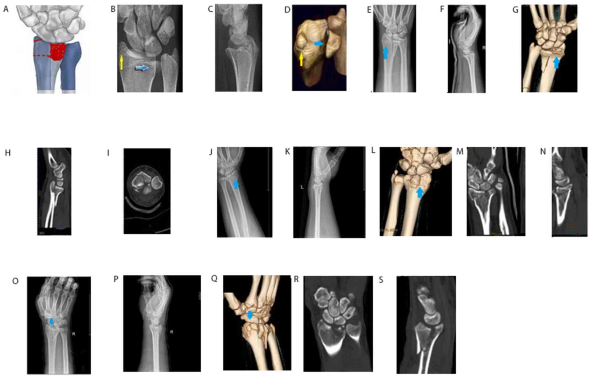
area of the radial column area and was described as follows: Type
I: Middle-column avulsion with fracture of the radial-column
articular surface; type II: Middle-column collapse with fracture of
the radial-column articular surface; type III: Middle-column
collapse with fracture of epiphysis of the radial column; type IV:
Mixed type, middle-column fracture with fractures of articular
surface as well as epiphysis of the radial column.
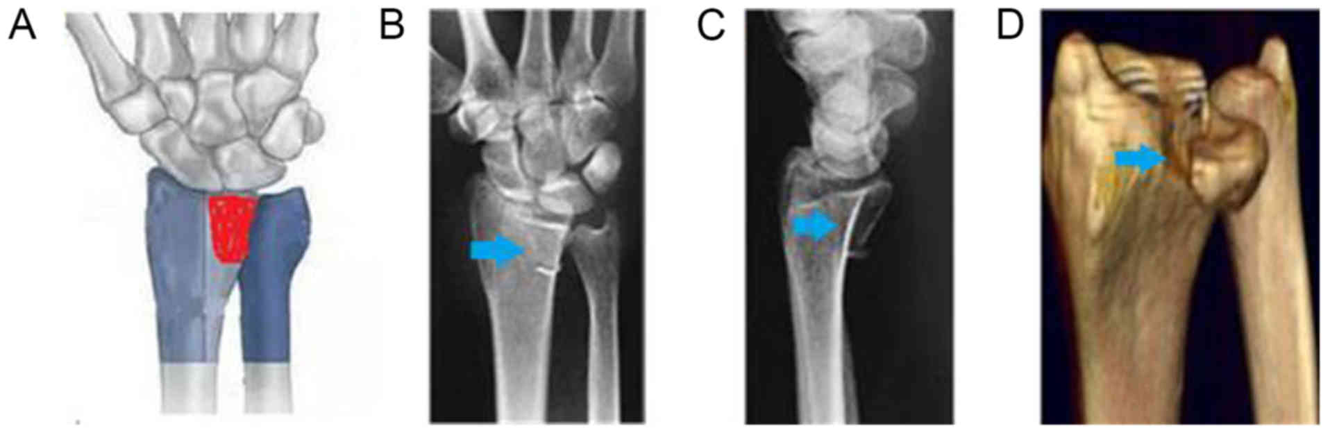
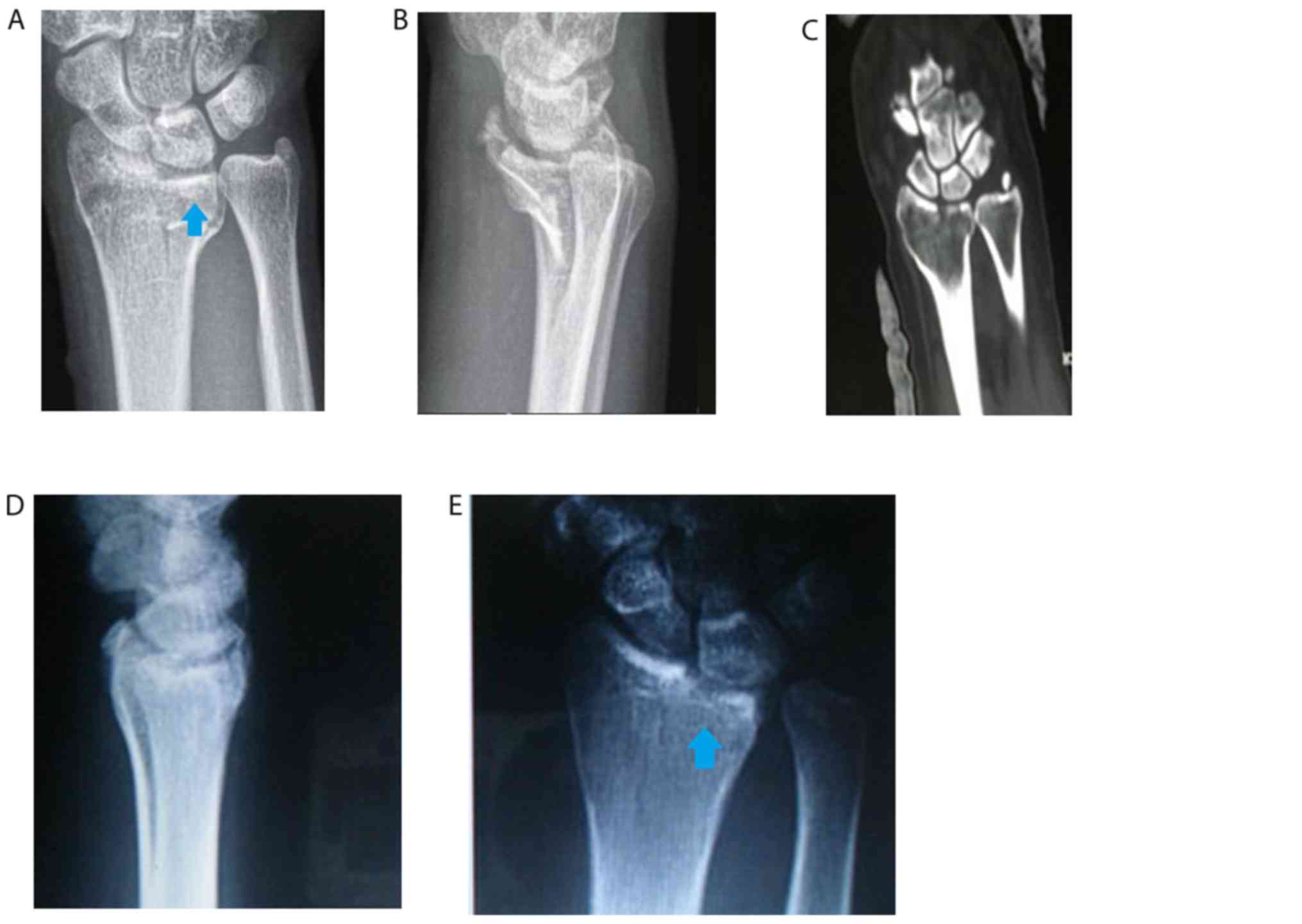
The type I and II fractures affected the articular
surface of the radial column, without affecting the metaphysis of
the radial column. Of note, no fracture was noted in the cortical
bone at the radial side of the metaphysis. These factures were
classified as type I with avulsion in the volar or dorsal edge of
the middle column (Fig. 3B-D), and
as type II for collapsed or comminuted fracture at the center or at
the volar and dorsal sides of the middle column (Fig. 3E-I).
The type III fractures of the patients affected the
metaphysis of the radial column, particularly the cortical bone at
the radial side of the metaphysis, without affecting the articular
surface of the radial column (Fig.
3J-N).
The type IV fractures affected the articular surface
and metaphysis of the radial column. In addition, the fracture line
of the radial column was usually horizontal and comminuted.
Fig. 3O-S displays images of
mixed-type fractures with fracture collapse of the middle column,
and a combination of epiphyseal and articular fractures of the
radial column.
Intra- and inter-observer agreement on
fracture classification
Three-Column Classification was taught to two senior
radiology residents. In this system the double-column die-punch
fractures (n=498) of the distal radius were classified as type
I–IV. The radiologists independently classified 100 cases of
double-column die-punch fractures as follows: i) Type I (n=10); ii)
type II (n=30); iii) type III (n=30); and iv) type IV (n=30). The
classification of the 100 cases of double-column die-punch
fractures was performed independently once more, three months after
study initiation. In cases of inconsistency between the fracture
types assigned by the radiologists, the decision of the most senior
radiologist was accepted. Cohen's kappa coefficients and quadratic
weighted kappa coefficients were subsequently determined (2,3).
Gartland and Werley score
The Gartland and Werley scoring system was used to
evaluate wrist and hand function (9). A score between 0 and 2 was regarded as
‘Excellent’; between 3 and 6 as ‘Good’; between 7 and 18 as ‘Fair’;
and >19 as ‘Poor’.
Statistical analysis
The inter- and intra-observer consistency were
analyzed using kappa statistics (SPSS software version 13.0; SPSS,
Inc.). The kappa coefficient ranged between −1 and +1. A kappa
coefficient of >0 was considered to indicate significant
consistency and improved consistency was associated with a larger
kappa value. Kappa coefficients that ranged between 0.00 and 0.20,
0.21 and 0.40, 0.41 and 0.60, 0.61 and 0.80, and 0.81 and 1.00
corresponded to low, fair, medium, relatively high and high
consistency, respectively.
Results
Distribution of fracture types
The incidence of single-column die-punch fractures
of the distal radius in 10,596 patients with distal radius
fractures was 0.70% (74/10,596), whereas the incidence of
double-column die-punch fractures of the distal radius was 4.70%
(498/10,596). Among the 498 patients diagnosed with double-column
die-punch fractures of the distal radius, 21 cases presented with
middle-column avulsion with radial-column articular surface
fracture (type I), whereas 135 cases exhibited middle-column
collapse with radial-column articular surface fracture (type II).
Furthermore, 130 cases revealed middle-column collapse with
fracture of the epiphysis of the radial column (type III), whereas
212 cases presented with mixed-type fractures (type IV).
Middle-column avulsion fractures involving the radial-column
articular surface (type I) were classified as type B (21 cases,
4.22%) according to the Müller-AO classification, whereas most
collapsed fractures of the middle column (type II and type III,
n=265) and the mixed-type (type IV, n=212) were classified as type
C (477 cases, 95.78%) according to the Müller-AO classification
(Table I). The fractures of all the
patients were able to be categorized using the aforementioned
classification system.
 | Table I.Distribution of fracture types and
consistency of fracture classification. |
Table I.
Distribution of fracture types and
consistency of fracture classification.
| Type I | Type II | Type III | Type IV | Inter-observer
agreement | Intra-observer
agreement |
|---|
| 21 (4.22) | 135 (27.11) | 130 (26.10) | 212 (42.57) | 0.810–0.861 | 0.830–0.876 |
Inter- and intra-observer agreement on
fracture classification
Inconsistency occurred in patients with minor
fractures, including a type IV fracture that was easily
misdiagnosed as type III fracture (Fig.
3J-S). In patients with obvious fractures, the inter-observer
agreement was optimal. In general, the intra-observer kappa
coefficient was between 0.810 and 0.861 and the inter-observer
kappa coefficient was between 0.830 and 0.876, with high
consistency (Table I).
Treatment outcomes vs. fracture
type
Among the 498 patients, 226 and 272 selected
conservative treatment or surgical treatment, respectively. After
clinical healing of the fractures, gradual rehabilitation exercise
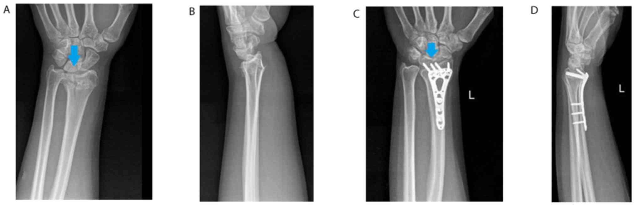
was taken. In the 13th month of the patients' follow-up, the wrist
function of 95.78% of the patients was rated as excellent or good
(n=477; Fig. 4), whereas that of 21
patients (4.22%) was rated as fair according to the Gartland-Werley
scoring system. This was mainly due to the development of
post-traumatic arthritis of the wrist as a result of inappropriate
therapy (Fig. 5). All of these
patients exhibited type IV and type III fractures. No patients with
a poor score were noted in the present study.
Discussion
Previous biomechanical studies have indicated that
the middle column of the distal radius is the hub and primary
load-bearing surface, which transmits an axial load to the wrist
and has a major role in mechanical conduction (10). The fracture line of the middle column
of the distal radius is usually vertical. The radial column of the
distal radius mainly stabilizes the wrist and controls rotation and
its fracture line is usually horizontal (11–14).
Different types of die-punch fractures may occur, depending on the
type of impact and stress, the position of the wrist, and the
effects of local anatomy and bone condition (2,5,11). A single-column die-punch fracture of
the middle column frequently occurs when the radiocarpal joint is
in a neutral position and the axial force is not severe. However,
single-column die-punch fractures of the distal radius are rare and
double-column die-punch fractures are commonly encountered in the
clinic (2). In the present study,
the incidence of single-column die-punch fractures of the distal
radius was 0.70%, whereas the incidence of double-column die-punch
fractures of the distal radius was 4.70%. According to the fracture
sites of the radial column noted on the radiographs, the
double-column die-punch fractures of the distal radius were divided
into the four following classes based on the Three-Colum
Classification system: Type I middle-column avulsion involving the
radial column articular surface; type II middle-column collapse
involving the radial column articular surface; type III
middle-column collapse involving epiphysis of the radial column;
and type IV mixed type. In the patients with type I fractures, the
axial force resulted in volar or dorsal edge fracture of the middle
column when the radiocarpal joint was in flexion or extension
position leading to articular fracture of the radial column due to
the associated rotational load. In patients with type II fractures,
the axial force resulted in fracture collapse of the middle column
when the radiocarpal joint was in a neutral position and the
associated rotational load resulted in articular fracture of the
radial column. Type III fractures occurred when the radiocarpal
joint was in a neutral position and during the incidence of
epiphysis fracture of the radial column as a result of ulnar
deviation of the position of the radiocarpal joint. Type IV
fractures were caused by a relatively large axial load or a
combination of the aforementioned factors. Therefore, the fracture
line in the radial column was usually horizontal and comminuted,
suggesting that the mechanisms underlying the different types of
die-punch fractures of the distal radius varied and depended on a
range of factors, including the size and nature of the axial load,
the associated rotational load and the position of the radiocarpal
joint. The results indicated that the Three-Column Classification
system reflected the mechanisms associated with the development of
different types of fracture.
The Melone classification II and Fernandez
classification III are established classifications of die-punch
fractures, which were denoted as type III fractures in the present
study (15,16). The Melone and Fernandez
classification systems have been extensively cited by previous
studies (17–21). Although this type of double-column
die-punch fracture is widely known, the description of
double-column die-punch fractures of the distal radius has not been
previously reported (10,22). Therefore, a lack of studies is
available regarding the mechanism of the occurrence and
classification of double-column die-punch fractures. In the present
study, 4 types of double-column die-punch fracture of the distal
radius were included as follows: 130 type III fractures, 156 type I
and II fractures and 212 type IV fractures. The incidence of the
mixed-type fractures (42.57%) was more common than that of the
other types (4.22, 27.11 and 26.10% for type I, II and III,
respectively).
A comprehensive fracture classification system must
include all fracture types and reflect the characteristics of the
fractures. In addition, the system must exhibit an optimal
performance (19,23,24). In
the present study, all of the 498 patients were successfully
grouped in the absence of missing data. The results indicated that
the Three-Column Classification system reflected the underlying
fracture mechanical mechanisms and the fracture sites. Furthermore,
it accurately presented all of the types of double-column die-punch
fracture, conforming to the principles of the AO fracture
classification.
Another purpose of the Three-Column Classification
is to provide a common language for the communication of fracture
severity (23,24). In the present study, the
classification of the fracture type was consistent in the vast
majority of the patients, with the exception of the classification
of several minor double column die-punch fractures. In general, the
intra- and inter-observer kappa coefficients were >0.80,
indicating that the consistency of the present classification
system was optimal.
A third purpose of the Three-Column Classification
system was to provide a reference for diagnosis, treatment
selection and prognostic evaluation (19,25).
According to the Müller-AO classification, a double-column
die-punch fracture is more severe than a single-column fracture, as
the majority of them are type C fractures, which usually have a
worse outcome and poor prognosis compared with those of type B
fractures (25–27). Therefore, type II, III and IV
fractures (AO type C) with a collapsing fracture in the middle
column are more severe and exhibit a poorer prognosis than type I
fractures (AO type B). In the present study, 21 cases (4.22%)
presented with a collapsing fracture in the middle column that
resulted in poor rehabilitation of the wrist due to poor reduction
of the displaced fracture. Optimal rehabilitation of the wrist was
present in 91.57% (n=456) of the subjects due to optimal reduction
and fixation of the displaced fracture, although all of these were
type IV and type III. The principal fractures that require
treatment are collapsing fractures of the middle-column radius that
are particularly noted in type IV and type III fractures. These
fragmented bone tissues are prone to inducing traumatic arthritis
of the wrist with inappropriate therapy. According to previous
studies, the surgical techniques used for die-punch fractures
involve the following: Reduction followed by depressed fixation for
avulsion fractures and reduction followed by bone graft support and
subsequent internal fixation for collapsing fractures (5,12,14,22).
This suggests that different types of fracture require different
treatment methods. Therefore, the Three-Column Classification
system applied in the present study reflects the characteristics
and severity of the fracture and may provide a reference for the
treatment and prognostic evaluation of patients.
The present study had certain limitations. First, it
included a relatively small number of cases. In addition, a small
number of misdiagnoses occurred. Furthermore, two types of DR
equipment were used in our hospital. In the present retrospective
study, DR images of 10,596 patients were included. Upon admission
to the hospital, the patients were randomly assigned to the two
different types of equipment. The difference noted between these
instruments may be further investigated in future studies.
In conclusion, the double-column die-punch fractures
of the distal radius were divided into four classes using the novel
classification system, named the Three Colum Classification. The
classification was mainly based on the different fracture sites of
the radial column noted from the radiographs. The advantages of
this novel classification are as follows: First, it comprises all
subtypes, while previously, only type III was included.
Furthermore, it reflects the mechanism of different types. In
addition, different subtypes have different outcomes and prognosis.
It also exhibited high consistency. Therefore, this classification
may provide a common, consistent and easy-to-use system and
reference values for clinical diagnosis, treatment and prognostic
evaluation of this type of disorder.
Acknowledgements
The authors would like to thank Dr Ying Yang of Wuxi
Ninth People's Hospital Affiliated to Soochow University for the
contribution of his cases to the study and Professor Yongjun Rui of
Wuxi Ninth People's Hospital Affiliated to Soochow University for
his photography and graphics assistance.
Funding
No funding was received.
Availability of data and materials
The datasets used and/or analyzed during the current
study are available from the corresponding author on reasonable
request.
Authors' contributions
DL and QY conceived the study. WT and YL designed
the study and drafted the manuscript. YL and DCL collected the data
and analyzed the data. QY and WT reviewed the manuscript. All
authors read and approved the final manuscript.
Ethics approval and consent to
participate
This study was approved by the Ethics Committees of
Liyang People's Hospital (Changzhou, China) and Wuxi Ninth People's
Hospital (Wuxi, China).
Patient consent for publication
All patients provided consent for the publication of
images.
Competing interests
The authors declare that they have no competing
interests.
References
|
1
|
Scheck M: Long-term follow-up of treatment
of comminuted fractures of the distal end of the radius by
transfixation with Kirschner wires and cast. J Bone Joint Surg Am.
44-A:337–351. 1962. View Article : Google Scholar : PubMed/NCBI
|
|
2
|
Ma Y, Yin Q, Rui Y, Gu S and Yang Y: Image
classification for Die-punch fracture of intermediate column of the
distal radius. Radiol Med. 122:928–933. 2017. View Article : Google Scholar : PubMed/NCBI
|
|
3
|
Zhang J, Ji XR, Peng Y, Li JT, Zhang LH
and Tang PF: New classification of lunate fossa fractures of the
distal radius. J Orthop Surg Res. 11:1242016. View Article : Google Scholar : PubMed/NCBI
|
|
4
|
Falcochio DF, Crepaldi BE, Trindade CA, da
Costa AC and Chakkour I: What is the best radiographic view for
‘Die Punch’ distal radius fractures? a cadaver model study. Rev
Bras Ortop. 47:27–30. 2015. View Article : Google Scholar : PubMed/NCBI
|
|
5
|
Sun YQ, Stephen M and Meinhard BP:
Surgical treatment of comminuted die-punch patellar fracture.
Orthopedics. 24:947–950. 2001.PubMed/NCBI
|
|
6
|
Zhang X, Hu C, Yu K, Bai J, Tian D, Xu Y
and Zhang B: Volar locking plate (VLP) versus non-locking plate
(NLP) in the treatment of die-punch fractures of the distal radius,
an observational study. Int J Surg. 34:142–147. 2016. View Article : Google Scholar : PubMed/NCBI
|
|
7
|
Rikli DA and Regazzoni P: Fractures of the
distal end of the radius treated by internal fixation and early
function. A preliminary report of 20 cases. J Bone Joint Surg Br.
78:588–592. 1996. View Article : Google Scholar : PubMed/NCBI
|
|
8
|
Inagaki K and Kawasaki K: Distal radius
fractures-Design of locking mechanism in plate system and recent
surgical procedures. J Orthop Sci. 21:258–262. 2016. View Article : Google Scholar : PubMed/NCBI
|
|
9
|
Lucas GL and Sachtjen KM: An analysis of
hand function in patients with colles' fracture treated by Rush rod
fixation. Clin Orthop Relat Res. 172–179. 1981.PubMed/NCBI
|
|
10
|
Almedghio S, Arshad MS, Almari F and
Chakrabarti I: Effects of ulnar styloid fractures on unstable
distal radius fracture outcomes: A systematic review of comparative
studies. J Wrist Surg. 7:172–181. 2018. View Article : Google Scholar : PubMed/NCBI
|
|
11
|
Anderson DD, Deshpande BR, Daniel TE and
Baratz ME: A three-dimensional finite element model of the
radiocarpal joint: Distal radius fracture step-off and stress
transfer. Iowa Orthop J. 25:108–117. 2005.PubMed/NCBI
|
|
12
|
Dwyer CL, Crosby NE, Cooney T, Seeds W and
Lubahn JD: Treating unstable distal radius fractures with a
nonspanning external fixation device: Comparison with volar locking
plates in historical control group. Am J Orthop (Belle Mead NJ).
46:E344–E352. 2017.PubMed/NCBI
|
|
13
|
Jose A, Suranigi SM, Deniese PN, Babu AT,
Rengasamy K and Najimudeen S: Unstable distal radius fractures
treated by volar locking anatomical plates. J Clin Diagn Res.
11:RC04–RC08. 2017.PubMed/NCBI
|
|
14
|
Peng F, Liu YX and Wan ZY: Percutaneous
pinning versus volar locking plate internal fixation for unstable
distal radius fractures: A meta-analysis. J Hand Surg Eur Vol.
43:158–167. 2018. View Article : Google Scholar : PubMed/NCBI
|
|
15
|
Melone CP Jr: Distal radius fractures:
Patterns of articular fragmentation. Orthop Clin North Am.
24:239–253. 1993.PubMed/NCBI
|
|
16
|
Fernandez DL: Fractures of the distal
radius: Operative treatment. Instr Course Lect. 42:73–88.
1993.PubMed/NCBI
|
|
17
|
Burnier M, Herzberg G and Izem Y:
Patient-accident-fracture (PAF) classification of distal radius
fractures. Hand Surg Rehabil. 35S:S34–S38. 2016.(In French).
View Article : Google Scholar : PubMed/NCBI
|
|
18
|
Chia B, Kozin SH, Herman MJ, Safier S and
Abzug JM: Complications of pediatric distal radius and forearm
fractures. Instr Course Lect. 64:499–507. 2015.PubMed/NCBI
|
|
19
|
Mathews AL and Chung KC: Management of
complications of distal radius fractures. Hand Clin. 31:205–215.
2015. View Article : Google Scholar : PubMed/NCBI
|
|
20
|
Mulders MA, Rikli D, Goslings JC and Schep
NW: Classification and treatment of distal radius fractures: A
survey among orthopaedic trauma surgeons and residents. Eur J
Trauma Emerg Surg. 43:239–248. 2017. View Article : Google Scholar : PubMed/NCBI
|
|
21
|
Qiu WJ, Li YF, Ji YH, Xu W, Zhu XD, Tang
XZ, Zhao HL, Wang GB, Jia YQ, Zhu SC, et al: The comparative risk
of developing postoperative complications in patients with distal
radius fractures following different treatment modalities. Sci Rep.
5:153182015. View Article : Google Scholar : PubMed/NCBI
|
|
22
|
Yamamoto K, Masaoka T, Shishido T and
Imakiire A: Clinical results of external fixation for unstable
Colles' fractures. Hand Surg. 8:193–200. 2003. View Article : Google Scholar : PubMed/NCBI
|
|
23
|
Kamphaus A, Rapp M, Wessel LM, Buchholz M,
Massalme E, Schneidmuller D, Roeder C and Kaiser MM: LiLa
classification for paediatric long bone fractures. Intraobserver
and interobserver reliability. Unfallchirurg. 118:326–335. 2015.(In
German). View Article : Google Scholar : PubMed/NCBI
|
|
24
|
Randsborg PH and Sivertsen EA:
Classification of distal radius fractures in children: Good inter-
and intraobserver reliability, which improves with clinical
experience. BMC Musculoskelet Disord. 13:62012. View Article : Google Scholar : PubMed/NCBI
|
|
25
|
Sonderegger J, Schindele S, Rau M and
Gruenert JG: Palmar multidirectional fixed-angle plate fixation in
distal radius fractures: Do intraarticular fractures have a worse
outcome than extraarticular fractures? Arch Orthop Trauma Surg.
130:1263–1268. 2010. View Article : Google Scholar : PubMed/NCBI
|
|
26
|
Bartl C, Stengel D, Bruckner T and Gebhard
F; ORCHID Study Group, : The treatment of displaced intra-articular
distal radius fractures in elderly patients. Dtsch Arztebl Int.
111:779–787. 2014.PubMed/NCBI
|
|
27
|
Chen C, Cai L, Zhang C, Wang J, Guo X and
Zhou Y: Treatment of die-punch fractures with 3D printing
technology. J Invest Surg. 31:385–392. 2018. View Article : Google Scholar : PubMed/NCBI
|