Introduction
Plants and associated derivatives are used widely in
skincare products. In particular, the lotus plant is native to
China and has been used in Chinese traditional medicine for
>1,000 years (1). According to
popular belief, lotus confers youthfulness and longevity, whereas
previous studies have reported that lotus extract possesses
hypoglycemic, antimicrobial, antipyretic (2), psychopharmacological (3) and anti-inflammatory properties
(4). Lotus extract has also been
used for skin whitening and anti-wrinkle treatment (5). Previous research has demonstrated the
antioxidant properties of various parts of the lotus plant,
including the leaf (6), stamen
(7) and rhizome (8). Lotus seed extract has been indicated to
exhibit anti-ischemic (9) and
antioxidant properties (10).
Furthermore, lotus seed embryos are commonly consumed as a tea
ingredient or ingested raw, as they are considered to exhibit
antiaging properties.
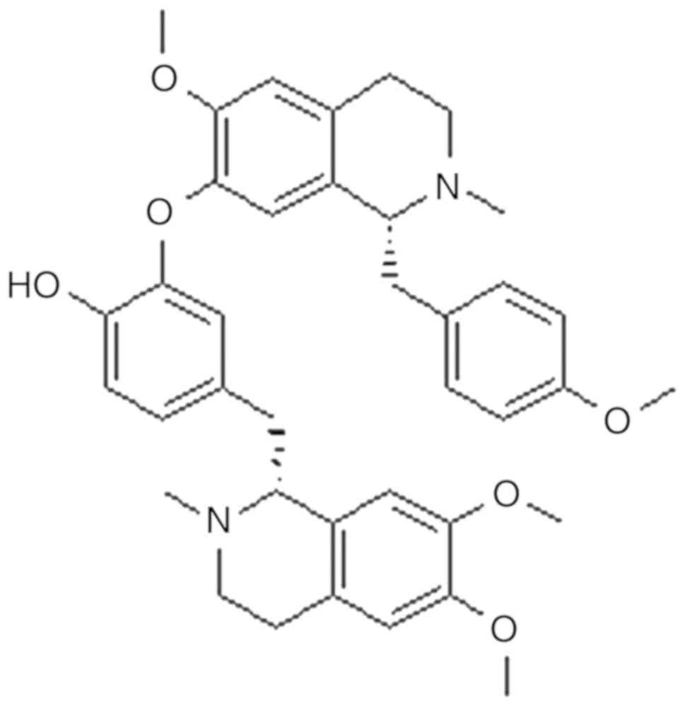
Neferine (Fig. 1) is
one of the major bisbenzylisoquinoline alkaloids, along with
liensinine and isoliensinine, and is derived from the green seed
embryo of the lotus plant (11).
Some of the pharmacological activities of neferine include
antiarrhythmic, antihypertensive (12), sedative (13) and anti-diabetic properties (14). A previous study has demonstrated the
anti-inflammatory and antioxidant actions of neferine (15). Neferine is being increasingly
investigated for its potential clinical applications; however, its
anti-photoaging effects have not yet been elucidated.
The skin is one of the most susceptible organs to
ultraviolet radiation (UVR), which is the main cause of photoaging
(16). Loss of skin tone, roughness,
resilience and dryness, irregular pigmentation and deep wrinkle
formation are the main characteristics of photoaged skin (17). It is well known that UV-B (280-315
nm) and UV-A (315-400 nm) can cause skin photoaging and photodamage
(18). The principal mode of action
by UVR is reactive oxygen species (ROS) generation (19), which is one of the main contributors
to UV-induced photoaging. ROS damage biological macromolecules,
including DNA, carbohydrates, lipids and proteins, and deplete
antioxidants, including superoxide dismutase (SOD) and glutathione
peroxidase (GPx) (20,21). Antioxidants scavenge free radicals
and aid the repair of cellular damage. A variety of antioxidants,
including vitamins C and E, selenium, soy isoflavones and
polyphenolic compounds, have been revealed to attenuate UV-induced
photoaging (22,23).
The aim of the present study was to analyze the
anti-photoaging activity of neferine by creating an in vivo
model of UV irradiation, in order to determine whether neferine
could replenish skin antioxidants including SOD and GPx, and
minimize skin wrinkles and collagen content loss upon UV
exposure.
Materials and methods
Reagents
Neferine was purchased from Pure One Biotechnology
Co., Ltd. SOD (cat. no. A001-1); GPx (cat. no. A005) and
bicinchoninic acid (BCA; cat. no. A045-3) assay kits were purchased
from the Nanjing Jiancheng Bioengineering Institute.
Animals
A total of 72 female Kunming mice (weight, 25-28 g;
age, 6-8 weeks) were purchased from the Experimental Animal Center,
School of Medicine, Xian Jiaotong University. The mice had access
to food and water ad libitum and were maintained in a 12-h
dark/light cycle with a controlled temperature of 24±2˚C and
humidity of 55±10%. All mice were allowed to acclimatize to their
new surroundings for 1 week prior to the experiments. The housing
conditions were maintained constant throughout the entirety of the
experimental study period. All animal experiments were approved and
conducted under the guidance of the International Association for
the Protection of Animal and Experimental Medicine and Laboratory
Animal Ethics Committee of Xian Jiaotong University School of
Medicine (Xian, China).
Preparation of neferine moisture base
cream
Two types of moisture base creams were prepared for
use in the current study.
Moisture base cream with neferine
Neferine (0.1%), ethanol (EtOH; 58.5%), propanediol
(20.0%), water (20%) and hydroxypropyl methyl cellulose (1.5%).
Moisture base cream without
neferine
EtOH (58.5%), propanediol (20.0%), water (20%),
hydroxypropyl methyl cellulose (1.5%).
UV exposure and neferine topical
treatment
UV irradiation was provided by Philips ACTINIC BL
TL-K 40W/10R and Philips TL 20W/01RS bulbs (Philips Lighting),
which were used to provide UV-A (315-400 nm; peak wavelength, 365
nm) and UV-B (280-315 nm; peak wavelength, 311 nm) using an array
of six lamps adjusted at a distance of 30 cm from the skin of the
mice. The minimal erythema dose (MED) was measured using a
Lutron-UV-340a meter (Lutron Electronic Enterprise Co., Ltd.). The
MED for UV-A and UV-B was calculated as 1,200 and 180
mJ/cm2, respectively. This dose was considered to be 1
MED. The dose was then increased from 1 to 4 MED towards week 12 of
the study. This method was applied as previously described by Li
et al (24). The skin on the
dorsal surface (1.5x1.5 inches) of all the mice was shaved three
times a week and depilated twice a week with Veet (Reckitt
Benckiser Group plc) for 12 weeks. Vehicle- and neferine
(0.1%)-treated groups were exposed to UVR five times a week (not on
Saturday or Sunday) for 12 weeks. The mice in the control group
were shaved and depilated twice a week with Veet (Reckitt Benckiser
Group, plc), but received no UV irradiation. As presented in
Table I, mice were divided into
three groups (n=6 mice/group): The neferine (0.1%) and vehicle
(without neferine) groups were topically treated with moisture base
cream with a brush applicator 30 min prior to every UV irradiation
treatment. The control group was left untreated.
 | Table IExperimental design of the study. |
Table I
Experimental design of the study.
| Groups | Shave | UV irradiation | Vehicle | Neferine
(0.1%) |
|---|
| Control | + | - | - | - |
| Vehicle | + | + | + | - |
| Neferine | + | + | + | + |
Evaluation of wrinkled appearance
One day prior to the end of the study period, mice
were anaesthetized and the dorsal skin was photographed using a
digital camera (Canon SX200 IS; Canon, Inc.). The photodamage was
assessed using visual scoring (Table
II), which was performed by two observers who were blinded to
the grouping, which was based on the protocols previously described
by Kong et al (20) and
Bissett et al (25). The skin
wrinkles of each mouse were further evaluated by the capture of
images using a light microscope (magnification, x100; Nikon
SM21500; Nikon Corporation).
| Table IIGrading scale for evaluation of skin
wrinkles. |
Table II
Grading scale for evaluation of skin
wrinkles.
| Grade | Description |
|---|
| 0 | Smooth skin without
any wrinkles; fine striations running down the length of the
body |
| 1 | Fine
striations |
| 2 | A few shallow
wrinkles; disappearance of all fine striations |
| 3 | Shallow wrinkles
across the dorsal skin |
| 4 | Deep and coarse
wrinkles with laxity |
| 5 | Increased wrinkle
depth |
| 6 | Surface marked by
severe wrinkles; development of lesions |
Hematoxylin and eosin (H&E)
staining and epidermal thickening analysis
At the end of experimental study, the mice were
anesthetized and sacrificed, following which the dorsal skin
tissues were collected. The tissues were treated with 10%
phosphate-buffered formaldehyde (pH 7.4) at room temperature for 24
h prior to staining. H&E staining was performed according to
standard protocols (26). Following
deparaffinization with 100% xylene for 3 min and 50:50 xylene/100%
EtOH for 3 min, and rehydration with 100% EtOH for 3 min and 95%
EtOH for 3 min, 4-µm skin sections were stained with hematoxylin
(Gill's IX) for 5 min at room temperature and subsequently rinsed
with water for 5 min. Slides were then treated with 5 dips in acid
alcohol (1% HCl in 70% EtOH) and subsequently rinsed with water.
Following treatment with ammonia (1 ml NH4OH in 1 l
water) and rinsing with water, the sections were stained with Eosin
Y solution for 1 min at room temperature, followed by dehydration
with graded alcohol (95% EtOH for 2 min; 100% EtOH for 2 min) and
clearing in xylene (50:50 xylene/100% EtOH for 2 min; 100% xylene
for 2 min). Stained slides were observed under a light microscope
(magnification, x200 and x400; Nikon DS-Ri1; Nikon Corporation) and
photographs of each specimen were captured at three randomly
selected locations. Epidermal thickness was evaluated by measuring
the distance from the stratum corneum to the basement membrane in
the interfollicular epidermis at five randomly selected positions.
Epidermal thickening was analyzed and quantified using Image J 1.36
analysis software (National Institutes of Health).
Masson's trichrome staining and
collagen content quantification
Mice were anesthetized and sacrificed at the end of
experimental study. Skin tissue samples (4 µm), after
deparaffinization and rehydration, were treated with Bouin's
solution at 56˚C for 15 min. After cooling the slides with water at
18-26˚C for 5 min, samples were stained using working Weigert's
iron hematoxylin solution for 5 min at room temperature.
Subsequently, slides were rinsed in deionized water and stained in
Biebrich scarlet-acid fuchsin for 5 min at room temperature and
rinsed in deionized water for 1 min. Slides were then treated with
working phosphotungstic/phosphomolybdic acid solution for 5 min at
room temperature and placed in Aniline Blue solution for 5 min at
room temperature. Slides were treated with acetic acid (1%) for 2
min at room temperature and dehydrated through a graded alcohol
series (70, 80, 90 and 100%), and cleared in xylene for 5 min.
Slides were observed under a light microscope (magnification, x200
and x400; Nikon DS-Ri1; Nikon Corporation) and each specimen was
photographed at three randomly selected locations. To evaluate the
dermal collagen density, quantification was conducted using Image J
1.36 software (National Institutes of Health) (27,28).
Collagen density was evaluated by measuring the distance from the
papillary layer to the upper layer of subcutaneous tissue at five
randomly selected positions. The relative % of collagen density was
then calculated using the following equation:
Scanning and transmission electron
microscopy
The dorsal skin samples were cut into small pieces
(~2x2x2 mm) and immediately fixed with 2.5% glutaraldehyde and 4%
paraformaldehyde for 2 h at 4˚C, washed with 0.1 M phosphate buffer
(PB) for 30 min at 4˚C, followed by post-fixation with 1% osmium
tetroxide for 2 h at 4˚C and washing 2 or 3 times with 0.1 M PB.
The skin fragments were dehydrated using a graded EtOH series (30%
EtOH in water 10 min, 50% EtOH in water 10 min, 70% EtOH in water
10 min, 90% EtOH in water 10 min and 100% EtOH). The samples were
immersed in a mixture of propylene oxide and Epon 812 overnight at
37˚C followed by polymerization embedding for 48 h at 60˚C. The
tissues were then cut into 1-3-µm sections and stained with Azure
ΙΙ or toluidine blue for 1 min on a 60˚C hotplate to identify and
observe the area of interest. The blocks were sectioned at 50-70 nm
using an LKB Ultratome. The ultra-thin sections were then stained
with uranyl acetate and lead hydroxide at room temperature for 10
min and observed using scanning electron microscopy (Hitachi
TM-1000; Hitachi, Ltd.) by probing the images with focused electron
beams. For transmission electron microscopy, mice dorsal skin
tissue samples were dehydrated through an ethanol series at 37˚C
and then transferred to propylene oxide followed by embedded in
epoxy resin at 60˚C overnight. Samples were observed using
transmission electron microscope (Hitachi H-7650; Hitachi,
Ltd.).
Assays of SOD and GPx activity
Dorsal skin samples (80 mg) were homogenized in
physiological saline at 4˚C and centrifuged at 13,000 x g for 15
min. The collected supernatant was used to measure the total SOD
and GPx activities using SOD and GPx assay kits according to
manufacturers' protocols. The absorbance of SOD and GPx was
measured spectrophotometrically (Multiskan; Thermo Fisher
Scientific, Inc.) at 512 and 405 nm, respectively.
Assay of protein concentration
Total protein concentration was measured according
to the BCA protein assay kit protocol. Briefly, the supernatant
obtained by the abovementioned method was centrifuged at 13,000 x g
for 15 min at 4˚C. The homogenate was then incubated for 30 min at
37˚C and the absorbance of SOD or GPx was measured at 560 nm
spectrophotometrically (Multiskan; Thermo Fisher Scientific,
Inc.).
Statistical analysis
Experimental values were analyzed using GraphPad
Prism 5 (GraphPad Software, Inc.). Analysis of data was carried out
by one-way ANOVA followed by Bonferroni's multiple comparisons
test. All data are expressed as the mean ± standard deviation.
P<0.05 was considered to indicate a statistically significant
difference.
Results
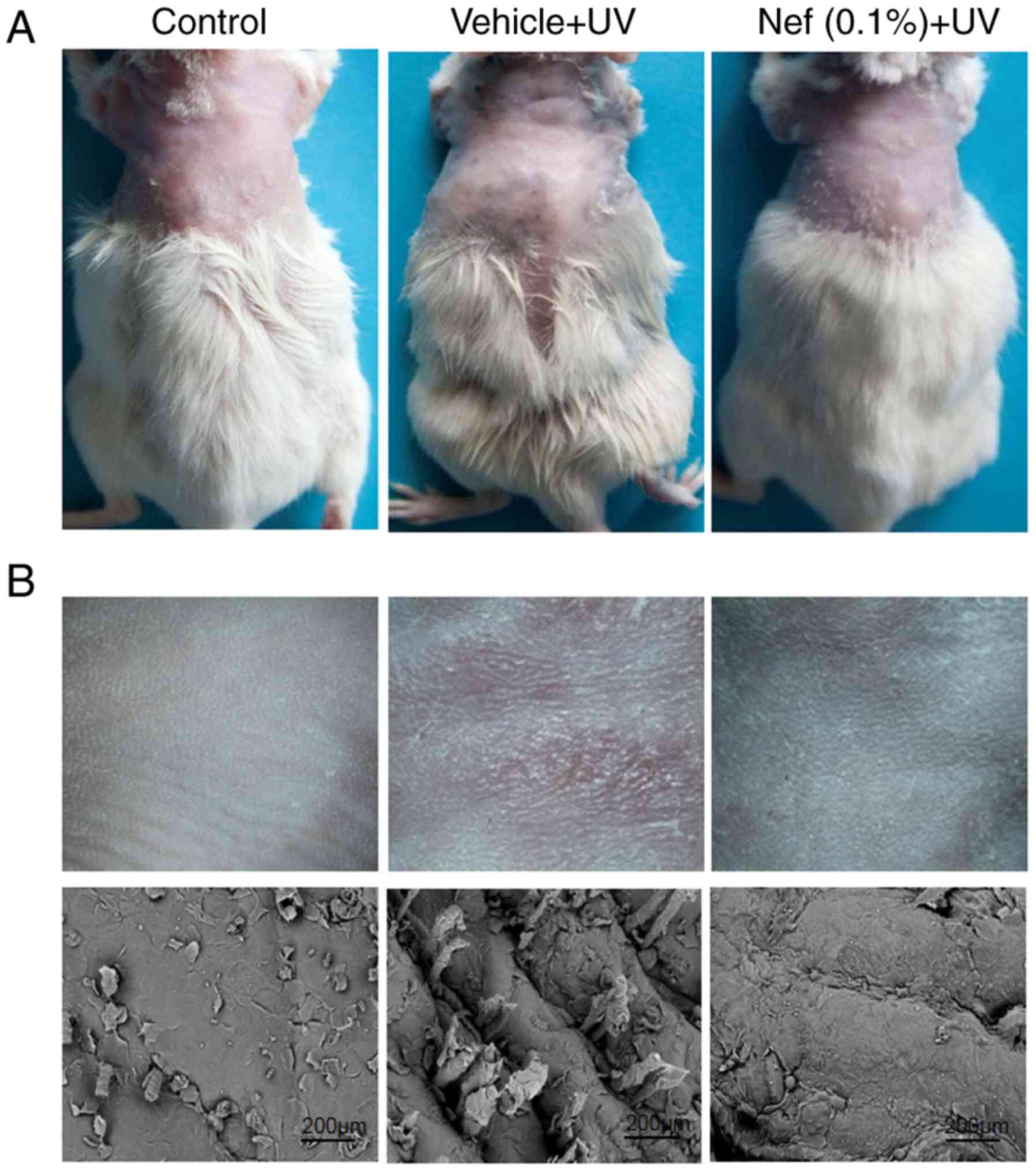
Neferine inhibits wrinkle
formation
During the 12-week duration of the current study,
the control group mice did not exhibit any skin changes, including
deep wrinkles, desquamation, and erythema or scaling. However,
there was a marked difference in the appearance of skin wrinkles
between the UV irradiated vehicle and neferine-treated groups
(Fig. 2). Mice in the
vehicle-treated group displayed dry, scaly skin with the formation
of deep wrinkles (visual scoring, 5). By contrast, the neferine-treated group
exhibited improved skin appearance and significantly diminished
wrinkles (visual scoring, 3; Fig.
2). Scanning electron microscopy analysis revealed deep
wrinkles in the vehicle-treated group, whereas less wrinkling was
observed in mice treated with neferine (Fig. 2B).
Neferine inhibits epidermal
hyperplasia
Epidermal hyperplasia is a histological
characteristic of photoaged skin and can be used as one of the
parameters to reflect the extent of UV-induced skin damage, since
epidermal thickening contributes to skin roughness (29-31).
As presented in Fig. 3, the control
group exhibited a low epidermal thickness compared with the vehicle
and neferine-treated groups (Fig.
3A). The vehicle-treated and Nef + UV groups exhibited
significantly increased epidermal thickening after 12 weeks of UV
irradiation when compared with the control group (P<0.05). In
mice treated with neferine, the epidermal thickness was
significantly reduced when compared with that in the
vehicle-treated group (P<0.05; Fig.
3B).
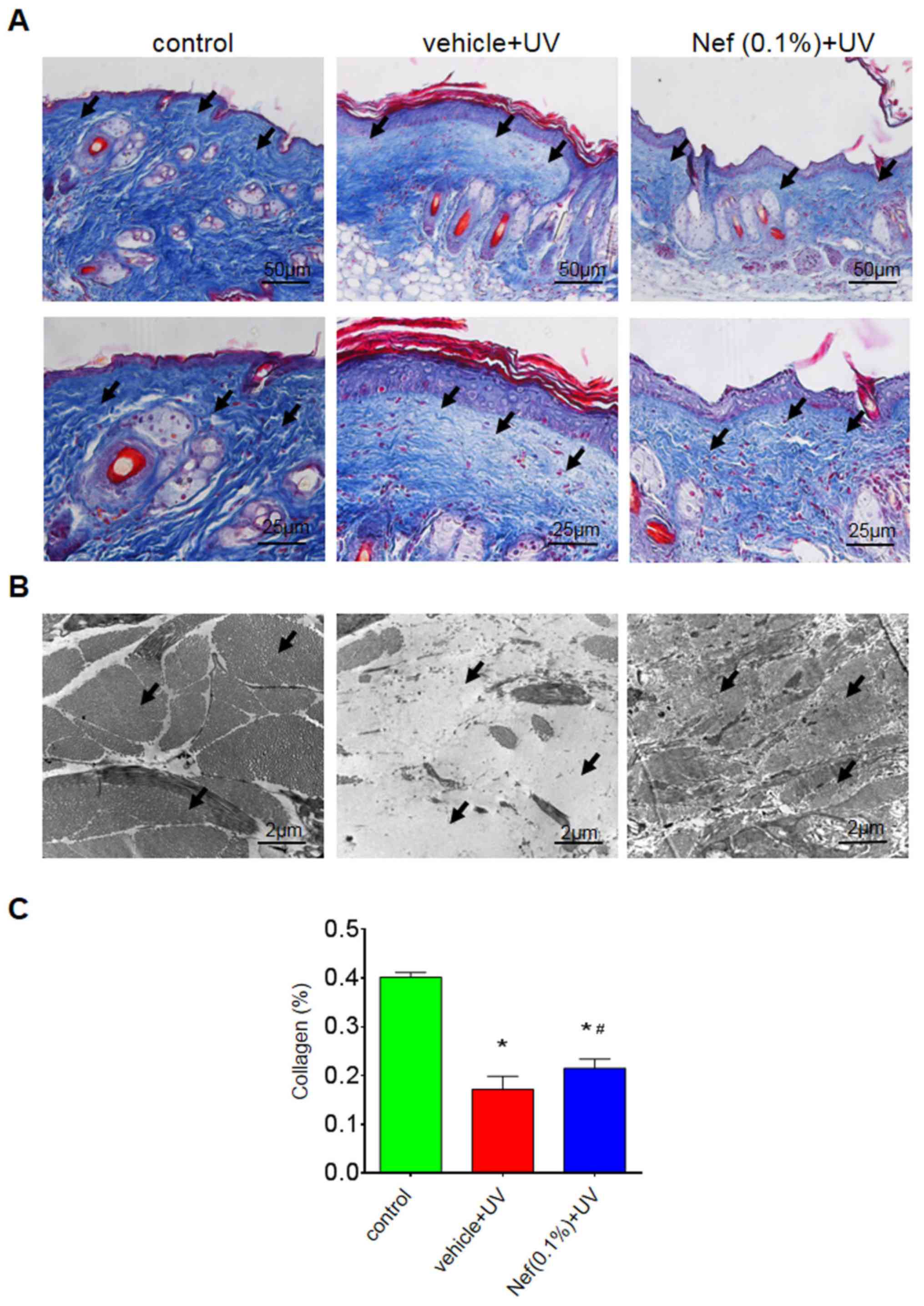
Neferine prevents dermal collagen
loss
The vehicle-treated group exhibited a marked
reduction in dermal collagen density compared with the control
group. However, skin pretreatment with neferine inhibited this
dermal collagen loss (Fig. 4).
Furthermore, the collagen fibers were less dense and somewhat
disorganized in the vehicle-treated group. In the neferine-treated
group, however, the collagen fibers were denser and more organized
(Fig. 4A).
Transmission electron microscopy revealed
disarrayed, broken and a reduced number of collagen fibers in the
vehicle-treated group compared with the control group, in which the
collagen fibers were intact and organized in bundles (Fig. 4B). By contrast, the neferine-treated
group displayed minimal collagen loss and structural alterations
(Fig. 4B). There was a significantly
reduced amount of collagen in the vehicle-treated group compared
with the control (*P<0.05); however, the UV-induced
reduction of collagen was significantly attenuated in the
neferine-treated group (#P<0.05; Fig. 4C).
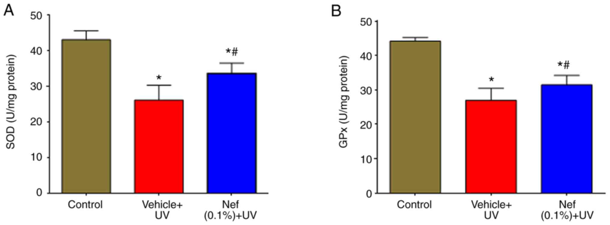
Neferine increases SOD and GPx
levels
The effects of neferine on UV-induced oxidative
stress were examined by measuring SOD and GPx levels. As presented
in Fig. 5, SOD and GPx activities
were significantly reduced in the UV-irradiated vehicle-treated
group compared with the control group (*P<0.05).
However, the decreased activities of SOD (Fig. 5A) and GPx (Fig. 5B) were significantly increased by the
topical application of neferine (#P<0.05).
Discussion
Natural phytochemicals are currently used in
cosmetics to replenish antioxidants, suppress collagen degradation
and reduce the harmful impact of UVR on the skin. The antioxidant
activity of certain natural compounds and their derivatives has
been reported to prevent skin aging (32). Natural compounds contain bioactive
substances, including anthocyanins, isoflavones and catechins,
which may exhibit antioxidant properties (33). It has also been previously suggested
that lotus seed embryos possess antiaging properties (34). Therefore, exploring the therapeutic
potential of the lotus seed embryo-derived compound neferine in
UV-induced photoaging may be beneficial. The present study
demonstrated the anti-photoaging effects of neferine on the skin of
mice that were exposed to UVR.
All cells have an antioxidant defence system. SOD,
GPx and catalase are naturally occurring antioxidants that serve a
crucial role in oxidative balance. SOD converts superoxide anions
into the substantially less toxic H2O2 and
O2. Its activity is an indirect measure of ROS
scavenging, which is essential for the dynamic balance of ROS in
the body. Glutathione (GSH) is the primary intracellular
non-enzymatic antioxidant defense against ROS and serves a central
role in the reduction of oxidative stress. GPx uses GSH as a
hydrogen donor to reduce H2O2 and
hydroperoxides to H2O and O2 (35). The imbalance between antioxidants and
free radicals, which is either caused by a decrease in antioxidants
or an increase in ROS, leads to cell damage and cell death
(36). A study has previously
demonstrated the photo-protective effect of neferine in
UVB-irradiated human epidermal keratinocytes, in which neferine
application increased SOD and GPx activities, and reduced
ROS-induced oxidative stress (37).
In the present study, it is indicated that the topical application
of neferine significantly increased SOD and GPx activities in mice
skin. However, a comparison between known antioxidants and neferine
was not performed in the present study, as the focus was on
neferine antioxidant activity. The antioxidant activity of neferine
may be associated with the presence of the hydroxyl group in its
structure. The photoprotective and anti-photoaging effects of
neferine were demonstrated to be associated with an increase in
antioxidant levels, which was associated with a reduction in
UV-induced oxidative stress and skin photoaging. However, the
effects of neferine and vehicle treatment alone were not evaluated
on non-irradiated mouse skin.
Photoaging is associated with an increase in
epidermal thickness and alterations in connective tissue
organization (38,39). In the present study, the topical
application of neferine reduced these effects, and was revealed to
minimize skin photo damage and the presence of wrinkles.
Atrophy of the dermal connective tissue,
particularly collagen fibers, is also associated with skin
photoaging. It has been demonstrated that collagen is mainly
responsible for maintaining the tensile strength and resistance of
the skin, and comprises up to 75% of the dry weight of the dermis
(29,40). Collagen is one of the major
components of the extracellular matrix and can be directly degraded
by UVR (41), which is an important
early event in the progression of wrinkle formation and skin
sagging (42). A number of studies
have investigated the role of matrix metalloproteinases (MMPs) in
skin aging. UV exposure has been demonstrated to induce MMP-1
expression in the mouse epidermis (39). Neferine has also been revealed to
reduce the expression of MMP-1 in UV-A-irradiated human dermal
fibroblasts (34). In the present
study, collagen density was indicated to be significantly decreased
after 12 weeks of UV irradiation. Observations from the present
study are consistent with the previous study, in which ICR mice
irradiated with UV light exhibited reduced collagen content and
skin photoaging (42). In addition,
neferine application to mice skin reduced UV-induced collagen
density loss in the present study, and it is hypothesized that this
may be associated with a reduction of MMP-1 expression. The
mechanisms underlying the anti-photoaging effects of neferine on UV
irradiation are associated with the inhibition of collagen
degradation and increased collagen synthesis, thereby causing the
reduction of wrinkles and skin sagging.
In conclusion, to the best of our knowledge, the
current study is the first to investigate the anti-photoaging
properties of neferine in vivo. Neferine application reduced
oxidative stress by restoring antioxidant levels, reducing collagen
degradation and the formation of skin wrinkles. However, further
studies are required to evaluate its anti-inflammatory properties
and underlying molecular pathways.
Acknowledgements
The authors would like to thank Dr Quanbao Wang,
Department Histopathology, First Affiliated Hospital of Xi'an
Jiaotong University (Xi'an, China), for his expert assistance in
producing histological slides and Professor Mingxia Chen,
Department of Electron Microscopy, Xi'an Jiaotong University
(Xi'an, China), for scanning and transmission electron
microscopy.
Funding
The present study was supported by grants from the
Shaanxi Provincial Natural Science Foundation research project
(grant no. 2014JQ4151) and the Science and Technology Commission of
Shaanxi Province (grant no. 2018SF-160).
Availability of data and materials
The datasets used and/or analyzed during the present
study are available from the corresponding author on reasonable
request.
Authors' contributions
AbK designed the study, performed experiments and
wrote the manuscript. AmK, HB and ZB contributed to designing the
study and revising the manuscript critically for important
intellectual content. The final version of the manuscript has been
read and approved by all authors.
Ethics approval and consent to
participate
The study was approved by the Committee of
International Association for the Protection of Animal and
Experimental Medicine and Laboratory Animal Ethics Committee of
Xian Jiaotong University, School of Medicine (Xian, China).
Patient consent for publication
Not applicable.
Competing interests
The authors declare that they have no competing
interests.
References
|
1
|
Poornima P, Weng CF and Padma VV:
Neferine, an alkaloid from lotus seed embryo, inhibits human lung
cancer cell growth by MAPK activation and cell cycle arrest.
Biofactors. 40:121–131. 2014.PubMed/NCBI View Article : Google Scholar
|
|
2
|
Mukherjee PK, Das J, Saha K, Giri SN, Pal
M and Saha BP: Antipyretic activity of Nelumbo nucifera
rhizome extract. Indian J Exp Biol. 34:275–276. 1996.PubMed/NCBI
|
|
3
|
Mukherjee PK, Saha K, Balasubramanian R,
Pal M and Saha BP: Studies on psychopharmacological effects of
Nelumbo nucifera gaertn. rhizome extract. J Ethnopharmacol.
54:63–67. 1996.PubMed/NCBI View Article : Google Scholar
|
|
4
|
Mukherjee PK, Saha K, Das J, Pal M and
Saha BP: Studies on the anti-inflammatory activity of rhizomes of
Nelumbo nucifera. Planta Med. 63:367–369. 1997.PubMed/NCBI View Article : Google Scholar
|
|
5
|
Kim T, Kim HJ, Cho SK, Kang WY, Baek H,
Jeon HY, Kim B and Kim D: Nelumbo nucifera extracts as
whitening and anti-wrinkle cosmetic agent. Korean J Chem
Engineering. 28:424–427. 2011.
|
|
6
|
Wu MJ, Wang L, Weng CY and Yen JH:
Antioxidant activity of methanol extract of the lotus leaf
(Nelumbo nucifera Gertn.). Am J Chin Med. 31:687–698.
2003.PubMed/NCBI View Article : Google Scholar
|
|
7
|
Jung HA, Kim JE, Chung HY and Choi JS:
Antioxidant principles of Nelumbo nucifera stamens. Arch
Pharm Res. 26:279–285. 2003.PubMed/NCBI View Article : Google Scholar
|
|
8
|
Cho EJ, Yokozawa T, Rhyu DY, Kim SC,
Shibahara N and Park JC: Study on the inhibitory effects of Korean
medicinal plants and their main compounds on the
1,1-diphenyl-2-picrylhydrazyl radical. Phytomedicine. 10:544–551.
2003.PubMed/NCBI View Article : Google Scholar
|
|
9
|
Kim JH, Kang M, Cho C, Chung HS, Kang CW,
Parvez S and Bae H: Effects of nelumbinis semen on contractile
dysfunction in ischemic and reperfused rat heart. Arch Pharm Res.
29:777–785. 2006.PubMed/NCBI View Article : Google Scholar
|
|
10
|
Rai S, Wahile A, Mukherjee K, Saha BP and
Mukherjee PK: Antioxidant activity of Nelumbo nucifera
(sacred lotus) seeds. J Ethnopharmacol. 104:322–327.
2006.PubMed/NCBI View Article : Google Scholar
|
|
11
|
Liu S, Wang B, Li XZ, Qi LF and Liang YZ:
Preparative separation and purification of liensinine,
isoliensinine and neferine from seed embryo of Nelumbo
nucifera GAERTN using high-speed counter-current
chromatography. J Sep Sci. 32:2476–2481. 2009.PubMed/NCBI View Article : Google Scholar
|
|
12
|
Qian JQ: Cardiovascular pharmacological
effects of bisbenzylisoquinoline alkaloid derivatives. Acta
Pharmacol Sin. 23:1086–1092. 2002.PubMed/NCBI
|
|
13
|
Sugimoto Y, Furutani S, Itoh A, Tanahashi
T, Nakajima H, Oshiro H, Sun S and Yamada J: Effects of extracts
and neferine from the embryo of Nelumbo nucifera seeds on
the central nervous system. Phytomedicine. 15:1117–1124.
2008.PubMed/NCBI View Article : Google Scholar
|
|
14
|
Pan Y, Cai B, Wang K, Wang S, Zhou S, Yu
X, Xu B and Chen L: Neferine enhances insulin sensitivity in
insulin resistant rats. J Ethnopharmacol. 124:98–102.
2009.PubMed/NCBI View Article : Google Scholar
|
|
15
|
Ling ZQ, Xie BJ and Yang EL: Isolation,
characterization, and determination of antioxidative activity of
oligomeric procyanidins from the seedpod of Nelumbo nucifera
Gaertn. J Agric Food Chem. 53:2441–2445. 2005.PubMed/NCBI View Article : Google Scholar
|
|
16
|
Fisher GJ, Wang ZQ, Datta SC, Varani J,
Kang S and Voorhees JJ: Pathophysiology of premature skin aging
induced by ultraviolet light. N Engl J Med. 337:1419–1428.
1997.PubMed/NCBI View Article : Google Scholar
|
|
17
|
Kligman LH and Kligman AM: The nature of
photoaging: Its prevention and repair. Photo Dermatol. 3:215–227.
1986.PubMed/NCBI
|
|
18
|
de Gruijl FR, van Kranen HJ and Mullenders
LH: UV-induced DNA damage, repair, mutations and oncogenic pathways
in skin cancer. J Photochem Photobiol B. 63:19–27. 2001.PubMed/NCBI View Article : Google Scholar
|
|
19
|
Tyrrell RM and Keyse SM: New trends in
photobiology. The interaction of UVA radiation with cultured cells.
J Photochem Photobiol B. 4:349–361. 1990.PubMed/NCBI View Article : Google Scholar
|
|
20
|
Kong SZ, Shi XG, Feng XX, Li WJ, Liu WH,
Chen ZW, Xie JH, Lai XP, Zhang SX, Zhang XJ and Su ZR: Inhibitory
effect of hydroxysafflor yellow a on mouse skin photoaging induced
by ultraviolet irradiation. Rejuvenation Res. 16:404–413.
2013.PubMed/NCBI View Article : Google Scholar
|
|
21
|
Scharffetter-Kochanek K, Brenneisen P,
Wenk J, Herrmann G, Ma W, Kuhr L, Meewes C and Wlaschek M:
Photoaging of the skin from phenotype to mechanisms. Exp Gerontol.
35:307–316. 2000.PubMed/NCBI View Article : Google Scholar
|
|
22
|
Jurkiewicz BA, Bissett DL and Buettner GR:
Effect of topically applied tocopherol on ultraviolet
radiation-mediated free radical damage in skin. J Invest Dermatol.
104:484–488. 1995.PubMed/NCBI View Article : Google Scholar
|
|
23
|
Pinnell SR: Cutaneous photodamage,
oxidative stress, and topical antioxidant protection. J Am Acad
Dermatol. 48:1–19. 2003.PubMed/NCBI View Article : Google Scholar
|
|
24
|
Li Z, Niu X, Xiao S and Ma H: Retinoic
acid ameliorates photoaged skin through RARmediated pathway in
mice. Mol Med Rep. 16:6240–6247. 2017.PubMed/NCBI View Article : Google Scholar
|
|
25
|
Bissett DL, Hannon DP and Orr TV: An
animal model of solar-aged skin: Histological, physical, and
visible changes in UV-irradiated hairless mouse skin. Photochem
Photobiol. 46:367–378. 1987.PubMed/NCBI View Article : Google Scholar
|
|
26
|
Guo Y, Wang L, Ma R, Mu Q, Yu N, Zhang Y,
Tang Y, Li Y, Jiang G, Zhao D, et al: JiangTang XiaoKe granule
attenuates cathepsin K expression and improves IGF-1 expression in
the bone of high fat diet induced KK-Ay diabetic mice. Life Sci.
148:24–30. 2016.PubMed/NCBI View Article : Google Scholar
|
|
27
|
Gaspar LR and Maia Campos PM: Rheological
behavior and the SPF of sunscreens. Int J Pharm. 250:35–44.
2003.PubMed/NCBI View Article : Google Scholar
|
|
28
|
Ouhtit A, Muller HK, Davis DW, Ullrich SE,
McConkey D and Ananthaswamy HN: Temporal events in skin injury and
the early adaptive responses in ultraviolet-irradiated mouse skin.
Am J Pathol. 156:201–207. 2000.PubMed/NCBI View Article : Google Scholar
|
|
29
|
Fujii T, Okuda T, Yasui N, Wakaizumi M,
Ikami T and Ikeda K: Effects of amla extract and collagen peptide
on UVB-induced photoaging in hairless mice. J Funct Foods.
5:451–459. 2013.
|
|
30
|
Kwon OS, Jung SH and Yang BS: Topical
administration of manuka oil prevents UV-B irradiation-induced
cutaneous photoaging in mice. Evid Based Complement Alternat Med.
2013(930857)2013.PubMed/NCBI View Article : Google Scholar
|
|
31
|
Tanaka YT, Tanaka K, Kojima H, Hamada T,
Masutani T, Tsuboi M and Akao Y: Cynaropicrin from Cynara scolymus
L. suppresses photoaging of skin by inhibiting the transcription
activity of nuclear factor-kappa B. Bioorg Med Chem Lett.
23:518–523. 2013.PubMed/NCBI View Article : Google Scholar
|
|
32
|
Moini H, Packer L and Saris NE:
Antioxidant and prooxidant activities of alpha-lipoic acid and
dihydrolipoic acid. Toxicol Appl Pharmacol. 182:84–90.
2002.PubMed/NCBI View Article : Google Scholar
|
|
33
|
Kaur C and Kapoor HC: Anti-oxidant
activity and total phenolic content of some Asian vegetables. Int J
Food Sci Technol. 37:153–161. 2002.
|
|
34
|
Khan A, Bai H, Shu M, Chen M, Khan A and
Bai Z: Antioxidative and antiphotoaging activities of neferine upon
UV-A irradiation in human dermal fibroblasts. Biosci Rep.
38(BSR20181414)2018.PubMed/NCBI View Article : Google Scholar
|
|
35
|
Jo WS, Yang KM, Park HS, Kim GY, Nam BH,
Jeong MH and Choi YJ: Effect of microalgal extracts of tetraselmis
suecica against UVB-induced photoaging in human skin fibroblasts.
Toxicol Res. 28:241–248. 2012.PubMed/NCBI View Article : Google Scholar
|
|
36
|
Sies H and de Groot H: Role of reactive
oxygen species in cell toxicity. Toxicol Lett. 64–65.
1992.PubMed/NCBI View Article : Google Scholar
|
|
37
|
Khan A, Bai H, Liu E, Chen M, Yu C, Wang
R, Khan A and Bai Z: Protective effect of neferine against
UV-B-mediated oxidative damage in human epidermal keratinocytes. J
Dermatolog Treat. 29:733–741. 2018.PubMed/NCBI View Article : Google Scholar
|
|
38
|
Smith JG Jr, Davidson EA, Sams WM Jr and
Clark RD: Alterations in human dermal connective tissue with age
and chronic sun damage. J Invest Dermatol. 39:347–350.
1962.PubMed/NCBI View Article : Google Scholar
|
|
39
|
Uitto J, Fazio MJ and Olsen DR: Molecular
mechanisms of cutaneous aging. Age-associated connective tissue
alterations in the dermis. J Am Acad Dermatol. 21:614–622.
1989.PubMed/NCBI
|
|
40
|
Hou H, Li B, Zhao X, Zhuang Y, Ren G, Yan
M, Cai Y, Zhang X and Chen L: The effect of pacific cod (Gadus
macrocephalus) skin gelatin polypeptides on UV
radiation-induced skin photoaging in ICR mice. Food Chem.
115:945–950. 2009.
|
|
41
|
Chiang HM, Lin TJ, Chiu CY, Chang CW, Hsu
KC, Fan PC and Wen KC: Coffea arabica extract and its constituents
prevent photoaging by suppressing MMPs expression and MAP kinase
pathway. Food Chem Toxicol. 49:309–318. 2011.PubMed/NCBI View Article : Google Scholar
|
|
42
|
Fan J, Zhuang Y and Li B: Effects of
collagen and collagen hydrolysate from jellyfish umbrella on
histological and immunity changes of mice photoaging. Nutrients.
5:223–233. 2013.PubMed/NCBI View Article : Google Scholar
|