Introduction
In humans the brain is the most sensitive organ to
oxygen, the cerebral tissue hypoxia or ischemia causes local damage
to cerebral tissues, and their function fails to be restored,
further leading to more severe cerebral dysfunction known as
cerebral ischemia-reperfusion injury (CIRI) (1-3).
CIRI is a complex physiological and pathological process, but it
has complicated and various pathogeneses mainly involving
inflammatory responses, Ca2+ overload, excessive
accumulation of free radicals, and excitatory amino acid toxicity
(4,5). Currently, there is no pathogenesis that
can elucidate the cause of CIRI. However, increasing number of
literature has found that neuronal apoptosis is closely associated
with the development and progression of CIRI, and hence, this
indicates that neuronal apoptosis in the brain could be a potential
treatment regimen for CIRI.
Rall and Sutherland first discovered the cyclic
adenosine monophosphate (cAMP)/protein kinase A (PKA) signaling
pathway in the 1990s, and they found through experiments that cAMP,
also known as adenosine 3',5'-cyclic monophosphate, is condensed
from an adenosine triphosphate by removing two phosphates (6). PKA is one of the crucial downstream
target genes regulated by cAMP and the most thoroughly researched
protein kinase as well, and it exerts varying physiological
functions in different tissues (7,8). Large
bodies of literature have proved that neuronal apoptosis modulated
by the cAMP/PKA signaling pathway, a vital neuronal transduction
pathway, is of great significance for organisms to exert cognitive,
learning and memory functions, and that this pathway has received
growing attention from researchers.
Lidocaine, a local anesthetic, can take effect only
at 1-3 min after administration, with the anesthetic effect lasting
for 1-3 h, and clinically, it is also used to treat arrhythmia and
serves as the preferred drug for ventricular tachycardia and tremor
(9,10). The incidence rate of adverse
reactions to lidocaine is ~6%, with the major nervous,
cardiovascular and allergic manifestations. Lidocaine has an exact
neuroprotective effect, but there are few studies on whether it can
exert the protective effect on the CIRI model rats through
regulating the cAMP/PKA signaling pathway.
Therefore, the rat CIRI model was established using
the suture-occluded method in this experiment to study the
influence of lidocaine on neuronal apoptosis in the brain of rats
and explore its mechanism of action, thereby providing an
experimental basis for the clinical treatment of CIRI with
lidocaine.
Materials and methods
Reagents
Lidocaine was purchased from Sigma-Aldrich; Merck
KGaA, enzyme-linked immunosorbent assay (ELISA) kit from
eBioscience, TRIzol solution from Shanghai Shiyi Biotech Inc.,
terminal deoxynucleotidyl transferase dUTP nick end labeling
(TUNEL) kit from Nanjing KeyGen Biotech Co., Ltd., bicinchoninic
acid (BCA) protein assay kit from Thermo Fisher Scientific, Inc.,
polymerase chain reaction (PCR) SuperMix kit from Beijing TransGen
Biotech Co., Ltd., cAMP, PKA and β-actin primers from Invitrogen;
Thermo Fisher Scientific, Inc. and rabbit anti-cAMP, PKA and
β-actin primary antibodies and horseradish peroxidase (HRP)-labeled
secondary antibodies from Beijing Biosynthesis Biotechnology Co.,
Ltd.
Instruments
Fluorescence inverted microscope was provided by
Olympus, low-temperature refrigerator by Qingdao Haier Co., Ltd.,
fluorescence quantitative PCR instrument by Applied Biosystems,
thermostatic water bath by Grant, low-temperature centrifuge by
Shenzhen Anke High-tech Co., Ltd., and western blotting
electrophoresis apparatus and transfer instrument by Bio-Rad
Laboratories.
Rats
A total of 30 healthy male Sprague-Dawley rats
weighing 200-240 g, aged 12-weeks were purchased from the
Laboratory Animal Center of Capital Medical University (Beijing,
China), and they were fed with standard granulated rat food in
clean environment and allowed to move freely. This study was
approved by the Animal Ethics Committee of Capital Medical
University Animal Center.
Methods
Preparation of rat CIRI models
In this experiment, the rat models were prepared
with reference to Longa's suture-occluded method, and the rats in
model group fasted for water and food 12 h preoperatively. After
being anesthetized using 10% chloral hydrate via intraperitoneal
injection at a dose of 300-350 mg/kg, the rats were fixed in the
supine position on an operating table, and the neck was cut open in
the middle to expose the common carotid artery, external carotid
artery and internal carotid artery. No rat exhibited signs of
peritonitis after the administration of 10% chloral hydrate. Then
the proximal ends of the external and common carotid arteries were
ligated, and a V-shaped incision was made at ~0.2 mm from the
common carotid artery. Subsequently, a nylon fishing line was
inserted to cause partial occlusion of the common carotid artery in
the brain of rats, with ~8 mm of the thread left outside, followed
by suture of the wound. After ischemia for 2 h, the thread was
pulled out. After modeling, the rats in lidocaine group were
intraperitoneally injected with 10 mg/kg lidocaine, while those in
control group with an equal dose of normal saline. Finally, the
neurological function score of rats in each group was evaluated and
recorded 24 h later based on Zea Longa's scoring criteria. Cervical
dislocation (after being anesthetized using 10% chloral hydrate at
a dose of 300 mg/kg) was used as the method of euthanasia. No rat
exhibited signs of peritonitis after the administration of 10%
chloral hydrate. We verified that the experimental animals (weighed
200-240 g) were dead by observing the breathing and heartbeat.
Determination of levels of serum
B-cell lymphoma-2 (Bcl-2) and Bcl-2 associated X protein (Bax) in
rats via ELISA
Serum was collected from each group of rats, let
stand for 1 h, and centrifuged at 2,500 x g at 4˚C for 10 min, and
the supernatant was aspirated. Each well was added with the
standard to plot the standard curves, and then with 30 µl of
buffer, 20 µl each of standard and sample, separately, and 50 µl of
antibody solution. The fluid was removed, and the resulting sample
and standard were cleaned by washing 3 times, adding 100 µl of
enzyme solution, and then reacted for 30 min, washed again, and
reacted with 100 µl of substrate solution for 15 min. The reaction
was terminated by adding 100 µl of stop solution. Finally, the
absorbance was measured using a microplate reader to calculate the
content of Bcl-2 and Bax.
Evaluation of neuronal apoptosis in
the brain of rats via TUNEL staining
The cerebral tissues of rats were embedded in
paraffin and sectioned, and the sections were transparentized using
xylene, soaked in ethanol solution at descending concentrations
each for 1 min, fixed in 4% paraformaldehyde, penetrated using 0.2%
Triton solution for 15 min, added with TUNEL staining solution,
incubated in the dark for 30 min, and added with antifade mounting
medium. Finally, the staining was observed under a fluorescence
microscope.
Measurement of messenger ribonucleic
acid (mRNA) levels in the cerebral tissues of rats via reverse
transcription (RT)-PCR
The cerebral tissues of rats in each group were
harvested, lysed with 1 ml of TRIzol solution and centrifuged at
10,500 x g at 4˚C for 10 min, and the supernatant was obtained.
According to the instructions of RT-PCR, complementary deoxyribose
nucleic acid (cDNA) was synthesized through RT, and then, PCR
amplification was performed, with the information of added primers
is shown in Table I. Subsequently,
3% gel electrophoresis was carried out. Finally, the bands were
observed under a gel imager.
 | Table IPrimer sequences of cAMP and PKA. |
Table I
Primer sequences of cAMP and PKA.
| Gene name | Sequence |
|---|
| cAMP |
5'-AGGTCCTCAGCTACAAGGAAG-3' |
|
5'-TCTTGAAGTCACAATCCTCTGGT-3' |
| PKA |
5'-CTAAAGCAGATCGAGCACACTC-3' |
|
5'-GCCACCAGCTACATACTCCA-3' |
| β-actin |
5'-CTCCATCCTGGCCTCGCTGT-3' |
|
5'-GCTGTCACCTTCACCGTTCC-3' |
Determination of cAMP and PKA protein
expression levels in cerebral tissues of rats using western
blotting
The cerebral tissues of rats were lysed using RIPA
lysis buffer containing protease inhibitor, and the concentration
of proteins was determined using BCA kit. The prepared samples were
loaded separately for sodium dodecyl sulphate-polyacrylamide gel
electrophoresis (SDS-PAGE) to separate proteins, and then the
proteins were transferred onto a polyvinylidene fluoride (PVDF)
membranes (Millipore), and blocked in 5% skim milk powder for 1 h,
followed by incubation with the corresponding rabbit anti-cAMP, PKA
and β-actin primary antibodies at 4˚C overnight. Subsequently, the
HRP-labeled secondary antibodies were added, and the bands were
detected using the chemiluminescence method. Finally, the optical
density was analyzed using ImageJ software (NIH).
Statistical analysis
Statistical Product and Service Solutions (SPSS)
20.0 software (IBM, Corp.) was used for statistical analysis.
Measurement data were expressed as mean ± standard deviation, and
two-sample t-test was performed for means. P<0.05 indicates that
the difference is statistically significant.
Results
The successful rat CIRI model
The rats in control group moved normally, and
compared with those in control group, the rats in model group
toppled rightward and circled to the right, with a higher
neurological function score. In comparison with those in model
group, few rats in lidocaine group circled to the right, with an
obvious improvement in behavioral symptoms and a substantial
decline in the neurological function score (Table II).
| Table IINeurobehavioral score of rats. |
Table II
Neurobehavioral score of rats.
| Group | Score |
|---|
| Control | 0.10±0.01 |
| Model |
3.12±0.25a |
| Lidocaine |
1.84±0.48b |
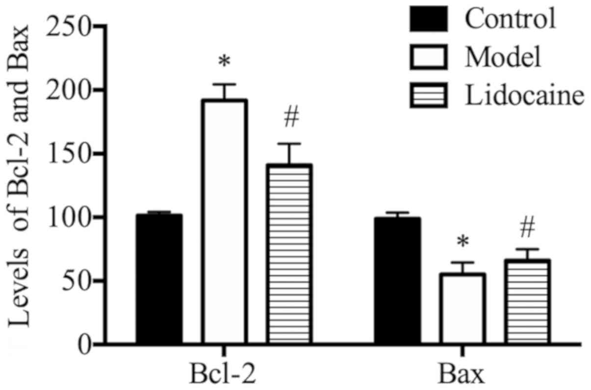
Lidocaine decreases the level of
Bcl-2, but increases that of Bax in the serum of CIRI rats
According to the ELISA results (Fig. 1), the rats in model group had a
higher level of Bcl-2, but an obviously lower level of Bax in the
serum than those in the control group (P<0.05), and compared
with those in the model group, the level of Bcl-2 was lower, but
that of Bax was evidently increased in the serum of rats in
lidocaine group (P<0.05).
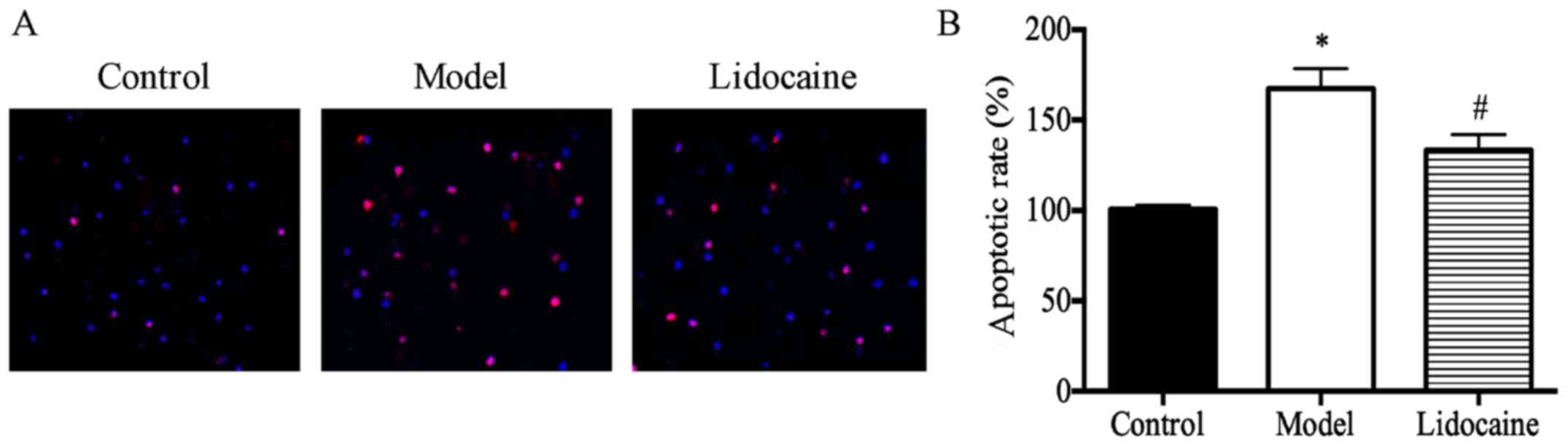
Lidocaine inhibits neuronal apoptosis
in the brain of CIRI rats
It was found through the analysis of TUNEL staining
results (Fig. 2A) that the rate of
neuronal apoptosis in the brain of rats in the model group was
substantially higher than that in the control group (P<0.05),
and that the rate in lidocaine group was obviously lower than that
in the model group (P<0.05) (Fig.
2B), suggesting that lidocaine can inhibit the neuronal
apoptosis in the brain of CIRI rats.
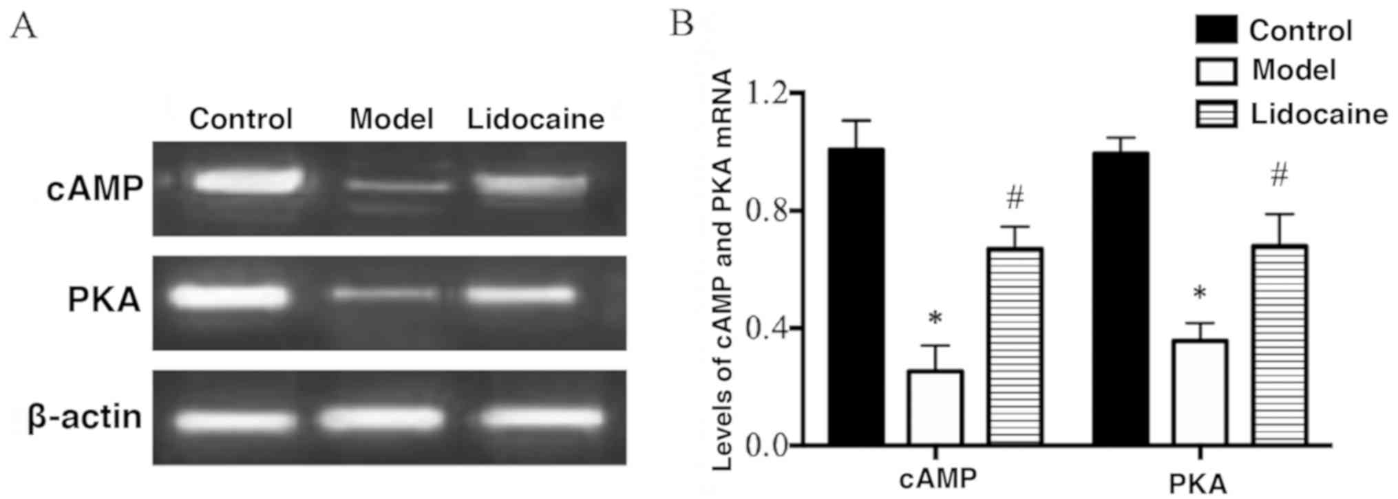
Lidocaine increases the mRNA levels of
cAMP and PKA in the cerebral tissues of CIRI rats
According to the results of RT-PCR (Fig. 3A), the mRNA levels of cAMP and PKA in
the cerebral tissues of rats in the model group were decreased
markedly compared with those in the control group (P<0.05), and
their mRNA levels in lidocaine group were remarkably increased in
comparison with those in the model group (P<0.05) (Fig. 3B), implying that lidocaine promotes
the mRNA expression of cAMP and PKA in the brain of CIRI rats.
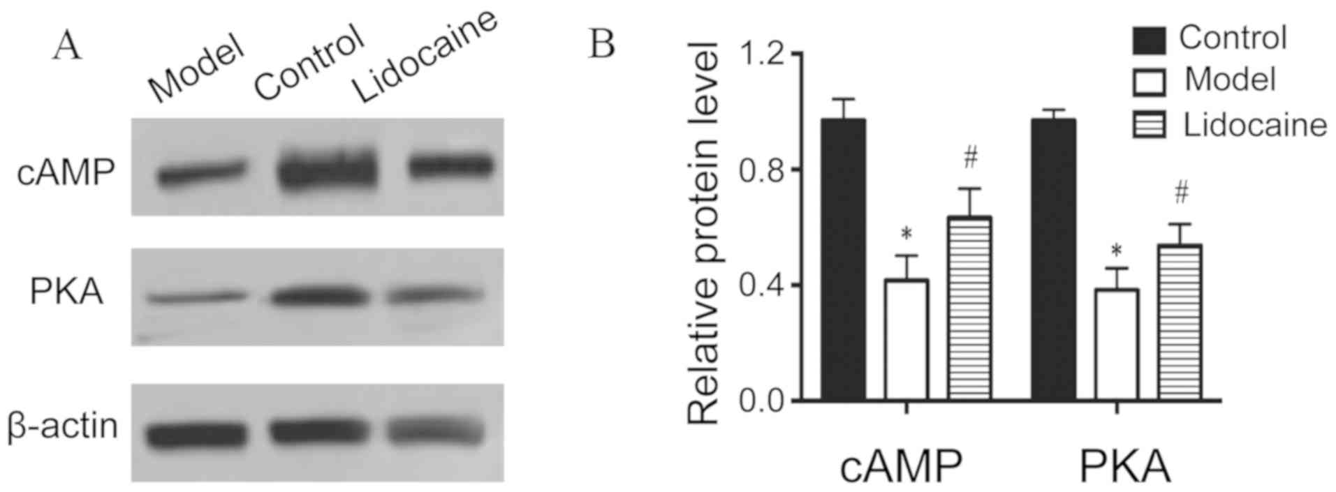
Lidocaine elevates protein levels of
cAMP and PKA in the cerebral tissues of CIRI rats
Based on the results of western blotting (Fig. 4A), the protein levels of cAMP and PKA
in the cerebral tissues of rats in the model group declined notably
in comparison with those in the control group (P<0.05), and the
protein levels in lidocaine group increased distinctly compared
with those in the model group (P<0.05) (Fig. 4B), illustrating that lidocaine
accelerates the protein expression of cAMP and PKA in the brain of
CIRI rats.
Discussion
CIRI is an ischemic cerebrovascular disease that
accounts for ~80-90% of the total cases, and its disability and
mortality rates are extremely high, seriously endangering human
health and posing heavy burden on the society and family (11). In cerebral ischemia, brain neurons
have energy metabolism disorders and acidosis, so that the
homeostasis in local cerebral tissues is damaged, and after
restoration of brain blood supply, cerebral function cannot return
to the original state, with progressive impairment (12,13).
CIRI is a complex pathological process, and a study found that
neuronal apoptosis in the brain is an important cause of
reperfusion injury (14). Therefore,
how to protect brain neuronal cells from IRI is a hotspot of
research.
Lidocaine (N-diethylaminoacetyl-2,6-dimethylaniline
and molecular formula of
C14H22N2O), is a common drug
clinically used for anesthesia (15). Increasing number of studies have
discovered that lidocaine can resist arrhythmia, prevent excessive
inflammatory responses, alleviate acute lung injury, inhibit
premature ejaculation and protect the brain (16,17).
Modern pharmacology of lidocaine, especially its protective effect
on cerebral tissues, has attracted increasingly extensive attention
from researchers. A previous study reported that lidocaine can
block the Na-K channel, and reduce the concentrations of
intracellular Na and K, thereby protecting the hypoxic neurons
(18). However, whether lidocaine
can be used to treat CIRI remains unclear, and its mechanism is
rarely reported.
Hence, in the present study, the rat CIRI model was
first prepared by the suture-occluded method, and 24 h later, the
neurological function score of rats was statistically analyzed in
each group. According to the statistics, the rats in the model
group had an obviously raised neurological function score and the
symptom of circling to the right, which were greatly decreased and
alleviated after treatment with lidocaine. Then the expression of
pro-apoptotic and anti-apoptotic factors in the serum of rats were
detected using ELISA, and as shown in Fig. 1, compared with those in the model
group, the expression of pro-apoptotic factor Bax was substantially
promoted by lidocaine, and that of anti-apoptotic factor Bcl-2 was
inhibited. Subsequently, neuronal apoptosis in the brain of rats
was detected via TUNEL staining in each group, and the results
showed that lidocaine considerably suppressed the neuronal
apoptosis in the brain. The regulatory mechanism of lidocaine was
further explored, as it was known through literature that the
cAMP/PKA signaling pathway plays a vital role in inhibiting
neuronal apoptosis. Once cells are stimulated externally, the
signaling molecules bind to the receptors on the surface of cell
membranes to form complexes, and then activate adenylate cyclases.
Ultimately, cAMP is generated, and it enters the nucleus and
directly activates RNA polymerases to promote the mRNA
transcription of the target genes (19,20).
Therefore, the mRNA and protein levels of cAMP and PKA in the
cerebral tissues of rats were determined using RT-PCR and western
blotting, in this study, and it was revealed that compared with the
model group, lidocaine group exhibited obviously raised mRNA and
protein levels of cAMP and PKA in the cerebral tissues of rats
(Figs. 3 and 4), suggesting that the mechanism by which
lidocaine protects the cerebral tissues of CIRI rats may be related
to the activation of the cAMP/PKA signaling pathway.
In conclusion, the results of this study demonstrate
that lidocaine substantially improves the neurological function
injury and promote the repair of neurological function in CIRI
rats, and that it can also inhibit the neuronal apoptosis in the
brain of rats by the mechanism of action that is probably
associated with the activation of the cAMP/PKA signaling pathway,
providing a novel experimental basis for the treatment of CIRI with
lidocaine.
Acknowledgements
Not applicable.
Funding
This study was supported by Beijing University of
Chinese Medicine Scientific Research Subject
(2016-JYB-JSMS-038).
Availability of data and materials
All data generated or analyzed during this study are
included in this published article.
Authors' contributions
YL, JZh, GL and AW designed the study and performed
the experiments, YL and JZa established the animal models, JZh and
FZ collected the data, GL and AW analyzed the data, YL, JZh, GL and
AW prepared the manuscript. All authors read and approved the final
manuscript.
Ethics approval and consent to
participate
This study was approved by the Animal Ethics
Committee of Capital Medical University Animal Center (Beijing,
China).
Patient consent for publication
Not applicable.
Competing interests
The authors declare they have no competing
interests.
References
|
1
|
Yang Z, Weian C, Susu H and Hanmin W:
Protective effects of mangiferin on cerebral ischemia-reperfusion
injury and its mechanisms. Eur J Pharmacol. 771:145–151.
2016.PubMed/NCBI View Article : Google Scholar
|
|
2
|
Wu XJ, Sun XH, Wang SW, Chen JL, Bi YH and
Jiang DX: Mifepristone alleviates cerebral ischemia-reperfusion
injury in rats by stimulating PPARγ. Eur Rev Med Pharmacol Sci.
22:5688–5696. 2018.PubMed/NCBI View Article : Google Scholar
|
|
3
|
Deng H, Zuo X, Zhang J, Liu X, Liu L, Xu
Q, Wu Z and Ji A: A lipoic acid protects against cerebral
ischemia/reperfusion-induced injury in rats. Mol Med Rep.
11:3659–3665. 2015.PubMed/NCBI View Article : Google Scholar
|
|
4
|
Zhang Y, Qiao L, Xu W, Wang X, Li H, Xu W,
Chu K and Lin Y: Paeoniflorin attenuates cerebral ischemia-induced
injury by regulating Ca(2+)/CaMKII/CREB signaling pathway.
Molecules. 22(359)2017.PubMed/NCBI View Article : Google Scholar
|
|
5
|
Yu W, Gao D, Jin W, Liu S and Qi S:
Propofol prevents oxidative stress by decreasing the ischemic
accumulation of succinate in focal cerebral ischemia-reperfusion
injury. Neurochem Res. 43:420–429. 2018.PubMed/NCBI View Article : Google Scholar
|
|
6
|
Forman MB, Gillespie DG, Cheng D and
Jackson EK: A novel adenosine precursor 2',3'-cyclic adenosine
monophosphate inhibits formation of post-surgical adhesions. Dig
Dis Sci. 59:2118–2125. 2014.PubMed/NCBI View Article : Google Scholar
|
|
7
|
Ould Amer Y and Hebert-Chatelain E:
Mitochondrial cAMP-PKA signaling: What do we really know? Biochim
Biophys Acta Bioenerg. 1859:868–877. 2018.PubMed/NCBI View Article : Google Scholar
|
|
8
|
Yang L: Neuronal cAMP/PKA signaling and
energy homeostasis. Adv Exp Med Biol. 1090:31–48. 2018.PubMed/NCBI View Article : Google Scholar
|
|
9
|
Berk T and Silberstein SD: The use and
method of action of intravenous lidocaine and its metabolite in
headache disorders. Headache. 58:783–789. 2018.PubMed/NCBI View Article : Google Scholar
|
|
10
|
Lancaster RJ, Wren K, Hudson A, Leavitt K,
Albala M and Tischaefer D: Intravenous lidocaine for chronic
neuropathic pain a systematic review addressing nursing care. Pain
Manag Nurs: Jul 30, 2019 (Epub ahead of print). doi:
10.1016/j.pmn.2019.06.008.
|
|
11
|
Zuo G, Zhang D, Mu R, Shen H, Li X, Wang
Z, Li H and Chen G: Resolvin D2 protects against cerebral
ischemia/reperfusion injury in rats. Mol Brain.
11(9)2018.PubMed/NCBI View Article : Google Scholar
|
|
12
|
Liao SL, Lin YW and Hsieh CL: Neuronal
regeneration after electroacupuncture treatment in
ischemia-reperfusion-injured cerebral infarction rats. Biomed Res
Int. 2017(3178014)2017.PubMed/NCBI View Article : Google Scholar
|
|
13
|
Shao X, Bao W, Hong X, Jiang H and Yu Z:
Identification and functional analysis of differentially expressed
genes associated with cerebral ischemia/reperfusion injury through
bioinformatics methods. Mol Med Rep. 18:1513–1523. 2018.PubMed/NCBI View Article : Google Scholar
|
|
14
|
Yaidikar L, Byna B and Thakur SR:
Neuroprotective effect of punicalagin against cerebral ischemia
reperfusion-induced oxidative brain injury in rats. J Stroke
Cerebrovasc Dis. 23:2869–2878. 2014.PubMed/NCBI View Article : Google Scholar
|
|
15
|
Dunn LK and Durieux ME: Perioperative use
of intravenous lidocaine. Anesthesiology. 126:729–737.
2017.PubMed/NCBI View Article : Google Scholar
|
|
16
|
Kirk LM, Brown SD, Luu Y, Ogle A, Huffman
J and Lewis PO: Beyond-use dating of lidocaine alone and in two
‘magic mouthwash’ preparations. Am J Health Syst Pharm.
74:e202–e210. 2017.PubMed/NCBI View Article : Google Scholar
|
|
17
|
Seah DS, Herschtal A, Tran H, Thakerar A
and Fullerton S: Subcutaneous lidocaine infusion for pain in
patients with cancer. J Palliat Med. 20:667–671. 2017.PubMed/NCBI View Article : Google Scholar
|
|
18
|
Arsyad A and Dobson GP: Lidocaine
relaxation in isolated rat aortic rings is enhanced by endothelial
removal: Possible role of Kv, KATP channels and A2a receptor
crosstalk. BMC Anesthesiol. 16(121)2016.PubMed/NCBI View Article : Google Scholar
|
|
19
|
Deb DK, Bao R and Li YC: Critical role of
the cAMP-PKA pathway in hyperglycemia-induced epigenetic activation
of fibrogenic program in the kidney. FASEB J. 31:2065–2075.
2017.PubMed/NCBI View Article : Google Scholar
|
|
20
|
Hu Y, Pan S and Zhang HT: Interaction of
Cdk5 and cAMP/PKA signaling in the mediation of neuropsychiatric
and neurodegenerative diseases. Adv Neurobiol. 17:45–61.
2017.PubMed/NCBI View Article : Google Scholar
|