Introduction
Fetal consequences of defective placentation with
poor vascular flow secondary to abnormal physiological
transformation of the spiral arteries are intrauterine growth
restriction, oligohydramnios, abruptio placentae, and adverse fetal
biophysical score. In this context, the fetuses resulting from
pre-eclampsia pregnancies have an increased risk of preterm birth
and unfavorable perinatal and neonatal prognosis.
In order to reduce these complications, expectation
management can be used, when the situation allows. The conditions
that impose emergency therapeutic behavior are divided into
maternal and fetal conditions. The maternal conditions include high
blood pressure values resistant to antihypertensive treatment
(>160/110 mmHg); persistent, treatment-resistant headache;
epigastralgia or pain in the right shoulder resistant to anti-algic
treatment; visual disorders, motor deficits or sensory disorders;
stroke; myocardial infarction; HELLP syndrome; newly developed
renal failure or worsening of renal function; pulmonary edema;
eclampsia; and suspected abruptio placentae or vaginal bleeding in
the context of the placenta previa (1-3).
Fetal conditions that require emergency therapeutic
action include a biophysical score of 4 or less; intrauterine fetal
death; minimal chances of fetal survival in the context of fetal
malformation incompatible with life or extreme prematurity; changes
in Doppler velocimetry with inverted end-diastolic flow of the
umbilical artery (4).
The decision to implement therapeutic intervention
is made after the complete clinical and paraclinical evaluation and
the determination of the risk/benefit ratio, both maternal and
fetal. More specifically, the biological evaluation should be
performed prior to obstetrical decision and should include:
Hemoleucogram; biochemical evaluation of renal function, liver
function, and markers of hemolysis; and urinary test for evaluation
of proteinuria. The fetal evaluation is based on the complete
obstetrical ultrasound examination, with assessment of fetal growth
and weight, respectively, as well as assessment of the volume of
the amniotic fluid and the fetal biophysical score along with the
Doppler velocimetry of the umbilical arteries, the mean cerebral
artery and the cerebroplacental ratio (5). Both prematurity and intrauterine growth
restriction are the fetal complications most often associated with
preeclampsia. Intrauterine growth restriction is a marker of fetal
distress and an important risk factor for fetal intrauterine
degradation, and the onset of complications of prematurity further
contribute to a less favorable prognosis (6).
The aim of the present study was to evaluate the
neonatal prognosis of preterm births with and without growth
restriction and term births with growth restriction in order to
improve decisional accuracy regarding the termination of
pregnancy.
Patients and methods
The purpose of this study was to comparatively
evaluate the neonatal evolution and the rate of short-term neonatal
complications of the preterm infants with normal weight for the
gestational age and of the preterm infants with low weight for the
gestational age, as well as of the term infants small for
gestational age (SGA). We carried out a retrospective study using
the database of the neonates of the Neonatology Clinic of the
Emergency University Hospital of Bucharest with a third degree
maternity ward, for a period of 3 years. The cases of preterm birth
were selected according to the World Health Organization (WHO)
definition of birth before 37 full weeks of gestation. Only live
newborn cases were selected and analyzed. The cases of premature
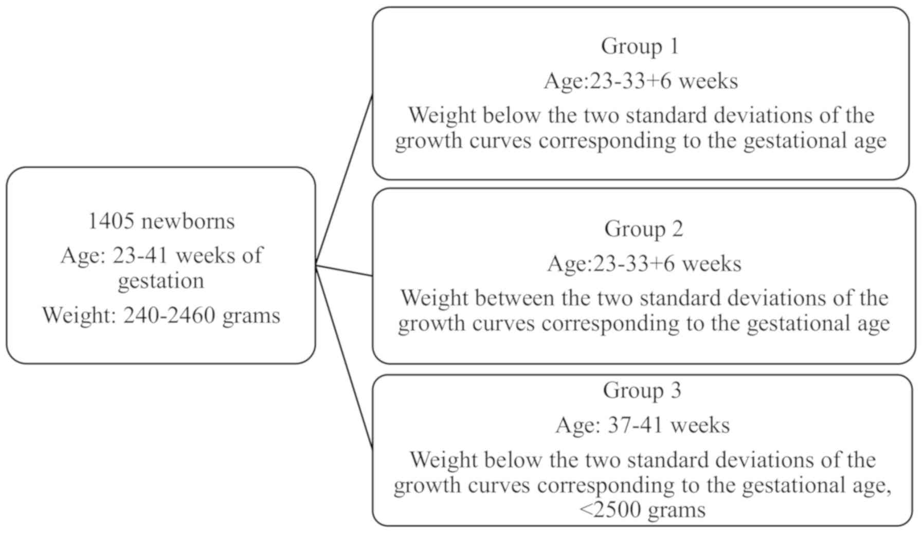
newborns were classified in two categories according to the birth
weight, namely with appropriate weight for the gestational age,
named group 1, including 78 cases with low weight according to the
gestational age corresponding to the international growth curves,
named group 2, including 1,121 cases. Therefore, the low weight
cases selected were those in which the weight at birth was below
the two standard deviations of the growth curves corresponding to
the gestational age. There were also cases of babies with low birth
weight born at term who were selected for the purpose of
comparative analysis of their neonatal prognosis (group 3 including
206 cases). The parameters analyzed were gestational weight and age
as directional criteria, fetal sex, mode of birth (cesarean or
spontaneous birth), and Apgar index at 1 min as a marker of
immediate postnatal fetal status. The obstetric features analyzed
included fetal presentation, the spectrum of hypertensive disorders
of pregnancy, fetal malformations, nuchal cord and true umbilical
cord knot. The immediate neonatal complications and neonatal
markers analyzed were: Cardiovascular arrest, acute respiratory
failure, ulcer-necrotic enterocolitis, hypoxia, respiratory
distress, cerebral edema, intraventricular hemorrhage, cerebral
hemorrhage, pulmonary hemorrhage, patent ductus arteriosus,
neonatal hypoglycemia, retinopathy, anemia, hemorrhagic disease,
disseminated vascular coagulation, hyaline membrane disease,
neonatal sepsis, need for neonatal intensive care and death. A
total of 1,405 subjects were analyzed and were divided into 3 study
groups: group 1 representing live, preterm infants with low weight
for gestational age; group 2 representing living newborns,
premature but with weight corresponding to the gestational age; and
group representing term newborns with low birth weight. The
cumulative and comparative analysis by frequency and sex were
analyzed in the first phase, following the cumulative and
comparative analysis of all the neonatal and obstetrical parameters
of the cases in the 3 group sand the statistical analysis regarding
the significant difference, or the frequency of the complications
analyzed, in the 3 study groups through the chi-square test. The
results were analyzed and interpreted according to the obtained
P-value; P<0.05 was considered to be statistically
significant.
The data collected retrospectively did not contain
personal information and only the ethics committee agreement of the
University Emergency Hospital of Bucharest was required and
obtained without the need of informed consent or the consent of the
patient/legal representative in the case of minors.
Results
Our study group included a total of 1,405 newborns
with a minimum gestational age of 23 weeks and a maximum of 41
weeks. Regarding the correspondent birth weight, the minimum value
recorded in the total study group was 240 grams and the maximum
4,260 grams. Fig. 1 shows the
division into groups, according to the working method. During the
duration of the study (3 years), a total of 1,199 premature infants
were registered and the majority (1,121 cases, 93.5%) had
appropriate weight for gestational age, and only 6.5% of the
preterm cases were classified as presenting reduced weight for the
gestational age. The frequency of term birth infants with low birth
weight for gestational age was ~2% of all births registered during
the three years of study. In the study group, the highest
proportion, group 2 (79.8%) had premature infants with normal
weight for the gestational age followed by group 3 with term
infants with low weight for the gestational age (14.7%), and group
1 with preterm infants with low weight for gestational age (5.6%)
(Table I). Regarding the frequency
per sex, the male sex, noted with 1 in Table II, predominated only in study group
2, the group of premature infants with normal weight for the
gestational age. The other two study groups were characterized by
low weight for gestational age with predominantly female
infants.
 | Table IFrequency by study groups. |
Table I
Frequency by study groups.
| Study groups | Frequency | % |
|---|
| Valid |
|
1 | 78 | 5.6 |
|
2 | 1,121 | 79.8 |
|
3 | 206 | 14.7 |
| Total | 1,405 | 100 |
| Table IIFrequency by sex in the study
groups. |
Table II
Frequency by sex in the study
groups.
| | Sex | |
|---|
| Group | Male | Female | Total |
|---|
| 1 | 37 | 41 | 78 |
| 2 | 573 | 548 | 1,121 |
| 3 | 85 | 121 | 206 |
| Total | 695 | 710 | 1,405 |
For each study group, the Apgar index was analyzed
in terms of median, maximum, and minimum. Thus, for all 3 study
groups, the following results were obtained for a 95% confidence
interval. Group 1 had a median Apgar index of 7, with a minimum
value of 1 and a maximum value of 9. Group 2 was characterized by a
median of the Apgar index of 8, a minimum value of 0, and a maximum
value of 10. Study group 3 was characterized by a median Apgar
index of 9, a minimum value of 1 and a maximum value of 10.
Regarding the mode of birth, for all the 3 study
groups, birth through cesarean section predominated (Table III). The greatest difference in
percentage was in the group of premature infants with low birth
weight for the gestational age, with a difference of more than 3
times the percentage (23%) for the spontaneous birth and for the
cesarean birth (77%).
| Table IIIManner of birth in the three study
groups. |
Table III
Manner of birth in the three study
groups.
| | 1 | 2 | 3 | Total | |
|---|
| Group | No. | % | No. | % | No. | % | No. | % | P-value |
|---|
| Spontaneous
birth | 18 | 23 | 370 | 33 | 87 | 42 | 475 | 34 | 0.004 |
| Cesarean birth | 60 | 77 | 751 | 67 | 119 | 58 | 930 | 66 | |
The smallest difference was obtained for the term
low birth cases with low weight for the gestational age, with a
difference of 16%, favoring birth through caesarean section. For
the entire study group, birth by cesarean section predominated, and
there was also a statistical significant difference between the 3
study groups, P=0.004.
As indicated in the working methods, obstetric and
neonatal parameters were independently analyzed both from a
descriptive point of view and based on the statistical differences
that exist between the three study groups.
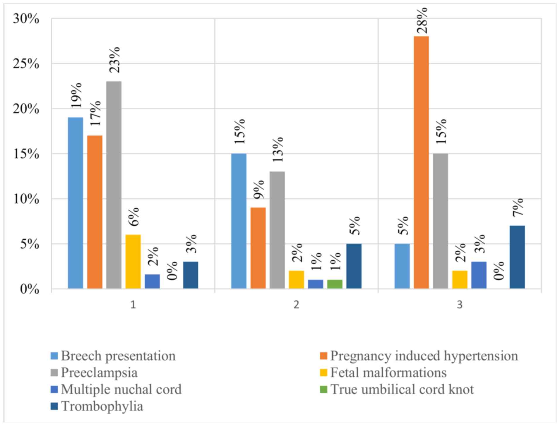
The incidence of obstetric aspects, namely fetal
presentation, obstetric pathology in the spectrum of hypertensive
pregnancy disorders, fetal malformations, multiple nuchal cord, or
true umbilical cord knot is summarized in Fig. 2.
The incidence of pelvic presentation predominates in
pregnancies with low gestational age, that is, in both normal and
low-weight premature pregnancies, and hypertensive pregnancy
pathology predominated in study groups characterized by low weight;
however, there was no statistical difference for the low birth
weight group in this study.
All the neonatal complications, including the
incidence of the complications within each study group, and the
existence or not of statistical significance for the study groups
are shown in Table IV.
| Table IVFrequency of neonatal complications in
the three study groups and their comparative statistical
analysis. |
Table IV
Frequency of neonatal complications in
the three study groups and their comparative statistical
analysis.
| | 1 | 2 | 3 | Total | |
|---|
| Neonatal
characteristic | No. | % | No. | % | No. | % | No. | % | P-value |
|---|
| Total | 78 | 6 | 1,121 | 80 | 206 | 15 | 1,405 | 100 | NA |
| Cardiovascular
arrest | 5 | 6 | 13 | 1 | 1 | 0 | 19 | 1 | <0.001 |
| Acute respiratory
failure | 19 | 24 | 87 | 8 | 5 | 2 | 111 | 8 | <0.001 |
| Ulcer-necrotic
enterocolitis | 10 | 13 | 10 | 1 | 0 | 0 | 20 | 1 | <0.001 |
| Hypoxia | 45 | 58 | 260 | 23 | 18 | 9 | 323 | 23 | <0.001 |
| Respiratory
distress | 23 | 29 | 147 | 13 | 0 | 0 | 170 | 12 | <0.001 |
| Cerebral edema | 5 | 6 | 27 | 2 | 0 | 0 | 32 | 2 | 0.004 |
| Intraventricular
hemorrhage | 22 | 28 | 114 | 10 | 3 | 1 | 139 | 10 | <0.001 |
| Pulmonary
hemorrhage | 9 | 12 | 38 | 3 | 0 | 0 | 47 | 3 | <0.001 |
| Patent ductus
arteriosus | 0 | 0 | 12 | 1 | 1 | 0 | 13 | 1 | 0.491 |
| Cerebral
hemorrhage | 7 | 9 | 49 | 4 | 1 | 0 | 57 | 4 | 0.003 |
| Neonatal
hypoglycemia | 21 | 27 | 157 | 14 | 9 | 4 | 187 | 13 | <0.001 |
| Retinopathy | 15 | 19 | 47 | 4 | 1 | 0 | 63 | 4 | <0.001 |
| Anemia | 25 | 32 | 101 | 9 | 5 | 2 | 131 | 9 | <0.001 |
| Hemorrhagic
disease | 6 | 8 | 28 | 2 | 1 | 0 | 35 | 2 | 0.002 |
| Disseminated
vascular coagulation | 2 | 3 | 1 | 0 | 0 | 0 | 3 | 0 | <0.001 |
| Neonatal
sepsis | 12 | 15 | 63 | 6 | 16 | 8 | 91 | 6 | 0.002 |
| Need for neonatal
intensive care | 24 | 31 | 94 | 8 | 4 | 2 | 122 | 9 | <0.001 |
| Death | 14 | 18 | 51 | 5 | 2 | 1 | 67 | 5 | <0.001 |
| Hyaline membrane
disease | 6 | 8 | 21 | 2 | 0 | 0 | 27 | 2 | <0.001 |
Analyzing each parameter separately, we obtained a
statistically significant difference, predominantly in preterm
infants with low weight for the gestational age, for the following
complications: Cardiovascular arrest occurred in 6% of the subjects
in group 1, with a significant difference of 1% in the incidence of
complications between groups 2 and 3; for acute respiratory
insufficiency, the difference in incidence was even greater, the
premature infants with low weight for the gestational age (24%
compared to 8 and 2% corresponding to the other groups); a
difference in the incidence for ulcer-necrotic enterocolitis (group
1, 13%; group 2, 1%; and group 3, 0%), having statistical
significance was otherwise obtained according to the collected
data; hypoxia also predominated among preterm infants: 58% of
infants with low weight for gestational age, 23% for premature
infants with weight corresponding to gestational age, and 9% for
term infants with low weight for gestational age.
Respiratory distress had an incidence of 29% in
group 1, significantly higher than group 2 (13%) and group 3 (0%),
and cerebral edema occurred only in premature cases, with an
incidence of 6% for those with low weight for gestational age and
2% for those with appropriate weight.
Neonatal complications, such as cerebral edema,
pulmonary hemorrhage, neonatal seizures and disseminated
intravascular coagulation, persistence of the arterial canal,
cerebral hemorrhage, hyaline membrane disease, and retinopathy had
a 0% incidence in the term neonatal group with low birth weight.
Each of these complications predominated in the group of preterm
newborns with intrauterine growth restriction, with the exception
of the persistence of the arterial canal, which predominated in
study group 2, with an incidence of 1%, and was absent in group 1.
For cerebral edema, pulmonary edema and disseminated intravascular
coagulation, the difference in incidence was statistically
significant. For group 3, complications such as intraventricular
hemorrhage and neonatal anemia presented minimum incidences of 1
and 2%, respectively. For these complications, the predominance was
also the highest in group 1 (premature infants with low weight for
the gestational age), namely 28 and 32%, respectively, which are
significantly higher percentages than those obtained in the other
study groups.
Regarding the incidence of neonatal infections, a
significant incidence was noted in all 3 study groups, with a
significant predominance among the group of premature infants with
low-weight for the gestational age (38%); whereas, for the
premature infants with the weight corresponding to the gestational
age, the incidence was 19%, similar to that of term newborns with
low weight for gestational age (16%). Regarding neonatal sepsis, a
higher incidence was obtained of infants with low birth weight for
gestational age (15%) and significant but smaller incidence was
obtained for the other 2 study groups (6 and 8%, respectively).
Regarding the need for neonatal intensive care, it
was 18% for group 1, 5% for group 2, and 1% for group 3, with a
statistically significant difference between the three groups.
Additionally, a statistically significant difference was obtained
for the rate of neonatal death, which predominated by 8% in preterm
infants with low weight for gestational age and was absent in term
infants with low weight for gestational age.
Discussion
Prematurity remains the leading cause of neonatal
morbidity and mortality. Intrauterine growth restriction is another
first-line cause of the adverse neonatal prognosis, both as a
single pathology or in association with prematurity. According to
previous studies, there is a concordant relationship between
preterm birth and intrauterine growth restriction (7). Intrauterine growth restriction shows
chronic fetal distress based on placental dysfunction, with
placental abnormal vascularization leading to hypoperfusion,
ischemia, and release of reactive oxygen species in the context of
oxidative stress.
Both prematurity and intrauterine growth restriction
have an increased incidence among pregnancies complicated with
preeclampsia (8), results that are
consistent with the data in the literature. In this context, we
decided to highlight the unfavorable short-term prognosis of
prematurity, as well as intrauterine growth restriction, in both
preterm and term infants with low birth weight.
According to the results obtained, the frequency of
premature births in the Emergency University Hospital of Bucharest,
a multidisciplinary hospital in which multiple complex cases are
addressed, was ~13%. The cases of complicated preterm infants with
intrauterine growth restriction accounted for 6.5% of all premature
births recorded in the clinic during the 3 years of study and ~1%
of all births. The cases of newborns with low birth weight for the
gestational age accounted for ~2% of all births recorded during the
study period. Thus, the data obtained are consistent with reports
in the literature; the proportion of intrauterine growth
restriction among preterm births is higher than that among term
births (7).
Another parameter that was consistent with the
published reports is the predominance of female fetal sex in cases
of intrauterine growth restriction (9,10) and
the predominance of male sex among premature births. However, the
difference was not significant; therefore, according to the
results, we can consider the following conclusions of the study by
Quiñones et al (11),
focusing mainly on the influence of fetal sex on the perinatal
prognosis of cases of intrauterine growth restriction: Fetal sex is
not associated with unfavorable perinatal prognosis in cases of
intrauterine growth restriction.
The purpose of the analysis of the values of the
Apgar index as a marker of the immediate neonatal adaptation was to
objectively highlight the difference of adaptation, especially in
the case of premature infants with low weight for gestational age
and those infants with weight corresponding to their gestational
age. The values obtained were not different from expectations, more
specifically, the adaptation of preterm infants with low weight for
the gestational age was the most deficient, with a median value of
7. Fig. 1 shows that there were no
values that deviated significantly from the value of the median,
with the distribution being proportional. However, the degree of
adaptation was higher in the group of premature infants with weight
corresponding to the gestational age, as expected. It appears that
the values of the Apgar index were higher in this group. However,
in the single cases of small Apgar index, the median assigned to
group 2 was 8. The most favorable adaptation was noted in the
newborns in group 3. Maximum values of the Apgar score were noted
in groups 2 and 3.
Birth by caesarean section predominated in all study
groups, with a statistically significant difference noted among the
3 groups. This is justified by the fact that birth by cesarean
section of premature infants is associated with lower neonatal
mortality (12), which is explained
by the possibility of early and promising neonatal intensive care
(13). Additionally, in this
context, it is worth mentioning that the birth weight is inversely
proportional to the rate of neonatal complications, in which the
impact of the vaginal birth decreases with increasing fetal weight
(14). Even though premature birth
is not an absolute indication of cesarean delivery, this mode of
birth provides a better prognosis for preterm infants by avoiding
prolonged labor and allowing for a less traumatic birth (14).
Regarding obstetric factors, it is not surprising
that we obtained a higher incidence of pelvic presentation among
preterm infants; however, the incidence of pelvic presentation
among low-weight newborns for gestational age was slightly higher
than that in the general population. The data at the general
population level show an incidence of caesarean section of 4-40%
among term births an 25-60% among premature births, which is
inversely proportional to the gestational age. As seen in Fig. 2, the incidence of pregnancy-induced
hypertension predominates in cases of growth restriction, both in
premature newborns and particularly in newborns with term growth
restriction. This situation is also characteristic of preeclampsia,
except that it prevails in preterm infants with intrauterine growth
restriction and low weight for gestational age. These results
confirm previous results of other studies, namely that placental
functional disorders belong to the group of progressive
multifactorial pathologies that present deteriorating signs and
symptoms over time.
Fetal malformations predominated in the group of
premature newborns with growth restriction, which is consistent
with published reports on the association of fetal malformations
with intrauterine growth restriction (15,16) and
prematurity (17). The umbilical
cord pathology, mainly the true cord knot, did not have a
significant association with intrauterine growth restriction, which
is consistent with the data in the literature; however, this
association has not been fully established and is still being
studied (18-20).
Regarding the multiple nuchal cord, a statistically significant
association has not yet been evidenced between these condition and
adverse neonatal prognosis (21).
According to the results obtained in this study, the incidence of
multiple nuchal cord was higher in the groups with growth
restriction.
Essentially, the purpose of the present study is to
show that intrauterine growth restriction, a condition closely
related to placental dysfunction, is a common diagnosis that is
associated with an increased risk of perinatal mortality and
morbidity. The fetal response consists in circulatory adaptations,
respectively brain-sparing reflected by the value of
cerebroplacental ratio, which has a better predictability index of
adverse outcomes especially in fetuses with intrauterine growth
restriction (22). The meta-analysis
published in 2016, aiming to evaluate the perinatal predictability
value of cerebroplacental ratio concluded that abnormal
cerebroplacental ratio is associated with increased rates of
unfavorable perinatal outcome, having a moderate-high specificity
and sensitivity (23). An abnormal
cerebroplacental ratio was associated with higher rates of need of
neonatal intensive care and neonatal complications and suggest a
poorer perinatal outcome of fetuses with intrauterine growth
restriction (23). Regarding the
normalization of the cerebroplacental ratio, the results of a
recent sub-analysis which started from the hypothesis that
normalization of this ratio associates with a poorer perinatal
outcome due to the loss of the compensatory mechanism of brain
sparing, showed that there is no additional worsening of the
perinatal prognosis given by this normalization (24). Recent studies have analyzed the
impact of the abnormal cerebroplacental ratio on neurodevelopmental
outcome in fetuses with intrauterine growth restriction. Meher
et al in their review suggested that the brain sparing
phenomenon has not only a protective benefit but is associated with
a poorer psychomotor development at one and two years caused by
implied cerebral hypoxia (25).
In the context of the results obtained, as well as
of the discussions regarding the advantages of the expectant
management for both short- and long-term outcomes, the decision
regarding choosing the most appropriate time for termination of the
pregnancy becomes even more difficult. Thus, each case should be
treated individually with a therapeutic behavior guided by the main
pathology but also the associated one in order to reduce the rate
of iatrogenic prematurity among the fetuses with intrauterine
growth restriction but also to offer them the best prognosis.
Further, with reference to Table IV, which contains the frequency and
comparative analysis of all the neonatal complications studied for
the 3 groups, one can observe the objective impact of intrauterine
growth restriction during the immediate neonatal period.
Thus, as discussed in the results, the highest
frequency of neonatal complications occurred in study group 1.
Statistical significance was obtained for the following
complications: Cardiovascular arrest (P<0.001), acute
respiratory failure (P<0.001), ulcer-necrotic enterocolitis
(P<0.001), hypoxia present in 58% of premature cases with growth
restriction and in 23% of cases of gestational age weight
(P<0.001), respiratory distress (P<0.001), cerebral edema
(P=0.004), intraventricular hemorrhage (P<0.001), cerebral
hemorrhage (P=0.003), pulmonary hemorrhage (P<0.001), neonatal
infection (P<0.001), hypoglycemia (P<0.001), retinopathy
(P<0.001), anemia (P<0.001), hemorrhagic disease (P=0.002),
disseminated intravascular coagulation (P<0.001), disease of
hyaline membranes (P<0.001), neonatal sepsis (P=0.002), need for
intensive neonatal therapy (P<0.001), and death
(P<0.001).
Intrauterine growth restriction is associated with
an increased risk of both antenatal and neonatal complications.
There is an increased negative impact on prognosis when fetuses
with intrauterine growth restriction are born premature. In this
study, newborns with low weight for gestational age had an
increased incidence in complications in comparison to newborns with
adequate weight, specifically: cardiovascular arrest, 0.1% in the
general population and 1% in the present study; acute respiratory
failure, 0.45% in the general population and 2% in the present
study (14); however, respiratory
distress, cerebral edema, ulcer-necrotic enterocolitis, pulmonary
hemorrhage, persistence of the arterial canal, cerebral hemorrhage,
seizures, retinopathy, hemorrhagic disease, disseminated
intravascular disease, and hyaline membrane disease were absent in
term infants with low birth weight for gestational age, indicating
that these newborns have a good neonatal adaptation by leaving an
environment already unfit for their well-being, i.e.
intrauterine.
In conclusion, intrauterine growth restriction can
occur both in the context of pre-existing chronic hypertension and
in the context of severe preeclampsia. In principle, preeclampsia
is so frequently associated with intrauterine growth restriction
that the latter has traditionally been included as a feature of
preeclampsia, regardless of any additional diagnostic criteria.
During the study, it was shown that preeclampsia is the main
condition that leads to the most severe cases of intrauterine
growth restriction. Immediate neonatal adaptation of preterm
neonate small for gestational age is more deficient (indicated by
lower Apgar index values) than for preterm neonates with
appropriate weight for gestational age; the adaptation of preterm
neonates, in turn, is more deficient than term newborns with
intrauterine growth restriction. The term newborns with
intrauterine growth restriction have a neonatal adaptation
comparable to that of the term newborns with weight corresponding
to the gestational age. Birth by caesarean section had an increased
incidence both in the cases of premature newborns with weight
corresponding to the gestational age and in the cases with
premature or term growth restriction. Gestational hypertension is a
major risk factor for intrauterine growth restriction without a
statistically significant difference between premature and term
births. Preeclampsia, on the other hand, is significantly
associated with prematurity and intrauterine growth restriction and
especially in cases presenting both conditions simultaneously.
Fetal malformations are a determinant factor of growth restriction,
but also of prematurity.
After analyzing the neonatal parameters of the 3
study groups, which included premature infants with low weight for
gestational age, preterm infants with weight corresponding to the
gestational age and term newborns with low weight for gestational
age, we can conclude that the growth restriction superimposed on
prematurity is associated the most unfavorable prognosis among all
the parameters.
Acknowledgements
Not applicable.
Funding
No funding was received.
Availability of data and materials
The datasets used and/or analyzed during the current
study are available from the corresponding author on reasonable
request.
Authors' contributions
FIR, DNa, CB and DNe collected, analyzed and
interpreted the patient data regarding the main neonatal
complications associated with prematurity and intrauterine growth
restriction. FF substantially contributed to the conception of the
study and the statistical analysis of the data. MMC contributed to
the interpretation of the results. NT and REB were involved in the
conception of the study, collected and interpreted the patient
data, and were major contributors in the writing of the manuscript.
All authors read and approved the final version of the
manuscript.
Ethics approval and consent to
participate
The data collected retrospectively did not contain
personal information and only the Ethics Committee agreement of the
University Emergency Hospital of Bucharest was required and
obtained without the need of informed consent or the consent of the
patient/legal representative in the case of minors.
Patient consent for publication
Not applicable.
Competing interests
The authors declare that they have no competing
interests.
References
|
1
|
Turcan N, Bohîlţea R, Neacsu A, Baros Al
and Cîrstoiu MM: The role of anticoagulant therapy in the
prevention of preeclampsia. Pharmacokinetic and pharmacodinamic
mechanisms. Rev Chim. 70:1424–1428. 2019. View Article : Google Scholar
|
|
2
|
Bumbu A, Nacer K, Bratu O, Berechet M,
Bumbu G and Bumbu B: Ureteral lesions in gynecological pathology.
In: Proceedings of the 14th National Congress of Urogynecology and
the National Conference of the Romanian Association for the Study
of Pain. Filodiritto Publisher, Eforie. pp82–89. 2017.
|
|
3
|
Nacer K, Bratu O, Berechet M, Bumbu G and
Bumbu A: Global surgical principles in the vaginal approach of
advanced pelvic organ prolapse. In: Proceedings of the 14th
National Congress of Urogynecology and the National Conference of
the Romanian Association for the Study of Pain. Filodiritto
Publisher, Eforie. pp172–180. 2017.
|
|
4
|
Trăistaru VA, Zamfirescu V, Burnei Rusu A,
Bohîlţea R, Nastasia Ş and Vlădăreanu R: Ultrasound markers in
early pregnancy predictive for preeclampsia. In: Proceedings of the
5th Romanian Congress of the Romanian Society of Ultrasound in
Obstetrics and Gynecology. pp593–597. 2017.
|
|
5
|
Trăistaru VA, Burnei Rusu A, Zamfirescu V,
Nastasia Ş, Bohîlţea R and Vlădăreanu R: Ultrasound markers in
early pregnancy predictive for preterm birth. In: Proceedings of
the 5th Romanian Congress of the Romanian Society of Ultrasound in
Obstetrics and Gynecology. pp598–602. 2017.
|
|
6
|
Ionescu AC, Popescu I, Banacu M and Matei
A: Is it possible to predict stillbirth in the third trimester? In:
Proceedings of the 5th Romanian Congress of the Romanian Society of
Ultrasound in Obstetrics and Gynecology. pp194–198. 2017.
|
|
7
|
Bukowski R, Gahn D, Denning J and Saade G:
Impairment of growth in fetuses destined to deliver preterm. Am J
Obstet Gynecol. 185:463–467. 2001.PubMed/NCBI View Article : Google Scholar
|
|
8
|
Bohîlţea R, Turcan N, Ionescu C, Toader O,
Nastasia S, Neculcea D, Movileanu I, Munteanu O and Cîrstoiu M: The
incidence of prematurity and associated short-term complications in
a multidisciplinary emergency hospital from Romania. Proceedings of
the 5th Romanian Congress of the Romanian Society of Ultrasound in
Obstetrics and Gynecology. pp105–112. 2017.
|
|
9
|
Spinillo A, Capuzzo E, Nicola S, Colonna
L, Iasci A and Zara C: Interaction between fetal sex and risk
factors for fetal growth retardation. Am J Obstet Gynecol.
171:1273–1277. 1994.PubMed/NCBI View Article : Google Scholar
|
|
10
|
Yunis KA, Beydoun H, Tamim H, Nassif Y and
Khogali M: National Collaborative Perinatal Neonatal Network. Risk
factors for term or near-term fetal growth restriction in the
absence of maternal complications. Am J Perinatol. 21:227–234.
2004.PubMed/NCBI View Article : Google Scholar
|
|
11
|
Quiñones JN, Stamilio DM, Coassolo KM,
Macones GA and Odibo AO: Is fetal sex associated with adverse
perinatal outcome in intrauterine growth restriction (IUGR)? Am J
Obstet Gynecol. 193:1233–1237. 2005.PubMed/NCBI View Article : Google Scholar
|
|
12
|
Bohîlţea RE, Zugravu CA, Neacsu A, Navolan
D, Berceanu C, Nemescu D, Bodean O, Turcan N, Baros Al and Cîrstoiu
MM: The prevalence of vitamin D deficiency and its obstetrical
effects. A prospective study on Romanian patients. Rev Chim.
70:1228–1233. 2019.
|
|
13
|
Dobson PC, Abell DA and Beischer NA:
Mortality and morbidity of fetal growth retardation. Aust NZ J
Obstet Gynaecol. 21:69–72. 1981.PubMed/NCBI View Article : Google Scholar
|
|
14
|
Ment LR, Oh W, Philip AG, Ehrenkranz RA,
Duncan CC, Allan W, Taylor KJ, Schneider K, Katz KH and Makuch RW:
Risk factors for early intraventricular hemorrhage in low birth
weight infants. J Pediatr. 121:776–783. 1992.PubMed/NCBI View Article : Google Scholar
|
|
15
|
Resnik R: Intrauterine growth restriction.
Obstet Gynecol. 99:490–496. 2002.PubMed/NCBI View Article : Google Scholar
|
|
16
|
Herghelegiu D, Ionescu CA, Pacu I,
Bohîlţea R, Herghelegiu C and Vladareanu S: Antenatal diagnosis and
prognostic factors of aneurysmal malformation of the vein of Galen.
A case report and literature review. Medicine (Baltimore).
96(e7483)2017.PubMed/NCBI View Article : Google Scholar
|
|
17
|
Purisch SE, DeFranco EA, Muglia LJ, Odibo
AO and Stamilio DM: Preterm birth in pregnancies complicated by
major congenital malformations: A population-based study. Am J
Obstet Gynecol. 199(287.e1-8.e8)2008.PubMed/NCBI View Article : Google Scholar
|
|
18
|
Marin JA, Calomfirescu M, Bohilţea R and
Ionescu Târgovişte C: Impact of maternal and placental pathology on
successful umbilical cord blood sampling and cryopreservation.
Gineco. 7:10–14. 2011.
|
|
19
|
Szczepanik ME and Wittich AC: True knot of
the umbilical cord: A report of 13 cases. Mil Med. 172:892–894.
2007.PubMed/NCBI View Article : Google Scholar
|
|
20
|
Schreiber H, Daikan Y, Arbib N, Markovitch
O, Berkovitz A and Biron-Shental T: Multiple nuchal cord loops and
neonatal outcomes. Am J Obstet Gynecol. 218(S91)2018.
|
|
21
|
Rubaltelli FF, Dani C, Reali MF, Bertini
G, Wiechmann L, Tangucci M and Spagnolo A: Italian Group of
Neonatal Pneumology. Acute neonatal respiratory distress in Italy:
A one-year prospective study. Acta Paediatr. 87:1261–1268.
1998.PubMed/NCBI View Article : Google Scholar
|
|
22
|
Bahado-Singh RO, Kovanci E, Jeffres A, Oz
U, Deren O, Copel J and Mari G: The Doppler cerebroplacental ratio
and perinatal outcome in intrauterine growth restriction. Am J
Obstet Gynecol. 180:750–756. 1999.PubMed/NCBI View Article : Google Scholar
|
|
23
|
Nassr AA, Abdelmagied AM and Shazly SA:
Fetal cerebro-placental ratio and adverse perinatal outcome:
Systematic review and meta-analysis of the association and
diagnostic performance. J Perinat Med. 44:249–256. 2016.PubMed/NCBI View Article : Google Scholar
|
|
24
|
Monteith C, Flood K, Mullers S,
Unterscheider J, Breathnach F, Daly S, Geary MP, Kennelly MM,
McAuliffe FM, O'Donoghue K, et al: Evaluation of normalization of
cerebro-placental ratio as a potential predictor for adverse
outcome in SGA fetuses. Am J Obstet Gynecol.
216(285.e1-285.e6)2017.PubMed/NCBI View Article : Google Scholar
|
|
25
|
Meher S, Hernandez-Andrade E, Basheer SN
and Lees C: Impact of cerebral redistribution on neurodevelopmental
outcome in small-for-gestational-age or growth-restricted babies: A
systematic review. Ultrasound Obstet Gynecol. 46:398–404.
2015.PubMed/NCBI View Article : Google Scholar
|