Introduction
Malignant gliomas are the most common primary tumors
observed in the central nervous system with a median survival time
of 12-15 months (1-3). At
present, the available treatment options are surgical resection and
adjuvant temozolomide (TMZ)-based chemotherapy combined with
radiotherapy (4,5). TMZ is a DNA-alkylating drug which has
improved overall survival in patients (6,7).
However, its efficacy is limited by the development of
chemotherapeutic resistance in tumors (8,9).
Therefore, identifying novel molecular mechanisms underlying TMZ
resistance may improve outcomes for patients.
The tripartite motif-containing (TRIM) family of
proteins contains more than 70 members and each member consists of
a conserved RING finger, B-box, and coiled-coil domains (10), These members are primarily involved
in important cellular biological processes (11,12).
TRIM31 is one member of the TRIM family which has been reported to
be involved in innate immunity and cancer development (13-20),
and TRIM31 expression was found to be upregulated in pancreatic
cancer tissues (20).
Mechanistically, TRIM31 confers gemcitabine resistance in
pancreatic cancer cells by activating the NF-κB signaling pathway
(20). However, the role of TRIM31
in governing TMZ resistance remains unclear and the exact mechanism
underlying its effects are unknown.
In the present study, TRIM31 was analyzed in glioma
tissues and we clarified the relationship between TRIM31
upregulation and certain clinicopathological features including
age, sex and WHO grade. Upregulation of TRIM31 enhanced TMZ
resistance in glioma cells in vitro. Mechanistically, the
PI3K/Akt pathway was determined to be involved in TRIM31-mediated
chemoresistance in glioma cells. The present study demonstrated
that overexpression of TRIM31 resulted in TMZ chemoresistance in
glioma and it may be possible to improve the chemosensitivity to
TMZ in patients with glioma by downregulating the expression of or
inhibiting TRIM31.
Materials and methods
Tissue samples
A total of 40 tissue samples and 8 cases of adjacent
normal brain tissues were collected at Department of Neurosurgery,
The Second Hospital of Shandong University (Shandong, China)
between June 2016 and March 2019. The samples ranged from 5 years
old to 72 years old, 27 males and 13 females with an average age of
45.3 years old. All samples were histopathologically and clinically
diagnosed and none of the patients received chemotherapy or
radiotherapy prior to obtaining the tissue samples. All samples
were frozen in liquid nitrogen and stored at -80˚C until further
use. Patient characteristics are presented in Table I. All patients provided written
informed consent and the study was approved by the Medical Ethics
Committee of the Second Hospital of Shandong University.
 | Table ICorrelations between TRIM31
expression and the clinicopathological characteristics of patients
with glioma. |
Table I
Correlations between TRIM31
expression and the clinicopathological characteristics of patients
with glioma.
| | TRIM31
expression | |
|---|
| Clinicopathological
characteristics | Number of patients
(n=40) | Low | High | P-value |
|---|
| Age | | | | 0.7462 |
|
<55 | 26 | 15 | 11 | |
|
≥55 | 14 | 9 | 4 | |
| Sex | | | | 0.5106 |
|
Male | 27 | 13 | 14 | |
|
Female | 13 | 8 | 5 | |
| WHO grade | | | | 0.007 |
|
I-II | 14 | 12 | 2 | |
|
III-IV | 26 | 10 | 16 | |
Cell culture and treatments
Normal human astrocytes (NHA) were obtained from
ScienCell Research Laboratories, Inc. LN229 cells were obtained
from American Type Culture Collection. U87, U251 and A172 cell
lines were purchased from The Cell Bank of Type Culture Collection
of the Chinese Academy of Sciences. U87 cells are a glioblastoma of
unknown origin which was authenticated by STR profiling, Cells were
cultured in DMEM (Thermo Fisher Scientific, Inc.) supplemented with
10% FBS (Biological Industries). The cells were cultured at 37˚C
with 5% CO2. TMZ (cat. no. T2577; Sigma-Aldrich; Merck
KGaA) was dissolved in DMSO (Sigma-Aldrich; Merck KGaA) at a stock
concentration of 200 mM and stored at -20˚C. A 5 mM solution of
LY294002 was purchased from MedChemExpress (cat. no. HY-10108),
after 10 h transfection, the glioma cells were incubated with
LY294002 for 12 h. Cells were harvested for further analysis.
Plasmid construction and cell
transfection
TRIM31 expression plasmids were kindly provided by
Professor Chengjiang Gao and Professor Lihui Han (Shandong
University School of Medicine, Shandong, China). TRIM31 small
interfering (si)RNAs were synthesized by Shanghai GenePharma Co.,
Ltd. And the sequences were as follows: siTRIM31,
5'-GGACCACAAAUCCCAUAAU-3'; and si-negative control (NC),
5'-UUCUCCGAACGUGUCACGU-3'. A172 and U251 cells were transfected
using Lipofectamine® 2000 (cat .no. 11668019,
Invitrogen; Thermo Fisher Scientific, Inc.) according to the
manufacturer's protocol. A total of 3x10⁵ A172 or U251
cells/well were seeded in 6-well plates and cultured overnight.
Lipofectamine® 2000 and opti-MEM (cat. no. 31985; Gibco;
Thermo Fisher Scientific, Inc.) were used for transient
transfection with 3 µg plasmid or 100 pmol siRNA. Subsequent
experiments were performed after 24 or 48 h of transfection.
Transfection efficiency was determined using reverse
transcription-quantitative (RT-q)PCR.
RT-qPCR
Total RNA was extracted using a RNeasy kit (cat. no.
DP430; Tiangen Biotech, Co., Ltd.) and 1 µg RNA was reverse
transcribed into cDNA using a First-strand Synthesis kit (cat. no.
KR116; Tiangen Biotech, Co., Ltd.). qPCR was performed on a
Mastercycler ep realplex (Eppendorf) with SuperReal Premix Plus
(cat. no. FP205; Tiangen Biotech, Co., Ltd.). The reverse
transcription temperature protocol was: 42˚C for 15 min and reverse
transcriptase inactivation 95˚C for 3 min. qPCR was performed using
10 µl 2x SuperReal Premix Plus, 20 ng cDNA template, 10 µM each
forward and reverse primers, and ddH2O. The
thermocycling conditions were: 95˚C for 15 min; followed by 40
cycles of denaturation at 95˚C for 10 sec, annealing at 60˚C for 25
sec and extension at 72˚C for 20 sec. The data were analyzed using
the 2-ΔΔCq method (21)
and normalized to the internal control. The sequence of the primers
were: TRIM31 forward, 5'-GGCAGATTCAAGAGCAG-3' and reverse,
5'-TCAGTGGAGGCAACATAG-3'; and β-actin forward,
5'-GGAAATCGTGCGTGACATTAA-3' and reverse,
5'-AGGAAGGAAGGCTGGAAGAG-3'.
Western blotting
Cells were lysed with 1 ml RIPA buffer (Beijing
Solarbio Science & Technology Co., Ltd.) supplemented with a
protease inhibitor (Beijing Solarbio Science & Technology Co.,
Ltd.) and centrifuged at 4˚C 12,000 x g for 15 min, and the
supernatant was collected. The protein concentration was measured
using a bicinchoninic acid protein assay kit (ABP Biosciences)
according to the manufacturer's protocol. Protein samples (20 µg)
were loaded on a 12% SDS gel and resolved using SDS-PAGE. Resolved
proteins were transferred onto PVDF membranes (EMD Millipore). The
PVDF membranes were blocked in 5% nonfat milk at room temperature
for 1 h and subsequently incubated with the following primary
antibodies at 4˚C overnight: anti-TRIM31 (cat. no. 12543-1-AP;
1:500; ProteinTech Group, Inc.), anti-p-AKT (cat. no. 4060,
1:1,000), AKT (cat. no. 4685; 1:1,000), anti-p53 (cat. no. 2524;
1:1,000) and GADPH (cat. no. 2118s; 1:1,000) all from Cell
Signaling Technology, Inc. The membranes were washed three times in
PBS-Tween (PBST) for 6 min. Membranes were incubated with a
secondary goat anti-rabbit immunoglobulin G (IgG) antibody
(1:5,000, OriGene Technologies, Inc.) at room temperature for 1 h.
Membranes were washed with PBST three times for 10 min. Signals
were visualized using a chemiluminescence fluorescent detection kit
(EMD Millipore) in the dark. Membranes were exposed in a gel
imaging system. The ratios of gray values of each band was
semi-quantified using Alpha Imager version 2011 (ProteinSimple).
All experiments were performed in triplicate.
Apoptosis assay
An AnnexinV/propidium iodide (PI) double staining
kit (cat. no. BB-4101; Bestbio) was used to measure apoptosis.
Cells were harvested using 0.25% trypsin following TMZ treatment,
washed twice with pre-cooled PBS and cells were resuspended in
binding buffer. 5 µl Annexin V/fluorescein isothiocyanate and 5 µl
PI was added. After incubation in the dark for 15 min at 4˚C, cells
were analyzed using BD flow cytometry. All experiments were
performed in triplicate.
Cell viability assay
Cell viability was measured using an MTT assay.
Cells were seeded in a 96-well plate at a density of
1x10³ cells/well in 100 µl of culture medium and
incubated at 37˚C with 5% CO2 for 24 h. After treatment
with different concentrations of TMZ for 48 h, 5 mg/ml MTT solution
(Beijing Solarbio Science & Technology Co., Ltd.) was added to
each group at the appropriate time and incubated for a further 4 h
at 37˚C in the dark, after which 200 µl DMSO was added to dissolve
the purple crystals. Cell viability was detected at 450 nm using a
microplate reader (Infinite M200 Pro; Tecan Group). IC50
was calculated using GraphPad Prism version 5.01 (GraphPad
Software, Inc.) by plotting a nonlinear regression curve fit using
log (inhibitor) vs. normalized response (variable slope).
Experiments were performed in triplicate.
Colony formation assay
A172 or U251 cells were plated in a 6-well plate at
a density of 100 cells/well and cultured in DMEM supplemented with
10% FBS. Cells were treated with the indicated agents for 10-14
days. Medium was replaced every 3 days. Cell colonies were gently
washed with PBS and fixed with 4% paraformaldehyde for 15 min at
room temperature, and subsequently stained with 0.1% crystal violet
for 20 min at room temperature. The stain was carefully washed
using running water and dried. The number of colonies with >50
cells were counted under a NikonTS-100F microscope light. Three
independent assays were performed for each condition.
Immunohistochemistry
Immunohistochemistry staining was performed using a
kit (cat. no. PV-9001; OriGene Technologies, Inc.). The sections
were deparaffinized with xylene, rehydrated and boiled in 0.01 M
citrate buffer (pH 6.0) for antigen retrieval. Hydrogen peroxide
was added to block endogenous peroxide activity, and the sections
were washed three times, and subsequently incubated with TRIM31
antibody (1:50) overnight at 4˚C. Tissues were washed three times
in PBS for 3 min each, after which the sections were incubated with
a secondary goat anti-rabbit IgG antibody at room temperature for
20 min. Sections were colored using a 3,3'-diaminobenzidine (cat.
no. ZLI-9018; OriGene Technologies, Inc.) at room temperature for
40 sec. Nuclei were counterstained with hematoxylin at room
temperature for 3-5 min. The intensity and the ratio of positive
cells in at least five separate fields were evaluated at x400
magnification under a Nikon90i microscope light. The intensity of
staining was scored between 1 and 4: 1, No staining; 2, weak
staining; 3, moderate staining; and 4, strong staining. The
proportion of cells stained were scored as follows: 1, 0-5%; 2,
6-50%; 3, 51-75%; and 4, 76-100%. The scores of the proportion of
cells stained and the intensity scores were multiplied to give a
final score between 1 and 16 and classed as follows: Negative, 1-4;
weakly positive, 5-8; moderately positive, 9-12; or strongly
positive, >12. The scores were evaluated by two pathologists who
were blinded to clinical data.
Statistical analysis
GraphPad Prism version 5.01 (GraphPad Software,
Inc.) was used for all statistical analyses. All experiments were
independently performed three times. Data are presented as the mean
± standard deviation. Comparisons between two groups were analyzed
using a Student's t-test. A one-way ANOVA with post hoc Tukey's
test was used to analyze differences between multiple groups. The
associations between TRIM31 levels and clinicopathological features
were analyzed using a Fisher's exact probability test. P<0.05
was considered to indicate a statistically significant
difference.
Results
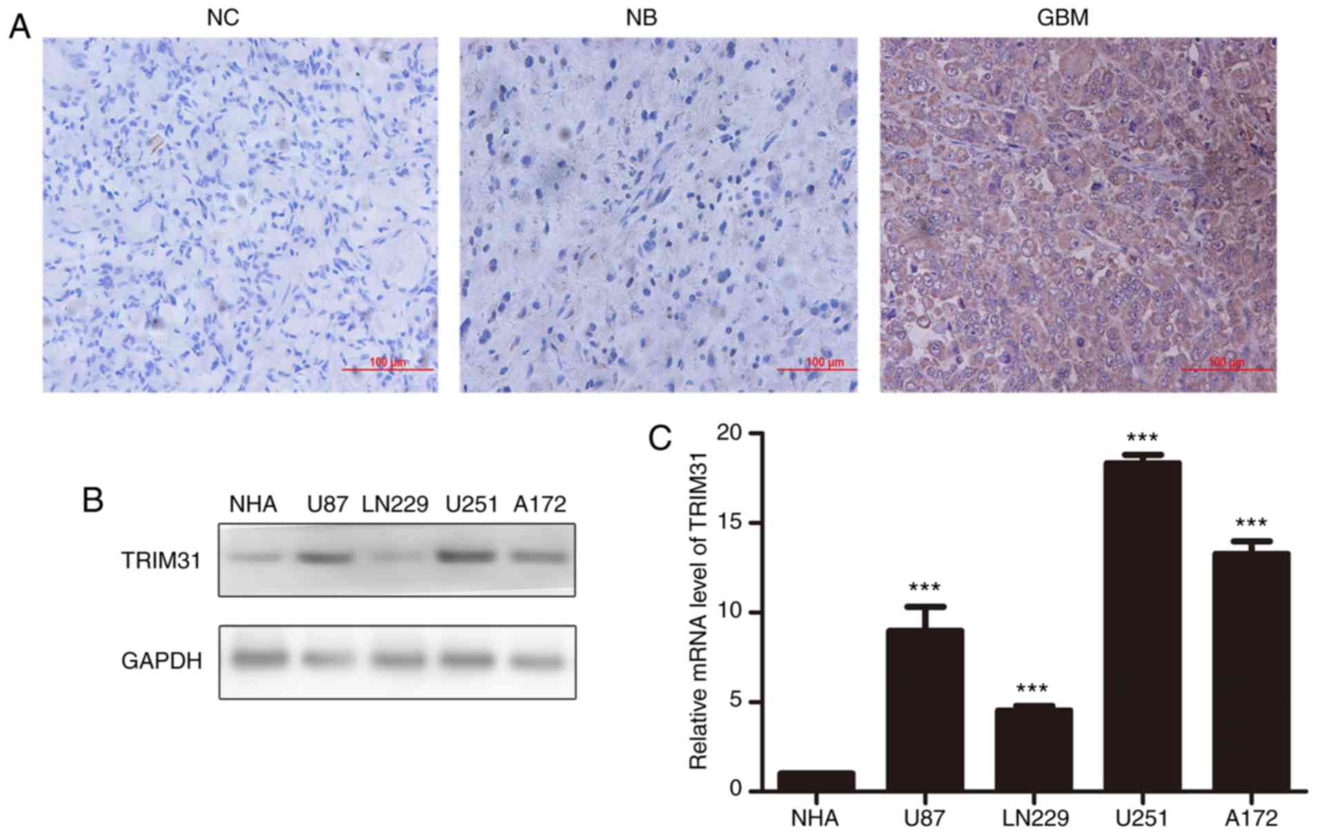
TRIM31 is upregulated in human glioma
tissues
To determine the role of TRIM31 in human glioma, its
expression in normal brain tissues and glioma tissues were examined
by RT-qPCR and immunohistochemistry assay. The results showed that
expression of TRIM31 was significantly upregulated in glioma
tissues compared with normal brain tissues and it was primarily
expressed in the cytoplasm of glioblastoma cells (Fig. 1A). Additionally, the expression of
TRIM31 in NHA, U87, LN229 U251 and A172 cell lines was examined.
TRIM31 expression was higher in U251 and A172 cells compared with
the other cell lines at both the protein and mRNA levels (Fig. 1B). Therefore, the U251 and A172 cell
lines were chosen for subsequent experiments. Subsequently, the
association between TRIM31 expression and clinicopathological
characteristics of patients with glioma were assessed. Analysis
showed that TRIM31 expression was associated with World Health
Organization grade. There was no significant difference between
TRIM31 expression and sex or age. Taken together, these findings
suggest that increased TRIM31 expression was associated with
aggressive clinical features of glioma.
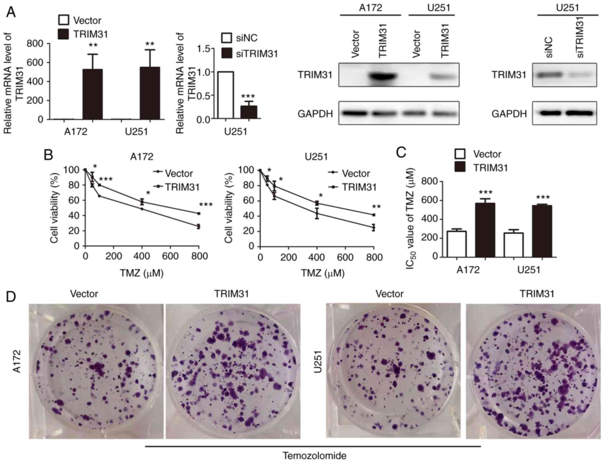
TRIM31 overexpression confers TMZ
resistance in glioma cells
To examine the potential role of TRIM31 in
chemoresistance in glioma cells, TRIM31 expression was knocked down
using siRNA or overexpressed in A172 and U251 cells. Overexpression
and knockdown efficiency were confirmed by RT-qPCR and western
blotting (Fig. 2A). The effect of
TRIM31 on the cell viability of A172 and U251 cells treated with
different concentrations of TMZ for 48 h was subsequently
determined. Cell proliferation was assessed using MTT assays and
they showed that TRIM31 overexpression increased cell viability
compared with the control group (Fig.
2B). The IC50 value of TMZ was significantly higher
in TRIM31 overexpressing cells than transfecting blank plasmids
cells (Fig. 2C). Colony formation
assays showed that TRIM31 overexpression resulted in increased
colony formation and exhibited greater clonogenic survival
following TMZ treatment compared with the control (Fig. 2D). These data suggest that TRIM31
enhanced TMZ resistance in glioma cells in vitro.
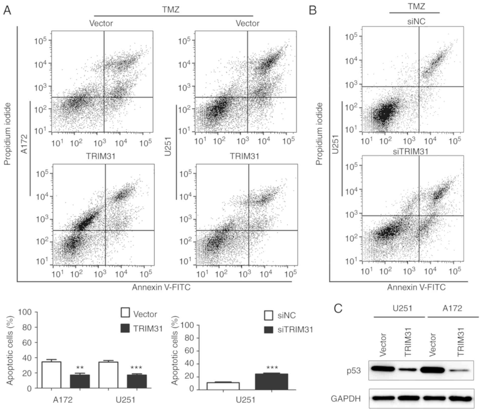
TRIM31 inhibits TMZ-induced glioma
cell apoptosis
To further investigate the effect of TRIM31 on
TMZ-induced apoptosis, apoptosis rates were measured by flow
cytometry analysis in A172 and U251 cells treated with TMZ for 48
h. A172 and U251 cells were transfected with empty vector or TRIM31
overexpression plasmid and treated with TMZ (100 µg/ml). The
results demonstrated that TRIM31 overexpression significantly
decreased the apoptotic proportion of cells compared with the
control cells (Fig. 3A). In U251
cells transfected with siNC or siTRIM31 and treated with TMZ (100
µg/ml), knockdown of TRIM31 increased the apoptotic proportion of
cells (Fig. 3B). Subsequently, p53
protein levels were detected in U251 and A172 cells treated with
TMZ, overexpressing TRIM31 decreased the expression of p53 protein
(Fig. 3C). These results showed
TRIM31 reduced TMZ-induced glioma cells apoptosis, which suggested
that TRIM31 decreased glioma cells sensitivity to TMZ.
The PI3K/Akt signaling pathway is
involved in TRIM31-mediated TMZ resistance of glioma cells
Activation of PI3K/Akt signaling is the primary
pathway which participates in glioblastoma growth and therapy
(22-29),
and activated Akt is indicative of increased signaling via this
pathway. Whether PI3K/Akt pathway was involved in TRIM31-mediated
chemoresistance of glioma cells was unknown. TRIM31 overexpression
upregulated the phosphorylation of AKT (Fig. 4A) compared with control cells treated
with TMZ. TRIM31 knockdown downregulated the phosphorylation of AKT
in U251 cells compared with control cells treated with TMZ
(Fig. 4B). These results suggested
that TRIM31 may activate the PI3K/Akt signal pathway in glioma
cells treated with TMZ.
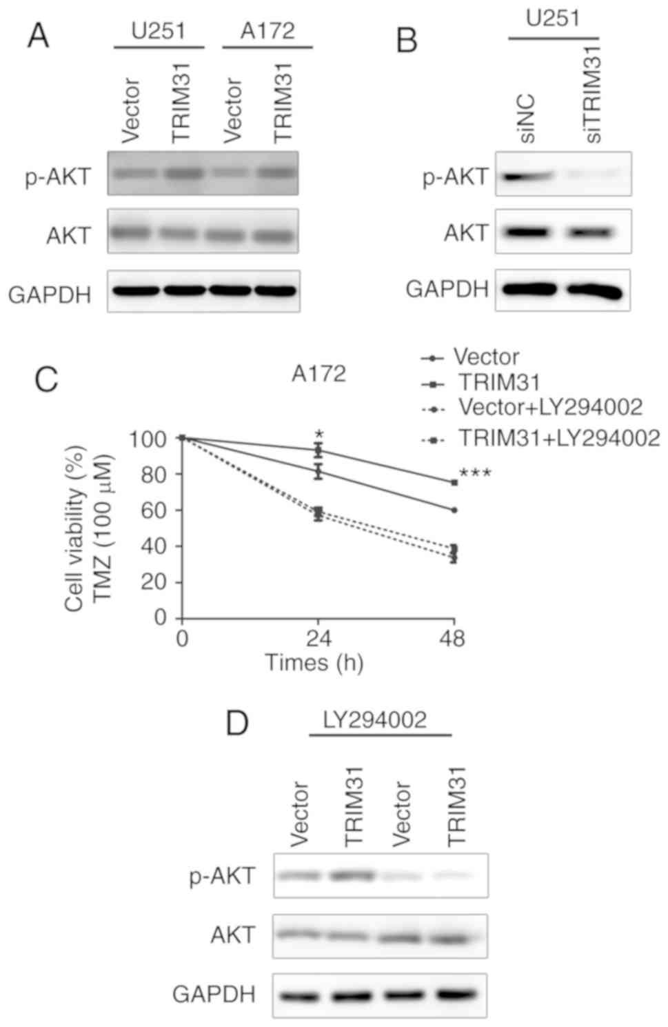 | Figure 4TRIM31 regulates TMZ resistance via
the PI3K/Akt signaling pathway. (A) Protein expression levels of
p-AKT and AKT in A172 and U251 cells transfected with the indicated
plasmids and treated with TMZ. (B) Protein expression levels of
p-AKT and AKT in U251 cells transfected with siTRIM31 and siNC and
treated with TMZ. (C) Cell viability of A172 cells transfected with
the TRIM31 and control plasmids and treated with the PI3K
inhibitor, LY294002, and TMZ. (D) Protein expression levels of
p-AKT and AKT in A172 cells transfected with the TRIM31 and control
plasmids and treated with the PI3K inhibitor, LY294002, and TMZ.
*P<0.05, ***P<0.001. Data are presented
as the mean ± standard deviation of three independent experiments.
TMZ, temozolomide; si, small interfering; NC, negative control;
TRIM31, tripartite motif-containing 31; p, phosphorylated. |
To further elucidate the mechanism by which TRIM31
induced chemoresistance in glioma cells, LY294002 was used to
inhibit PI3K/Akt activation in U251 and A172 cells. Inhibition of
PI3K/Akt signaling decreased glioma cell viability and abolished
the effects of TRIM31 expression (Fig.
4C). LY294002 also reduced the expression of p-AKT induced by
TRIM31 overexpression in A172 cells treated with TMZ (Fig. 4D). The PI3K/Akt inhibitor abolished
the effects of TRIM31 on p-AKT upregulation. These data suggest
that TRIM31 regulated TMZ resistance through the PI3K/Akt
activation and p-AKT upregulation.
Discussion
TMZ chemoresistance is a significant challenge faced
during glioma recurrence which results in treatment failure
(8,9), Therefore, enhancement of sensitivity of
the tumor to TMZ may improve the prognosis of patients with
gliomas.
TRIM31, is a member of the TRIM family of proteins,
which are been involved in various cellular processes (13-20).
Recently, studies have demonstrated that TRIM31 serves an important
role in the development of various types of cancer (15-20).
A recent study reported that TRIM31 promoted progression of
hepatocellular carcinoma through the mTORC1-HIF1α pathway by
directly targeting the TSC1-TSC2 complex for degradation (19). Notably, TRIM31 has also been found to
be involved in the development of chemotherapeutic resistance in
cancer cells. For instance, TRIM31 overexpression conferred
gemcitabine resistance in pancreatic cancer cells via the NF-κB
signaling pathway (20). It has also
been reported to participate in the development of drug resistance
in ovarian cancer (30). In the
present study, it was demonstrated that TRIM31 expression was
upregulated in glioblastoma (GBM) tissues and TRIM31 expression was
significantly correlated with tumor grade, indicating that TRIM31
functions as a tumor oncogene in glioma progression. The data
together suggested that TRIM31 may be associated with
chemoresistance in glioblastoma cells.
To clarify the biological function of TRIM31 in
affecting TMZ sensitivity in glioblastoma cells, stable TRIM31
overexpression and knockdown models were created. MTT assays
demonstrated that overexpression of TRIM31 increased cell viability
and the effects were augmented by treatment with TMZ in a
dose-dependent manner. TRIM31 overexpression combined with TMZ
promoted cell growth and resulted in reduced apoptosis of glioma
cells compared with untransfected cells treated with TMZ. The
IC50 value was used as a measure of TMZ chemoresistance
in GBM cells, thus increased IC50 generally corresponded
to increased clinical chemoresistance to TMZ. TRIM31 overexpression
significantly increased the IC50 values of TMZ.
Furthermore, TRIM31 overexpression significantly increased colony
formation ability in vitro compared with the TMZ treated
untransfected cells. Similarly, flow cytometry analysis showed that
combined TRIM31 overexpression and TMZ treatment resulted in
significantly reduced apoptosis compared with TMZ treated
untransfected cells. Conversely, TRIM31 knockdown increased
apoptosis. The results of the present study suggested that
overexpression of TRIM31 in glioblastoma cells induced resistance
to TMZ. The tumor suppressor protein p53 serves various functional
roles in the cell by regulating responses to several cellular
stresses and it is inactivated in a variety of cancer cells via
point mutations (31,32). p53 regulates tumor proliferation and
apoptosis synergistically (33-35).
p53 mimetic agents designed to stabilize the p53wt sensitize glioma
cells to TMZ (36). In the present
study, upregulation of TRIM31 significantly reduced the expression
of p53, these results suggested that decreased p53 expression may
account for the inhibitory role of TRIM31 in TMZ-induced glioma
cell apoptosis.
Numerous studies have demonstrated a close
association between the PI3K/Akt pathway and TMZ resistance in
gliomas (37-41).
Akt is a primary regulator of PI3K-initiated signaling and its
activation contributes to chemoresistance (42,43).
Therefore, it was determined whether the PI3K/Akt signaling pathway
was involved in TRIM31 induced TMZ resistance of glioma cells.
Firstly, the results showed that TRIM31 overexpression
significantly increased the expression of p-Akt in glioma cells,
which indicated that the biological effects of TRIM31 were partly
mediated by the PI3K/Akt pathway. LY294002, a PI3K inhibitor, was
used to further confirm this assumption. TRIM31 failed to
upregulate cell viability when treated with LY294002, and the cell
viability was reduced to levels similar to TMZ treated cells and in
the TRIM31 overexpressing cells. The results indicated that PI3K
inhibition decreased p-Akt protein expression and abolished the
effects of TRIM31 on p-Akt upregulation, suggesting that TRIM31
regulated TMZ resistance possibly via the PI3K/Akt pathway.
In the present study, upregulation of TRIM31
resulted in an increase of p-AKT and a decrease of p53 expression,
which demonstrated that TRIM31 induced chemoresistance in gliomas
to TMZ via the PI3K/Akt/p53 signaling. The E3 ubiquitin ligase has
been reported to catalyze the polyubiquitin of p53 and trigger the
degradation of p53 in hepatocellular carcinoma cells (44). In glioma cells, whether TRIM31
directly targeted p53 for ubiquitin-mediated degradation or if
TRIM31 bound adaptors involved in the PI3K/Akt pathway remains
unclear.
In conclusion, the present study showed that TRIM31
mediated TMZ sensitivity via the PI3K/Akt signaling pathway in
glioblastoma. The results present a possible molecular mechanism
underlying TMZ resistance and proposed a novel strategy to
potentially improve the therapeutic outcomes of glioblastoma
treatment.
Acknowledgements
The authors would like to thank Professor Chengjiang
Gao (Key Laboratory of Infection and Immunity of Shandong Province
and Department of Immunology, The School of Basic Medical Sciences,
Shandong University, Jinan, China) for providing valuable
suggestions.
Funding
The present study was supported by grants from the
Natural Science Foundation of Shandong Province (grant no.
2013ZRE27073) and China Natural Science Foundation (grant no.
81771270).
Availability of data and materials
All the datasets generated and analyzed during the
present study are included in this manuscript.
Authors' contributions
MF and XZ performed the experiments. ZD, RZ and BL
collected and analyzed the data. JQ and YJ conceptualized the study
design and analyzed the data, drafted and reviewed the manuscript
and supervised the entire study. CW and QP designed the study,
revised the manuscript and provided material support. All the
authors have read and approved the final manuscript.
Ethics approval and consent to
participate
All patients provided written informed consent for
their tissues to be used for clinical research. The present study
was approved by the Medical Ethics Committee of the Second Hospital
of Shandong University (Jinan, China).
Patient consent for publication
Not applicable.
Competing interests
The authors declare that they have no competing
interests.
References
|
1
|
Omuro A and DeAngelis LM: Glioblastoma and
other malignant gliomas: A clinical review. JAMA. 310:1842–1850.
2013.PubMed/NCBI View Article : Google Scholar
|
|
2
|
Gladson CL, Prayson RA and Liu WM: The
pathobiology of glioma tumors. Annu Rev Pathol. 5:33–50.
2010.PubMed/NCBI View Article : Google Scholar
|
|
3
|
Tanase C, Albulescu R, Codrici E, Popescu
ID, Mihai S, Enciu AM, Cruceru ML, Popa AC, Neagu AI, Necula LG, et
al: Circulating biomarker panels for targeted therapy in brain
tumors. Future Oncol. 11:511–524. 2015.PubMed/NCBI View Article : Google Scholar
|
|
4
|
Van Meir EG, Hadjipanayis CG, Norden AD,
Shu HK, Wen PY and Olson JJ: Exciting new advances in
neuro-oncology: The avenue to a cure for malignant glioma. CA
Cancer J Clin. 60:166–193. 2010.PubMed/NCBI View Article : Google Scholar
|
|
5
|
Wen PY and Kesari S: Malignant gliomas in
adults. N Engl J Med. 359:492–507. 2008.PubMed/NCBI View Article : Google Scholar
|
|
6
|
Stupp R, Hegi ME, Mason WP, van den Bent
MJ, Taphoorn MJ, Janzer RC, Ludwin SK, Allgeier A, Fisher B,
Belanger K, et al: Effects of radiotherapy with concomitant and
adjuvant temozolomide versus radiotherapy alone on survival in
glioblastoma in a randomised phase III study: 5-year analysis of
the EORTC-NCIC trial. Lancet Oncol. 10:459–466. 2009.PubMed/NCBI View Article : Google Scholar
|
|
7
|
Minniti G, De Sanctis V, Muni R, Filippone
F, Bozzao A, Valeriani M, Osti MF, De Paula U, Lanzetta G,
Tombolini V and Maurizi Enrici R: Radiotherapy plus concomitant and
adjuvant temozolomide for glioblastoma in elderly patients. J
Neurooncol. 88:97–103. 2008.PubMed/NCBI View Article : Google Scholar
|
|
8
|
Chamberlain MC: Temozolomide: Therapeutic
limitations in the treatment of adult high-grade gliomas. Expert
Rev Neurother. 10:1537–1544. 2010.PubMed/NCBI View Article : Google Scholar
|
|
9
|
Prasad G, Sottero T, Yang X, Mueller S,
James CD, Weiss WA, Polley MY, Ozawa T, Berger MS, Aftab DT, et al:
Inhibition of PI3K/mTOR pathways in glioblastoma and implications
for combination therapy with temozolomide. Neuro Oncol. 13:384–392.
2011.PubMed/NCBI View Article : Google Scholar
|
|
10
|
Sardiello M, Cairo S, Fontanella B,
Ballabio A and Meroni G: Genomic analysis of the TRIM family
reveals two groups of genes with distinct evolutionary properties.
BMC Evol Biol. 8(225)2008.PubMed/NCBI View Article : Google Scholar
|
|
11
|
Kimsa MW, Strzalka-Mrozik B, Kimsa MC,
Mazurek U, Kruszniewska-Rajs C, Gola J, Adamska J and Twardoch M:
Differential expression of tripartite motif-containing family in
normal human dermal fibroblasts in response to porcine endogenous
retrovirus infection. Folia Biol (Praha). 60:144–151.
2014.PubMed/NCBI
|
|
12
|
Hatakeyama S: TRIM family proteins: Roles
in autophagy, immunity, and carcinogenesis. Trends Biochem Sci.
42:297–311. 2017.PubMed/NCBI View Article : Google Scholar
|
|
13
|
Liu B, Zhang M, Chu H, Zhang H, Wu H, Song
G, Wang P, Zhao K, Hou J, Wang X, et al: The ubiquitin E3 ligase
TRIM31 promotes aggregation and activation of the signaling adaptor
MAVS through Lys63-linked polyubiquitination. Nat Immunol.
18:214–224. 2017.PubMed/NCBI View
Article : Google Scholar
|
|
14
|
Song H, Liu B, Huai W, Yu Z, Wang W, Zhao
J, Han L, Jiang G, Zhang L, Gao C and Zhao W: The E3 ubiquitin
ligase TRIM31 attenuates NLRP3 inflammasome activation by promoting
proteasomal degradation of NLRP3. Nat Commun.
7(13727)2016.PubMed/NCBI View Article : Google Scholar
|
|
15
|
Sugiura T and Miyamoto K: Characterization
of TRIM31, upregulated in gastric adenocarcinoma, as a novel RBCC
protein. J Cell Biochem. 105:1081–1091. 2008.PubMed/NCBI View Article : Google Scholar
|
|
16
|
Li H, Zhang Y, Zhang Y, Bai X, Peng Y and
He P: TRIM31 is downregulated in non-small cell lung cancer and
serves as a potential tumor suppressor. Tumour Biol. 35:5747–5752.
2014.PubMed/NCBI View Article : Google Scholar
|
|
17
|
Wang H, Yao L, Gong Y and Zhang B: TRIM31
regulates chronic inflammation via NF-κB signal pathway to promote
invasion and metastasis in colorectal cancer. Am J Transl Res.
10:1247–1259. 2018.PubMed/NCBI
|
|
18
|
Li H, Zhang Y, Hai J, Wang J, Zhao B, Du L
and Geng X: Knockdown of TRIM31 suppresses proliferation and
invasion of gallbladder cancer cells by down-regulating MMP2/9
through the PI3K/Akt signaling pathway. Biomed Pharmacother.
103:1272–1278. 2018.PubMed/NCBI View Article : Google Scholar
|
|
19
|
Guo P, Ma X, Zhao W, Huai W, Li T, Qiu Y,
Zhang Y and Han L: TRIM31 is upregulated in hepatocellular
carcinoma and promotes disease progression by inducing
ubiquitination of TSC1-TSC2 complex. Oncogene. 37:478–488.
2018.PubMed/NCBI View Article : Google Scholar
|
|
20
|
Yu C, Chen S, Guo Y and Sun C: Oncogenic
TRIM31 confers gemcitabine resistance in pancreatic cancer via
activating the NF-κB signaling pathway. Theranostics. 8:3224–3236.
2018.PubMed/NCBI View Article : Google Scholar
|
|
21
|
Livak KJ and Schmittgen TD: Analysis of
relative gene expression data using real-time quantitative PCR and
the 2(-Delta Delta C(T)) method. Methods. 25:402–408.
2001.PubMed/NCBI View Article : Google Scholar
|
|
22
|
Zhang B, Liu Y, Li Y, Zhe X, Zhang S and
Zhang L: Neuroglobin promotes the proliferation and suppresses the
apoptosis of glioma cells by activating the PI3K/AKT pathway. Mol
Med Rep. 17:2757–2763. 2018.PubMed/NCBI View Article : Google Scholar
|
|
23
|
Ping YF, Yao XH, Jiang JY, Zhao LT, Yu SC,
Jiang T, Lin MC, Chen JH, Wang B, Zhang R, et al: The chemokine
CXCL12 and its receptor CXCR4 promote glioma stem cell-mediated
VEGF production and tumour angiogenesis via PI3K/AKT signalling. J
Pathol. 224:344–354. 2011.PubMed/NCBI View Article : Google Scholar
|
|
24
|
Wei Y, Jiang Y, Zou F, Liu Y, Wang S, Xu
N, Xu W, Cui C, Xing Y, Liu Y, et al: Activation of PI3K/Akt
pathway by CD133-p85 interaction promotes tumorigenic capacity of
glioma stem cells. Proc Natl Acad Sci USA. 110:6829–6834.
2013.PubMed/NCBI View Article : Google Scholar
|
|
25
|
Zhang P, Chen XB, Ding BQ, Liu HL and He
T: Down-regulation of ABCE1 inhibits temozolomide resistance in
glioma through the PI3K/Akt/NF-κB signaling pathway. Biosci Rep.
38:2018.PubMed/NCBI View Article : Google Scholar
|
|
26
|
Burris HA III: Overcoming acquired
resistance to anticancer therapy: Focus on the PI3K/AKT/mTOR
pathway. Cancer Chemother Pharmacol. 71:829–842. 2013.PubMed/NCBI View Article : Google Scholar
|
|
27
|
Li X, Wu C, Chen N, Gu H, Yen A, Cao L,
Wang E and Wang L: PI3K/Akt/mTOR signaling pathway and targeted
therapy for glioblastoma. Oncotarget. 7:33440–33450.
2016.PubMed/NCBI View Article : Google Scholar
|
|
28
|
Dimitrova V and Arcaro A: Targeting the
PI3K/AKT/mTOR signaling pathway in medulloblastoma. Curr Mol Med.
15:82–93. 2015.PubMed/NCBI View Article : Google Scholar
|
|
29
|
Cruceru ML, Enciu AM, Popa AC, Albulescu
R, Neagu M, Tanase CP and Constantinescu SN: Signal transduction
molecule patterns indicating potential glioblastoma therapy
approaches. Onco Targets Ther. 6:1737–1749. 2013.PubMed/NCBI View Article : Google Scholar
|
|
30
|
Wei Z, Liu Y, Wang Y, Zhang Y, Luo Q, Man
X, Wei F and Yu X: Downregulation of Foxo3 and TRIM31 by miR-551b
in side population promotes cell proliferation, invasion, and drug
resistance of ovarian cancer. Med Oncol. 33(126)2016.PubMed/NCBI View Article : Google Scholar
|
|
31
|
Freed-Pastor WA and Prives C: Mutant p53:
One name, many proteins. Genes Dev. 26:1268–1286. 2012.PubMed/NCBI View Article : Google Scholar
|
|
32
|
Bieging KT, Mello SS and Attardi LD:
Unravelling mechanisms of p53-mediated tumour suppression. Nat Rev
Cancer. 14:359–370. 2014.PubMed/NCBI View
Article : Google Scholar
|
|
33
|
Chen SR, Cai WP, Dai XJ, Guo AS, Chen HP,
Lin GS and Lin RS: Research on miR-126 in glioma targeted
regulation of PTEN/PI3K/Akt and MDM2-p53 pathways. Eur Rev Med
Pharmacol Sci. 23:3461–3470. 2019.PubMed/NCBI View Article : Google Scholar
|
|
34
|
Toledo F and Wahl GM: Regulating the p53
pathway: In vitro hypotheses, in vivo veritas. Nat Rev Cancer.
6:909–923. 2006.PubMed/NCBI View
Article : Google Scholar
|
|
35
|
Zhang Z, Li M, Wang H, Agrawal S and Zhang
R: Antisense therapy targeting MDM2 oncogene in prostate cancer:
Effects on proliferation, apoptosis, multiple gene expression, and
chemotherapy. Proc Natl Acad Sci USA. 100:11636–11641.
2003.PubMed/NCBI View Article : Google Scholar
|
|
36
|
Hermisson M, Klumpp A, Wick W, Wischhusen
J, Nagel G, Roos W, Kaina B and Weller M: O6-methylguanine DNA
methyltransferase and p53 status predict temozolomide sensitivity
in human malignant glioma cells. J Neurochem. 96:766–776.
2006.PubMed/NCBI View Article : Google Scholar
|
|
37
|
Bleau AM, Hambardzumyan D, Ozawa T,
Fomchenko EI, Huse JT, Brennan CW and Holland EC: PTEN/PI3K/Akt
pathway regulates the side population phenotype and ABCG2 activity
in glioma tumor stem-like cells. Cell Stem Cell. 4:226–235.
2009.PubMed/NCBI View Article : Google Scholar
|
|
38
|
Li M, Liang RF, Wang X, Mao Q and Liu YH:
BKM120 sensitizes C6 glioma cells to temozolomide via suppression
of the PI3K/Akt/NF-κB/MGMT signaling pathway. Oncol Lett.
14:6597–6603. 2017.PubMed/NCBI View Article : Google Scholar
|
|
39
|
Mueller S, Phillips J, Onar-Thomas A,
Romero E, Zheng S, Wiencke JK, McBride SM, Cowdrey C, Prados MD,
Weiss WA, et al: PTEN promoter methylation and activation of the
PI3K/Akt/mTOR pathway in pediatric gliomas and influence on
clinical outcome. Neuro Oncol. 14:1146–1152. 2012.PubMed/NCBI View Article : Google Scholar
|
|
40
|
Huang BS, Luo QZ, Han Y, Huang D, Tang QP
and Wu LX: MiR-223/PAX6 axis regulates glioblastoma stem cell
proliferation and the chemo resistance to TMZ via regulating
PI3K/Akt pathway. J Cell Biochem. 118:3452–3461. 2017.PubMed/NCBI View Article : Google Scholar
|
|
41
|
Wu Y, Dong L, Bao S, Wang M, Yun Y and Zhu
R: FK228 augmented temozolomide sensitivity in human glioma cells
by blocking PI3K/AKT/mTOR signal pathways. Biomed Pharmacother.
84:462–469. 2016.PubMed/NCBI View Article : Google Scholar
|
|
42
|
Cioce M, Canino C, Goparaju C, Yang H,
Carbone M and Pass HI: Autocrine CSF-1R signaling drives
mesothelioma chemoresistance via AKT activation. Cell Death Dis.
5(e1167)2014.PubMed/NCBI View Article : Google Scholar
|
|
43
|
Yang H, He L, Kruk P, Nicosia SV and Cheng
JQ: Aurora-A induces cell survival and chemoresistance by
activation of Akt through a p53-dependent manner in ovarian cancer
cells. Int J Cancer. 119:2304–2312. 2006.PubMed/NCBI View Article : Google Scholar
|
|
44
|
Guo P, Qiu Y, Ma X, Li T, Ma X, Zhu L, Lin
Y and Han L: Tripartite motif 31 promotes resistance to anoikis of
hepatocarcinoma cells through regulation of p53-AMPK axis. Exp Cell
Res. 368:59–66. 2018.PubMed/NCBI View Article : Google Scholar
|