Open Access

Posterior fossa assessment in the axial view of the head at 11-14 weeks of gestation in normal and aneuploid fetuses

  • Authors:
    • Mona Elena Zvanca
    • Alexandra Munteanu
    • Mihaela Bot
    • Aida Petca
    • Dragos Nemescu
  • View Affiliations

  • Published online on: May 26, 2020     https://doi.org/10.3892/etm.2020.8793
  • Pages: 2470-2474
  • Copyright: © Zvanca et al. This is an open access article distributed under the terms of Creative Commons Attribution License.

Metrics: Total Views: 0 (Spandidos Publications: | PMC Statistics: )
Total PDF Downloads: 0 (Spandidos Publications: | PMC Statistics: )


Abstract

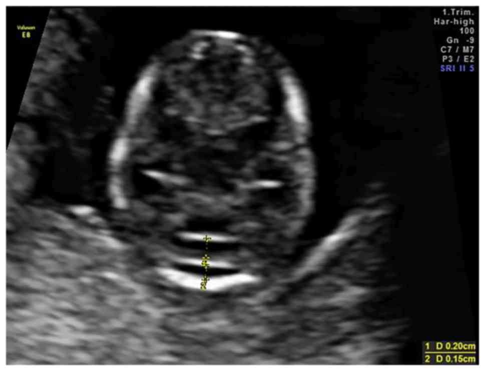
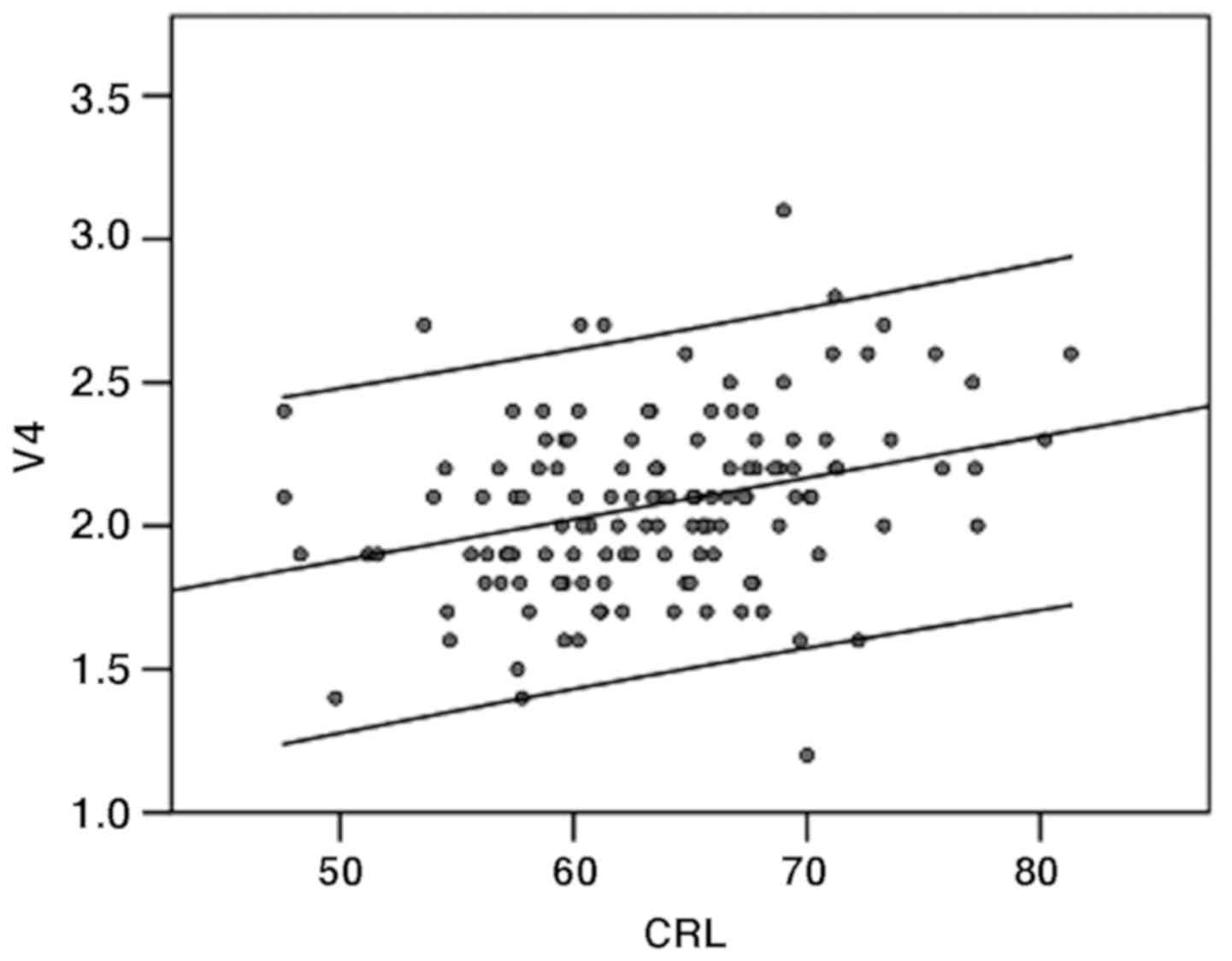
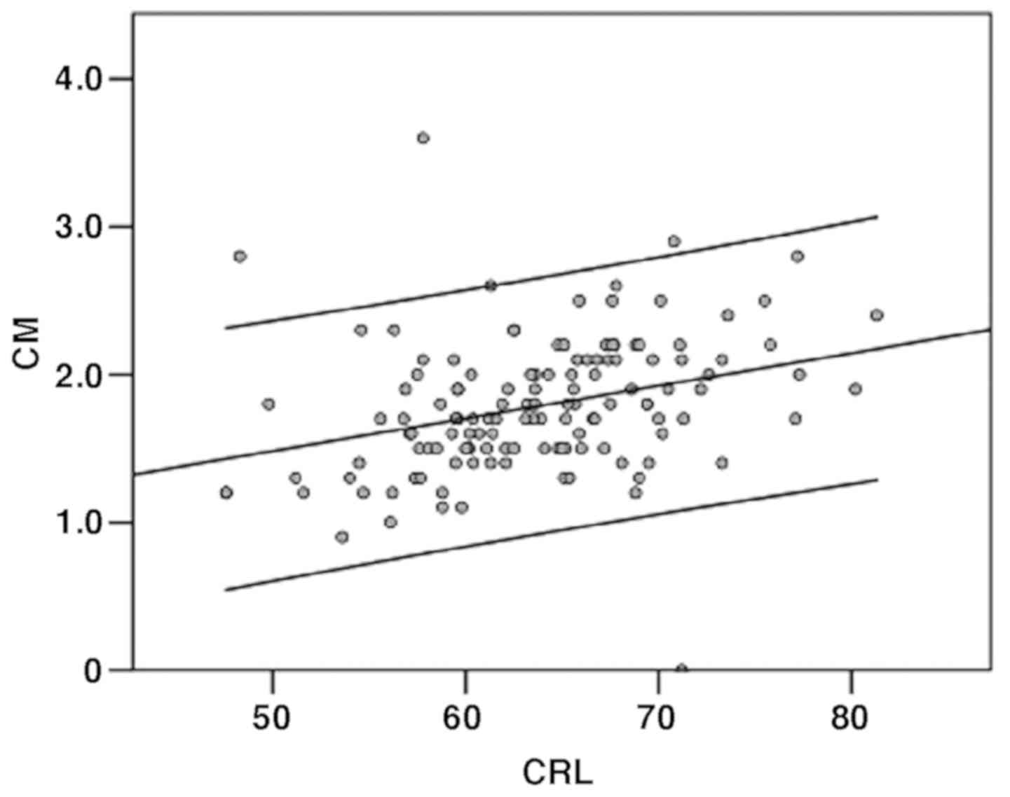
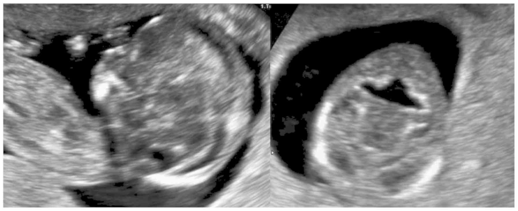
Posterior fossa ultrasound appearance may offer clues for brain anomalies as early as the first trimester. The purpose of the study was to find an easy, reproducible method to examine the posterior fossa. From January 2017 to March 2018, 132 consecutive pregnancies presenting for first‑trimester screening, were selected at 11‑14 weeks' gestation. An oblique axial view of the fetal head was routinely achieved with visualization of the posterior fossa, wherein the cisterna magna (CM) and the fourth ventricle (V4) anteroposterior diameter was measured. Moreover, 81 patients had a follow‑up scan at 19‑24 weeks, and the CM and transverse cerebellar diameter (TCD) were measured. Normal ranges were established at 11-14 weeks for CM and V4 according to crown‑rump length (CRL). The 50th centile for CM ranges from 1.2 mm to 2.3 mm at a CRL between 45 and 85 mm. The V4 50th centile ranges from 1.8 to 2.4 mm. A positive correlation was found between the first-trimester and second‑trimester CM diameter and between the first‑trimester V4 and second-trimester TCD. There is an inverse correlation between the first-trimester CM and second-trimester TCD. The measurements of different components of the posterior fossa in the first trimester cannot predict the size of CM and TCD in the second trimester. The presence of the three hypoechoic structures (cerebral peduncles, V4, and CM) separated by two hyperechoic lines is easy to see and measure.
View Figures
View References

Related Articles

Journal Cover

September-2020
Volume 20 Issue 3

Print ISSN: 1792-0981
Online ISSN:1792-1015

Sign up for eToc alerts

Recommend to Library

Copy and paste a formatted citation
x
Spandidos Publications style
Zvanca ME, Munteanu A, Bot M, Petca A and Nemescu D: Posterior fossa assessment in the axial view of the head at 11-14 weeks of gestation in normal and aneuploid fetuses. Exp Ther Med 20: 2470-2474, 2020
APA
Zvanca, M.E., Munteanu, A., Bot, M., Petca, A., & Nemescu, D. (2020). Posterior fossa assessment in the axial view of the head at 11-14 weeks of gestation in normal and aneuploid fetuses. Experimental and Therapeutic Medicine, 20, 2470-2474. https://doi.org/10.3892/etm.2020.8793
MLA
Zvanca, M. E., Munteanu, A., Bot, M., Petca, A., Nemescu, D."Posterior fossa assessment in the axial view of the head at 11-14 weeks of gestation in normal and aneuploid fetuses". Experimental and Therapeutic Medicine 20.3 (2020): 2470-2474.
Chicago
Zvanca, M. E., Munteanu, A., Bot, M., Petca, A., Nemescu, D."Posterior fossa assessment in the axial view of the head at 11-14 weeks of gestation in normal and aneuploid fetuses". Experimental and Therapeutic Medicine 20, no. 3 (2020): 2470-2474. https://doi.org/10.3892/etm.2020.8793