Introduction
Spinal fusion is a surgical procedure used to treat
several types of spinal disease, including spinal disc herniation,
spondylolisthesis and spinal stenosis (1), which is extensively performed
worldwide. The number of lumbar spinal fusion surgeries
approximately quadrupled between 1992-2013 in the United States,
which led to a significant increase in medical care enrollees, from
0.3-1.1 per 1,000(2). A previous
study reported that spinal fusion surgery accounts for the highest
total aggregate hospital costs compared with any other surgical
procedure performed in the United States medical care institution,
accounting for $12.8 billion in 2011(3). With the increase in the aging
population and the prevalence of degenerative spinal diseases, the
number of spinal surgeries are predicted to continue increasing. Of
the different types of spinal fusion techniques, posterolateral
fusion (PLIF) with autogenous bone graft is considered the gold
standard for lumbar spinal fusion (4).
Autogenous bone grafting has osteogenic,
osteoinductive and osteoconductive properties (5). Furthermore, it is histocompatible,
osteointegrative and does not pose the risk of disease transmission
or immune rejection (6).
Corticocancellous morcellized fragments and corticocancellous
struts from the iliac crest or the laminar process (locally) are
commonly used autologous grafts for PLIF (7). However, the fusion rates of these
autologous grafts have not yet been fully investigated. Therefore,
the present study aimed to prospectively compare the fusion rates
and effectiveness of corticocancellous structural autograft and
morcellized fragments autograft used in lumbar PLIF, for the
treatment of patients with stenosis. The primary outcome
measurements included the PLIF rate, radio density and dimensions
of the PLIF mass on both sides, which were assessed using X-ray at
3, 6 and 12 months postoperatively. Changes in bilateral bone
fusion bridges were assessed by CT according to the Lenke CT fusion
measurement criteria (4). The
present study suggested that corticocancellous structural autograft
is more effective for earlier resorption and stabilization of
patients undergoing PLIF, compared with that of the morcellized
fragments autograft.
Patients and methods
Study design
The prospective study was designed to evaluate the
radiological changes of bone fusion mass in 135 patients with
degenerative lumbar stenosis, who underwent PLIF surgery between
January 2013 and January 2016 in Honghui Hospital, Xi'an Jiaotong
Unviersity, School of Medicine (Xi'an, China). The present study
was approved by the Ethics Committee of Honghui Hospital (approval
no. 201000919) and performed according to the 2010 CONSORT
guidelines (http://www.consort-statement.org). Written informed
consent was provided by all patients prior to the start of the
study.
Inclusion and exclusion criteria
Patients with revision surgery (instrumental
failure, including screw fixation, rods fixation or fusion
failure), sagittal imbalance and scoliosis or patients with
pulmonary comorbidity or severe cardiac complications were excluded
from the present study. A total of 72 men and 63 women were
recruited, with an age range of 50-80 years (mean age, 65.7 years)
and the following up period is between January 2013 and January
2018 (24±2.1 months). All patients included in the present study
underwent one segment PLIF with pedicle screw fixation. The patient
demographics are presented in Table
I.
 | Table IPatient demographics. |
Table I
Patient demographics.
| Characteristic | Measurements |
|---|
| Sex | Total number of
patients, n |
|
Male | 72 |
|
Female | 63 |
| Mean age, years
(range) | 65.7 (50-80) |
| Diagnoses | Total number of
patients, na |
|
Back
pain | 89 |
|
Leg
pain | 125 |
| Numbness of lower
limb | 92 |
| Intermittent
claudication | 135 |
| | Mean (range) |
| ODI | 39.6 (34.0-41.0) |
| VAS (back) | 5.3 (0.0-8.0) |
| VAS (leg) | 7.8 (0.0-9.0) |
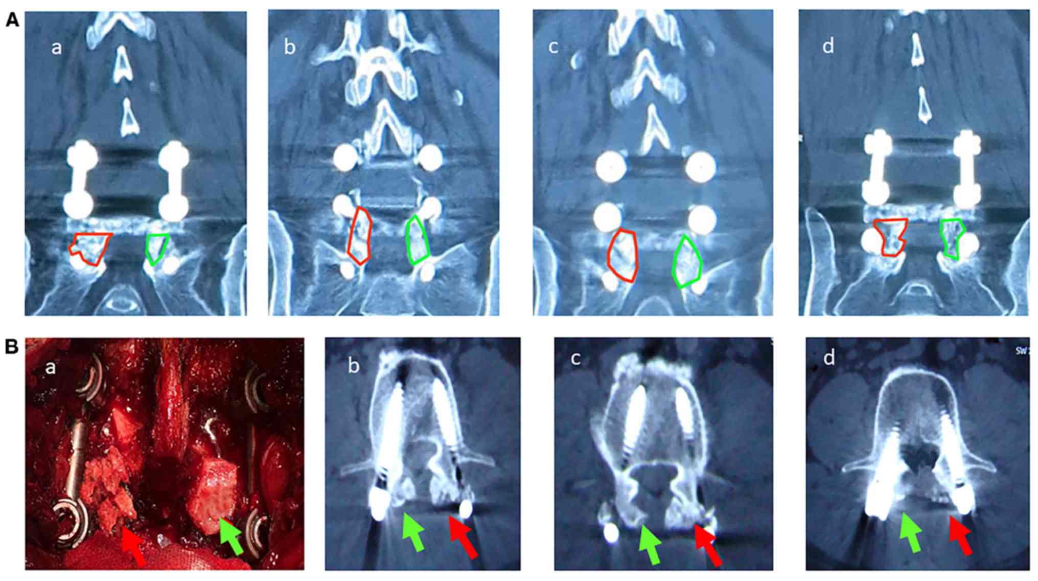
Surgical procedure and bone graft
Corticocancellous struts (group 1) and
corticocancellous morcellized fragments (group 2) surgeries were
performed within the same patient in each patient in the present
study. Briefly, a 3- to 6-inch long incision was made in the
midline of the back, and the left and right lower back muscles
(erector spinae) were stripped of the lamina on both sides, at
multiple levels. The lamina was removed via laminectomy on approach
to the spine, in order to visualize the nerve roots. Subsequently,
the facet joints, which lie directly above the nerve roots, were
trimmed to provide more space for the nerve roots. The nerve roots
were extended to one side while the disc space was cleared of all
material. A cage made of allograft bone, or posterior lumbar
interbody cages with bone graft was subsequently inserted into the
disc space to allow efficient bone growth, between the vertebral
bodies (2,5). The facet joints were decorticated and
bone grafting was performed by connecting each facet joint with the
local autologous corticocancellous struts (group 1) or
corticocancellous morcellized fragments (group 2). The amount of
autologous bone graft was equal for both groups (group 1; 4.5
cm3 local autologous corticocancellous morcellized
fragments and group 2; 3x1.5x1 cm local autologous
corticocancellous struts).
Radiographic analysis
Lumbar spinal CT (SOMATOM® Perspective;
Siemens Healthineers) scans and X-rays (Polydoros 80/100; Siemens
Healthineers) were performed at 3, 6 and 12 months,
postoperatively. Quantitative image density of bone mass fusion
from the AP X-rays was analyzed in both groups, as previously
described (8). Briefly, the mean
radio density on the X-rays was calculated using the picture
archiving and communication system, which outlines the frame of
bone fusion mass and titanium rod and via bone fusion mass that
divides the titanium rod (bone fusion mass/titanium rod).
Furthermore, the bilateral bone fusion mass dimensions were
measured using the ImageJ software (version 1.52; National
Institutes of Health). Bone graft volume was determined using the
axial 1-mm CT scans at 3 and 12 months postoperatively. A total of
three continuous CT images (100 kilovoltage/115 milliampere
seconds) were assessed for bone mass fusion according to the Lenke
CT fusion measurement criteria for PLIF (Table II), as previously described
(9). The bone fusion mass results
were divided into two groups, definitely fused and definitely not
fused, according to the reported hierarchical combination of the
fusion criteria, which were confirmed (Table II) and bilaterally compared between
groups 1 and 2.
| Table IILenke classification of posterolateral
fusion success. |
Table II
Lenke classification of posterolateral
fusion success.
| Grade | Description |
|---|
| Grade A | Solid, with the
presence of bilateral trabeculated stout fusion masses |
| Grade B | Possibly solid, with
the presence of a unilateral large fusion mass and a contralateral
small fusion mass |
| Grade C | Probably not solid,
with the presence of a bilateral small fusion mass |
| Grade D | Not solid, with the
presence of bone graft reabsorption or obvious bilateral
pseudarthrosis |
Statistical analysis
Statistical analysis was performed using GraphPad
Prism software (version 8; GraphPad Software, Inc.). The Linear
Mixed Model was used to statistically analyze the radiological
differences between radio density and dimensions of PLIF mass in
both groups, as previously described (9). Briefly, grey scale images from all
groups were analyzed using ImageJ software (version 1.52; National
Institutes of Health). Subsequently, McNemar's test was used to
compare differences in fusion rates between the groups (9,10).
P<0.05 was considered to indicate a statistically significant
difference.
Results
Demographics information of patients
included in the present study and fusion rate
The results demonstrated that spinal fusion was
completely achieved in patients with corticocancellous morcellized
fragments (group 1), as follows: In four patients at 3-months
postoperatively, in 27 patients at 6-months postoperatively and in
40 patients at 12-months postoperatively (Table III). However, 88 patients in group
1 failed to exhibit complete spinal fusion, whereby the fusion mass
was detected using X-rays and CT scans, but a definite fusion was
not achieved. Conversely, bone fusion was completely achieved in
patients with corticocancellous struts (group 2), as follows: In 37
patients at 3-months postoperatively, in 70 patients at 6-months
postoperatively and in 92 patients at 12-months postoperatively.
However, 36 patients in group 2 failed to exhibit complete spinal
fusion (Table III). The overall
fusion rate was significantly higher in group 1 (71.9%; 92/128)
compared with group 2 (31.3%; 40/128) (P<0.05; Table III).
| Table IIIFusion rate of follow-up data. |
Table III
Fusion rate of follow-up data.
| Time, months
(postoperatively) | Group | Grade A, n (%) | Grade B, n (%) | Grade C, n (%) | Grade D, n (%) |
|---|
| 3 | 1 | 42 (32.8) | 51 (39.8) | 31 (24.2) | 4 (3.1) |
| | 2 | 11 (8.6) | 29 (22.7) | 51 (39.8) | 37 (28.9) |
| 6 | 1 | 22 (17.2) | 33 (25.8) | 46 (35.9) | 27 (21.1) |
| | 2 | 7 (5.5) | 19 (14.8) | 32 (25.0) | 70 (54.7) |
| 12 | 1 | 21 (16.4) | 25 (19.5) | 42 (32.8) | 40 (31.3) |
| | 2 | 5 (3.9) | 12 (9.4) | 19 (14.8) | 92 (71.9) |
Radiographic analysis results
Radiographic analysis included X-rays and CT scans,
where mean density and dimensions of bone fusion masses were
determined using the lumbar spine AP images. The mean radio
densities of group 2 (0.5067±0.01581, 0.6102±0.01322 and
0.6739±0.01553) were significantly higher than the mean densities
of group 1 (0.301±0.01741, 0.3991±0.02081 and 0.4907±0.01079) at 3,
6 and 12 months postoperatively, respectively. Similarly, the
dimensions of the fusion masses were significantly higher in group
2 (470.0±5.627, 410.0±6.205 and 351±6.991 mm2) compared
with group 1 (420.3±5.332, 332.0±4.031 and 261±6.011
mm2) at 3, 6 and 12 months postoperatively, respectively
(P<0.05; Figs.
1-2; Table III).
Bone fusion success was evaluated via CT scanning at
3, 6 and 12 months postoperatively. The mean volumes were
significantly higher in group 2 (4.970±0.02739, 4.281±0.0211 and
3.191±0.0341 cm3) compared with group 1 (4.609±0.02981,
3.610±0.01991 and 2.330±0.01881 cm3) at 3, 6 and 12
months postoperatively, respectively (P<0.05; Fig. 1). This finding suggests that during
bone graft incorporation, the bone graft was initially partially
resorbed and was subsequently remodeled.
Discussion
Spine fusion is a surgical procedure used to treat
different types of spinal disease, including severe spine trauma,
spinal infection, spinal deformities and spinal degenerative
diseases (11). With the rapid
progression of surgical techniques and broadening indications,
there has been a rapid increase in spinal fusion surgery (12). However, several factors may lead to
the failure of solid fusion, such as pseudarthrosis, which is a
major iatrogenic complication (6).
Thus, the present study investigated fusion for corticocancellous
structural autograft vs. morcellized fragments autograft in
patients who underwent decompressive single level lumbar
laminectomy and one segment PLIF with pedicle screw fixation. To
the best of our knowledge, the present study was the first to
compare the fusion rates of two types of structural allografts used
for PLIF. The results demonstrated that the corticocancellous
structural autograft had a better fusion rate in patients with PLIF
compared with the morcellized autograft.
The results of the present study demonstrated that
during bone graft incorporation, the bone graft is initially partly
resorbed and subsequently remodeled. However, this resorptive phase
may weaken the bone graft, particularly during the initial months
postoperatively. Several factors may affect the fusion rate.
Previous studies have reported that decreased bone graft volume
decreases the mass, which consolidates into a thick bone mass and
in turn fails to significantly increase the fusion mass (5,7,13). Conversely, increasing the bone graft
volume has been demonstrated to induce extensive bone resorption,
which in turn decreases the bone matrix for new bone construction,
resulting in failure of spinal fusion (14). Thus, the bone graft volume and
structural changes essentially determine the fusion rate success.
The present study investigated the differences in the fusion rate
between the local autologous corticocancellous struts and
corticocancellous morcellized fragments, using the same volume.
The results demonstrated simultaneous (at 3 months)
and short-term (at 6 months) fusion rates in group 2 (28.9 and
54.7%) and group 1 (3.1 and 21.1%), respectively. These results
suggested that the autologous corticocancellous strut is a better
choice for patients undergoing PLIF for earlier lumbar fusion.
Furthermore, the overall fusion rates at 12 months were 71.9 and
31.3% in groups 2 and 1, respectively. A previous study reported
that when autologous iliac bone was used for PLIF, the fusion rate
was increased from 40 to 98% (15).
The fusion rate in group 1 in the present study was consistent with
this previously reported range. However, the fusion rate in group 2
was significantly lower compared with that in group 1 and with that
of the previous study. Comparisons between groups 1 and 2 in the
present study indicated that local autologous corticocancellous
struts attenuates the risk of pseudoarthrosis.
The present study is not without limitations.
Firstly, all surgeries were performed by two surgeons from the same
institution (Honghui Hospital). Secondly, the follow-up period was
relatively short. Thirdly, the present study only examined patients
who underwent one-level PLIF.
In conclusion, the results of the present study
demonstrated that the short-term fusion rates were higher with
corticocancellous structural autografts compared with morcellized
fragments autografts for PLIF procedures. Thus, corticocancellous
structural autografts may be developed as a safe and effective
clinical algorithm by surgeons to provide optimal bone fusion in
patients undergoing posterolateral lumbar fusion.
Acknowledgements
Not applicable.
Funding
The present study was funded by the Shaanxi Social
Development Science and Technology Project (grant no. 2016SF-227)
and the Xi'an Social Development Science and Technology Project
(grant no. 2017TTSSF/YX009).
Availability of data and materials
The datasets used and/or analyzed during the current
study are available from the corresponding author on reasonable
request.
Authors' contributions
XY, DH and BH designed the present study and
analyzed the data. DW, LY and YH analyzed the data. XY and BH
acquired the data, while BH and XY drafted the initial manuscript.
All authors have read and approved the final manuscript.
Ethics approval and consent to
participate
The present study was approved by the Ethics
Committee of Honghui Hospital (approval no. 201000919) and
performed according to the 2010 CONSORT guidelines (http://www.consort-statement.org). Written
informed consent was provided by all patients prior to the
commencement of the study.
Patient consent for publication
Not applicable.
Competing interests
The authors declare that they have no competing
interests.
References
|
1
|
Rizkalla N, Zane NR, Prodell JL, Elci OU,
Maxwell LG, DiLiberto MA and Zuppa AF: Use of intravenous
acetaminophen in children for analgesia after spinal fusion
surgery: A randomized clinical trial. J Pediatr Pharmacol Ther.
23:395–404. 2018.PubMed/NCBI View Article : Google Scholar
|
|
2
|
Liu JJ, Raskin JS, Hardaway F, Holste K,
Brown S and Raslan AM: Application of lean principles to
neurosurgical procedures: The case of lumbar spinal fusion surgery,
a literature review and pilot series. Oper Neurosurg (Hagerstown).
15:332–340. 2018.PubMed/NCBI View Article : Google Scholar
|
|
3
|
Wahlman M, Häkkinen A, Dekker J, Marttinen
I, Vihtonen K and Neva MH: The prevalence of depressive symptoms
before and after surgery and its association with disability in
patients undergoing lumbar spinal fusion. Eur Spine J. 23:129–134.
2014.PubMed/NCBI View Article : Google Scholar
|
|
4
|
Lu VM, Ho YT, Nambiar M, Mobbs RJ and Phan
K: The perioperative efficacy and safety of antifibrinolytics in
adult spinal fusion surgery: A systematic review and meta-analysis.
Spine. 43:E949–E958. 2018.PubMed/NCBI View Article : Google Scholar
|
|
5
|
Ohashi M, Hirano T, Watanabe K, Katsumi K,
Shoji H, Mizouchi T and Endo N: Bone mineral density after spinal
fusion surgery for adolescent idiopathic scoliosis at a minimum
20-year follow-up. Spine Deform. 6:170–176. 2018.PubMed/NCBI View Article : Google Scholar
|
|
6
|
Lewis SJ, Arun R, Bodrogi A, Lebel DE,
Magana SP, Dear TE and Witiw C: The use of fusion mass screws in
revision spinal deformity surgery. Eur Spine J. 23 (Suppl
2):181–186. 2014.PubMed/NCBI View Article : Google Scholar
|
|
7
|
Rushton A, Staal JB, Verra M, Emms A,
Reddington M, Soundy A, Cole A, Willems P, Benneker L, Masson A, et
al: Patient journey following lumbar spinal fusion surgery (LSFS):
Protocol for a multicentre qualitative analysis of the patient
rehabilitation experience (FuJourn). BMJ Open.
8(e020710)2018.PubMed/NCBI View Article : Google Scholar
|
|
8
|
Choi JH, Jang JS, Yoo KS, Shin JM and Jang
IT: Functional limitations due to stiffness after long-level spinal
instrumented fusion surgery to correct lumbar degenerative flat
back. Spine. 43:1044–1051. 2018.PubMed/NCBI View Article : Google Scholar
|
|
9
|
Liu G, Tan JH, Yang C, Ruiz J and Wong HK:
A Computed tomography analysis of the success of spinal fusion
using ultra-low dose (0.7 mg per facet) of recombinant human bone
morphogenetic protein 2 in multilevel adult degenerative spinal
deformity surgery. Asian Spine J. 12:1010–1016. 2018.PubMed/NCBI View Article : Google Scholar
|
|
10
|
Imagama S, Ando K, Kobayashi K, Ishikawa
Y, Nakamura H, Hida T, Ito K, Tsushima M, Matsumoto A, Morozumi M,
et al: Efficacy of early fusion with local bone graft and
platelet-rich plasma in lumbar spinal fusion surgery followed over
10 years. Global Spine J. 7:749–755. 2017.PubMed/NCBI View Article : Google Scholar
|
|
11
|
Ling T, Liu L, Yang X, Qiang Z, Hu X and
An Y: Revision surgery for spinal tuberculosis with secondary
deformity after treatment with debridement, instrumentation, and
fusion. Eur Spine J. 24:577–585. 2015.PubMed/NCBI View Article : Google Scholar
|
|
12
|
Yamato Y, Hasegawa T, Kobayashi S, Yasuda
T, Togawa D, Yoshida G, Banno T, Oe S, Mihara Y and Matsuyama Y:
Treatment strategy for rod fractures following corrective fusion
surgery in adult spinal deformity depends on symptoms and local
alignment change. J Neurosurg Spine. 29:59–67. 2018.PubMed/NCBI View Article : Google Scholar
|
|
13
|
Jain A, Sponseller PD, Kebaish KM and
Mesfin A: National trends in spinal fusion surgery for scheuermann
kyphosis. Spine Deform. 3:52–56. 2015.PubMed/NCBI View Article : Google Scholar
|
|
14
|
Senker W, Gruber A, Gmeiner M, Stefanits
H, Sander K, Rössler P and Pflugmacher R: Surgical and clinical
results of minimally invasive spinal fusion surgery in an
unselected patient cohort of a spinal care unit. Orthop Surg.
10:192–197. 2018.PubMed/NCBI View
Article : Google Scholar
|
|
15
|
Wortham TC, Rice AN, Gupta DK and Goode V:
Implementation of an obstructive sleep apnea protocol in the
postanesthesia care unit for patients undergoing spinal fusion
surgery. J Perianesth Nurs. 34:739–748. 2019.PubMed/NCBI View Article : Google Scholar
|