Introduction
Breast cancer is one of the most common malignant
tumors among women. In recent years, the rising incidence of breast
cancer which has exceeded that of cervical cancer has attracted the
attention of medical societies (1).
Due to the hidden nature of breast cancer symptoms at early stage,
most patients come to the hospital at the middle or late stage of
breast cancer, missing the best treatment opportunity. Early
diagnosis and treatment are essential for better prognosis
(2). The preferred diagnostic method
for breast cancer is color Doppler ultrasonography. Ultrasound can
show the internal structure and blood flow in breast masses, and
can reveal the shape, boundary and activity of the lesions. In
addition, ultrasound can distinguish solid and cystic masses
(3). However, its application has
certain limitations as the distinction between small lesions and
some benign and malignant masses is very difficult. In recent
years, with the development of molecular biology techniques, more
tumor molecular markers related to breast cancer have been
discovered. The evaluation of the expression levels of these
markers in the serum of patients can provide precious information
related to the occurrence, invasion and metastasis of breast cancer
(4). Among these markers,
carbohydrate antigen 153 (CA153), carcinoembryonic antigen (CEA)
and tumor specific growth factor (TSGF) are closely related to
breast cancer occurrence and their detection could be used as an
auxiliary examination method for breast cancer diagnosis. Although
the combined diagnostic method could improve the accuracy of breast
cancer diagnosis, no standard diagnostic method has been developed.
In the present study, color Doppler ultrasound combined with the
detection of serum markers CA153, CEA and TSGF was used in the
diagnosis of breast cancer, aiming to provide future reference for
the early diagnosis of breast cancer.
Patients and methods
Clinical data
From January 2017 to June 2018, a total of 103
patients with breast cancer admitted to Yantaishan Hospital
(Yantai, China) were enrolled in the study and served as the breast
cancer group. All cases were confirmed by pathological and
histological examinations. Patients were all females, 35-79 years
of age, with an average age of 56.32±12.47 years. Regarding tumor
location, there were 63 patients with tumors in the left breast, 37
patients with tumors in the right breast and 3 patients with
bilateral breast cancer. According to the tumor-node-metastasis
classification standard of breast cancer published by the Union for
International Cancer Control (5),
there were 26 cases at stage I, 35 cases at stage II, 30 cases at
stage III and 12 cases at stage IV. The histopathological types of
cancer included carcinoma in situ, invasive ductal
carcinoma, invasive lobular carcinoma and medullary carcinoma.
Furthermore, 50 patients with benign breast lesions were selected
as the benign lesion group. All patients in the benign lesion group
were females, 33-80 years of age, with an average age of
55.32±13.01 years. Canceration was excluded by pathological
histology, including breast fibroadenoma, breast papilloma, breast
hyperplasia and breast cyst. There was no statistically significant
difference between the two groups in terms of sex, age and other
general characteristics (P>0.05).
Inclusion criteria for breast cancer patients: i)
All patients met the criteria of the breast cancer staging system
(6) as confirmed by pathological
examinations; ii) patients had not been diagnosed before with
breast cancer and had not received radiotherapy, chemotherapy or
other endocrine therapy before treatment; iii) patients underwent
color Doppler ultrasound and serum CA153, CEA and TSGF
examinations. Exclusion criteria: Patients with related tumor
diseases or liver and kidney dysfunction. The study was approved by
the Ethics Committee of Yantaishan Hospital. Patients who
participated in the study had complete clinical data. Signed
written informed consents were obtained from the patients and/or
guardians.
Color Doppler ultrasound
examination
Color Doppler ultrasound diagnosis was based on the
Breast Imaging-Reporting and Data System developed by the American
College of Radiology for the evaluation of breast cancer imaging
(7). Philips iU22 (Philips
Healthcare) was the diagnostic instrument used and the probe
frequency was 5-12 MHz. Patients were instructed to lie in supine
position, lift their arms and place their hands behind the head in
order to fully expose the bilateral breasts, supraclavicular fossa
and bilateral axillary regions. A probe was used to scan radially
along the areola with the nipple as the center. At the same time,
the supraclavicular fossa and bilateral armpits were scanned.
Firstly, the location, shape, size, boundary, internal echo,
calcification, correlation with surrounding tissues, axillary lymph
node metastasis and other acoustic images were observed. Next, the
morphology and distribution of blood flow signals inside and around
the focus were examined and hemodynamic parameters were measured
(8).
Serum tumor markers
Specimen collection and
pretreatment
A total of 4 ml of cubital venous blood were
collected from all patients in both groups, on an empty stomach at
7 a.m., before treatment and 3 months after treatment. After
autocoagulation at 25˚C, the blood samples were centrifuged at
2,264 x g for 20 min at 25˚C and the upper serum was collected for
examination. Lipids and hemolysis were excluded from all
specimens.
Determination of serum CA153 and CEA
expression levels
An electrochemiluminescence immunoassay was used for
the detection of CA153 and CEA expression levels, following
the principles of double antibody sandwich. The instrument used was
Roche Elecsys 2010 with CA153 (cat. no. 03045838122) and CEA (cat.
no. 11731629322) diagnostic reagents provided by Roche Applied
Science. Step 1: Each sample was firstly diluted (1:10) with
Elecsys universal diluent. CA153 and CEA antigens formed immune
sandwich complexes with biotinylated anti-CA153 and CEA monoclonal
antibodies and ruthenium-labeled anti-CA153 and CEA antibodies in
the reagent, respectively. Step 2: Streptavidin-coated particles
were automatically added by Elecsys 2010, allowing the complexes to
bind to the particles through the reaction of biotin with
streptavidin. Step 3: The reaction mixture was sucked into the
cells and the particles were attracted to the electrode by a
magnet. The free substance was washed away by a cleaning solution
and the immune complex generated chemiluminescence after voltage
was applied to the electrodes. Chemiluminescence activity was
measured by a photomultiplier tube (Roche Applied Science). Step 4:
The standard curve was calibrated by the instrument via a 2-point
calibration. The measurement results were automatically detected
from the standard curve. When the sample exceeded the linear range,
the instrument automatically diluted the sample using a universal
diluent. The whole process was fully automatic and the actual serum
dosage in each experiment was 20 µl.
TSGF detection
Chemical colorimetric assay was performed using a
Hitachi 7600-110E (Hitachi, Ltd.) with TSGF kit (cat. no. A0229)
provided by Newland Biotech. All operation steps were carried out
following strictly the manufacturer's instructions. Step 1: TSGF
reagents, calibrators and quality control materials were positioned
as designated. Step 2: The samples were placed at the specified
position to perform the test. After the completion of the test, the
results were obtained based on the standard curve.
Result determination
With pathological diagnosis as the gold standard,
the cases in the breast cancer group with consistent results of
ultrasound and pathological diagnosis were determined as true
positives. The cases with inconsistent results (misdiagnosis,
missed diagnosis and uncertainty) were determined as false
negatives. In the benign lesion group, the cases with consistent
results of ultrasound and pathological diagnosis were determined as
true negatives, whereas the cases with inconsistent results
(misdiagnosis, uncertainty) were determined as false positive. When
one or more items in the combined detection (parallel test) group
were positive, the case was determined as positive. If all items
were negative, the case was determined as negative.
Statistical analysis
SPSS 19.0 software (IBM Corp.) was used for the
statistical analysis of the data. Measured data were expressed as
the mean ± standard deviation (mean ± SD) and the comparison of the
mean values between groups was made using independent samples
t-test. The comparison of the counting data was made using
χ2 test. Receiver operating characteristics (ROC) curve
analysis of CA153, CEA and TSGF was also performed and the optimal
cut-off values for CA153, CEA and TSGF in diagnosing breast cancer
were determined according to the maximum Youden index. P<0.05
was considered to indicate a statistically significant
difference.
Results
Results of color Doppler
ultrasonography in the breast cancer and the benign lesion
group
A total of 103 cases of breast cancer were examined
by color Doppler ultrasound. The pathological results were
consistent with the ultrasound results in 80 cases (confirmed in 80
cases) and inconsistent in 23 cases (misdiagnosed in 8 cases,
missed in 3 cases, uncertain in 12 cases). In the benign lesion
group (n=50), the pathological results were consistent with the
ultrasound results in 42 cases (confirmed in 42 cases) and
inconsistent in 8 cases (misdiagnosed in 3 cases and uncertain in 5
cases).
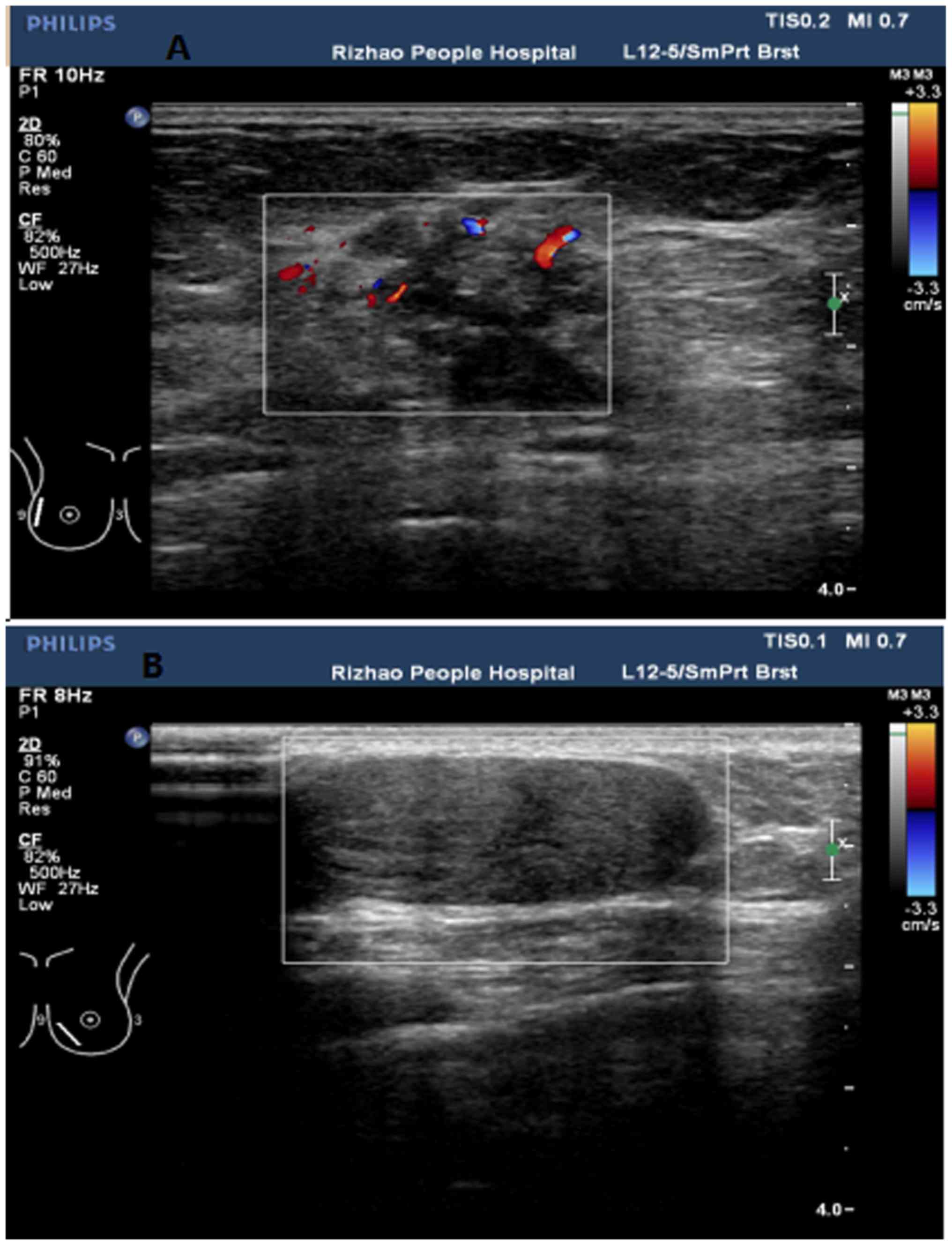
Color Doppler ultrasound detection showed that there
were statistically significant differences between the two groups
in terms of tumor boundary, morphology, internal echo,
calcification, peak blood flow velocity (Vmax),
resistance index (RI), pulsatility index (PI) and blood flow
classification (0, I, II, III) (P<0.05; Tables I and II). Color Doppler ultrasound images of
breast cancer showed the unclear boundaries of the tumors, the
tumors' irregular shape, inhomogeneous internal echo,
calcification, as well as the increase of Vmax, RI and
PI of blood flow. The internal blood flow of the tumors was also
shown to be rich. Representative ultrasonic images are shown in
Fig. 1.
 | Table IComparison of color Doppler
ultrasonography features between patients with breast cancer and
patients with benign lesions [n (%)]. |
Table I
Comparison of color Doppler
ultrasonography features between patients with breast cancer and
patients with benign lesions [n (%)].
| | Boundary | Internal echo | Morphology | Calcification |
|---|
| Group | Clear | Unclear | Homogeneous | Inhomogeneous | Regular | Irregular | No | Yes |
|---|
| Breast cancer group
(n=103) | 18
(17.48)a | 85
(82.52)a | 25
(24.27)a | 78
(75.73)a | 16
(15.53)a | 87
(84.47)a | 14
(13.59)a | 89
(86.41)a |
| Benign lesion group
(n=50) | 40 (80.00) | 10 (20.00) | 35 (70.00) | 15 (30.00) | 38 (76.00) | 12 (24.00) | 41 (82.00) | 9 (18.00) |
| Table IIComparison of color Doppler ultrasound
blood flow features between patients with breast cancer and
patients with benign lesions [mean ± SD, n (%)]. |
Table II
Comparison of color Doppler ultrasound
blood flow features between patients with breast cancer and
patients with benign lesions [mean ± SD, n (%)].
| | | | | Blood flow signal
classification |
|---|
| Group | Vmax
(cm/sec) | RI | PI | 0 | I | II | III |
|---|
| Breast cancer group
(n=103) |
25.63±3.26a |
0.82±0.05a |
1.65±0.31a | 6 (5.83)a | 15
(14.56)a | 27
(26.21)a | 55
(53.40)a |
| Benign lesion group
(n=50) | 13.69±2.01 | 0.61±0.03 | 1.02±0.21 | 25 (50.00) | 16 (32.00) | 8 (16.00) | 1 (2.00) |
Comparison of serum tumor markers
between the breast cancer and the benign lesion group
The serum levels of tumor markers CA153, CEA and
TSGF in the patients with breast cancer were significantly higher
than those in the patients with benign lesions (P<0.01; Table III). The expression levels of these
tumor markers in patients with high-stage cancer (III, IV),
recurrence and metastasis of breast cancer were significantly
higher than those with low-stage cancer (I, II), no recurrence and
no metastasis. The expression levels of CA153, CEA and TSGF after
breast cancer treatment were significantly lower than those before
treatment (P<0.01; Table
IV).
| Table IIIComparison of the expression levels of
serum tumor markers between patients with breast cancer and
patients with benign lesions (mean ± SD). |
Table III
Comparison of the expression levels of
serum tumor markers between patients with breast cancer and
patients with benign lesions (mean ± SD).
| Group | CAl53 (U/ml) | CEA (ng/ml) | TSGF (U/ml) |
|---|
| Breast cancer group
(n=103) |
72.61±23.40a |
36.24±16.25a |
157.69±46.72a |
| Benign lesion group
(n=50) | 14.67±8.76 | 2.36±0.82 | 50.24±15.61 |
| Table IVComparison of CA153, CEA and TSGF
serum levels in patients with different clinical features in the
breast cancer group (mean ± SD). |
Table IV
Comparison of CA153, CEA and TSGF
serum levels in patients with different clinical features in the
breast cancer group (mean ± SD).
| Group | Cases | CAl53 (U/ml) | CEA (ng/ml) | TSGF (U/ml) |
|---|
| Clinical stage | | | | |
|
I+II | 61 | 46.92±12.29 | 23.58±10.12 | 120.53±36.12 |
|
III+IV | 42 |
110.83±36.12a |
55.64±23.14a |
212.09±70.21a |
| Distant
metastasis | | | | |
|
No | 91 | 56.83±15.64 | 26.54±11.15 | 128.72±40.15 |
|
Yes | 12 |
196.17±67.23a |
110.48±36.57a |
378.33±97.26a |
| Recurrence | | | | |
|
No | 82 | 19.37±9.21 | 5.62±2.73 | 96.31±22.14 |
|
Yes | 21 |
96.49±43.27a |
47.37±18.87a |
203.39±66.42a |
| Treatment | | | | |
|
Before | 103 | 72.61±23.40 | 36.24±16.25 | 157.69±46.72 |
|
After | 103 |
34.21±20.15a |
13.52±6.82a |
107.82±32.97a |
Comparison of the clinical value of
color Doppler ultrasound, CA153, CEA, TSGF single and combined
detection in the diagnosis of breast cancer
ROC curve analysis revealed that the area under the
curve (AUC) of CA153, CEA and TSGF in the diagnosis of breast
cancer was 0.812, 0.807 and 0.843, respectively, and the maximum
Youden index was 0.568, 0.521 and 0.560, respectively. The best
cut-off values were 25.00 U/ml for CA153, 3.40 ng/ml for CEA and
70.00 U/ml for TSGF. The consistency of the results of
ultrasonography, CA153, CEA, TSGF single and combined detection
with the results of the pathological diagnosis is presented in
Table V. The sensitivity, accuracy
and negative predictive value of color Doppler ultrasound combined
with serum CA153, CEA and TSGF detection in the diagnosis of breast
cancer were 95.15, 90.20 and 88.89% respectively, which were
significantly higher than those of any single examination
(P<0.01; Table VI).
| Table VAssociation of color Doppler
ultrasound, CA153, CEA, TSGF single and combined detection results
with pathological diagnosis (n). |
Table V
Association of color Doppler
ultrasound, CA153, CEA, TSGF single and combined detection results
with pathological diagnosis (n).
| | | Pathological
diagnosis |
|---|
| Detection | Detection
result | Positive | Negative |
|---|
| Ultrasound | Positive | 80 | 8 |
| | Negative | 23 | 42 |
| CA153 | Positive | 66 | 4 |
| | Negative | 37 | 46 |
| CEA | Positive | 64 | 5 |
| | Negative | 39 | 45 |
| TSGF | Positive | 68 | 5 |
| | Negative | 35 | 45 |
| Combined
detection | Positive | 98 | 10 |
| | Negative | 5 | 40 |
| Table VIComparison of the clinical value of
color Doppler ultrasound, CA153, CEA, TSGF single and combined
detection in the diagnosis of breast cancer [% (ratio)]. |
Table VI
Comparison of the clinical value of
color Doppler ultrasound, CA153, CEA, TSGF single and combined
detection in the diagnosis of breast cancer [% (ratio)].
| Detection | Sensitivity | Specificity | Accuracy | Positive predictive
value | Negative predictive
value |
|---|
| Ultrasound | 77.67 (80/103) | 84.00 (42/50) | 79.74
(122/153) | 90.91 (80/88) | 64.62 (42/65) |
| CA153 | 64.08 (66/103) | 92.00 (46/50) | 73.20
(112/153) | 94.29 (66/70) | 55.42 (46/83) |
| CEA | 62.14 (64/103) | 90.00 (45/50) | 71.24
(109/153) | 92.75 (64/69) | 53.57 (45/84) |
| TSGF | 66.02 (68/103) | 90.00 (45/50) | 73.86
(113/153) | 93.15 (68/73) | 56.25 (45/80) |
| Combined
detection | 95.15
(98/103)a | 80.00 (40/50) | 90.20
(138/153)a | 90.74 (98/108) | 88.89
(40/45)a |
| χ2 | 39.652 | 1.515 | 20.892 | 1.808 | 18.873 |
| P-value | <0.001 | 0.341 | <0.001 | 0.771 | 0.001 |
Discussion
Results obtained in a previous study (9) have shown that the early diagnosis of
breast cancer can assist early clinical intervention and improve
the patients' long-term survival rate and overall quality of life.
Considering the number of women affected by breast cancer, to find
a simple and effective early screening method for the early
diagnosis and treatment of breast cancer is essential.
Ultrasound examination is the first choice for
breast disease screening. This method, which is the most widely
used breast cancer screening method, is suitable for all age groups
and is convenient, non-invasive, non-radioactive, repeatable, and
with good patient compliance. In particular, Doppler high-frequency
ultrasound has high resolution of details and can clearly display
the location, shape, size, boundary, internal structure, echo,
calcification and other conditions of the lesion. In addition,
Doppler high-frequency ultrasound can clearly visualize the
surrounding tissues determining invasion (10). With the increase of probe frequency
and the application of blood flow imaging technology, Doppler
high-frequency ultrasound can clearly reflect hemodynamic
information by detecting tumor angiogenesis and peripheral blood
flow characteristics of lesions to further identify benign and
malignant breast masses (11),
especially cystic and solid masses (12). For dense breast masses, the location
and shape can be clearly shown (13). However, ultrasound examination has
certain limitations, such as low sensitivity to calcifications and
limited ability to accurately identify calcification at the early
stages of breast cancer (14).
Ultrasound examination has also low sensitivity to the diagnosis of
carcinoma-in situ and breast cancer with maximum mass
diameter <1 cm. As the ultrasound image of a small mass is not
easy to obtain and there is no obvious typical signs of breast
cancer, a small mass can be easily misidentified as a benign
lesion. Furthermore, some benign masses are indistinguishable from
malignant tumors in terms of image characteristics due to
degeneration and ischemia of tissues around the lesions and
internal structural disorder of the masses. Moreover, in some cases
it has been difficult to confirm some unusual ultrasound images
(15). Therefore, there is always a
probability of misdiagnosis and missed diagnosis when Doppler
ultrasound is used alone. The results of the present study showed
that the sensitivity and accuracy of color Doppler ultrasound in
the diagnosis of breast cancer were 77.67 and 79.74%, respectively
which were not satisfactory.
Tumor markers are molecules secreted by tumor cells
or produced by the interaction between tumor and host cells during
carcinogenesis. The occurrence or level variation of the tumor
markers reflects the existence of a tumor. Such markers can be
detected in tissues or body fluids (16). During cell canceration a dramatic
increase in the serum levels of these markers is observed, as tumor
markers often appear before clinical symptoms. As an in
vitro diagnostic method, tumor marker detection is a low cost
method with low risk. In addition, tumor marker detection is
simple, rapid, quantitative and dynamic and is a commonly used
method for the detection of malignant tumors, early diagnosis and
prognosis monitoring. However, the results obtained by this method
can be affected by various in vitro and in vivo
factors, as well as experimental errors. Detection of tumor markers
is prone to false positives and false negatives, and therefore the
results should be judged with caution.
CA153 is a variant glycoprotein on the surface of
breast cells. During malignancy, the activities of salivary enzymes
and proteases on the cell membrane are enhanced and the
cytoskeleton is destroyed, leading to a fall in cell surface
antigen as they are released into the blood stream. The release of
CA153 into the blood increases the expression level of this marker
in the peripheral blood (17). CA153
is currently used as the most classical tumor marker for screening
breast cancer. It has been reported (18) that 80% of patients with breast cancer
metastasis have a high level of serum CA153 and the CA153 level has
been reported to be positively correlated with the recurrence and
metastasis of breast cancer. Currently, in clinical practice, CA153
is the preferred indicator for monitoring the disease condition.
However, there are a few limitations associated with the use of
this marker. For example, the level of CA153 in the peripheral
blood does not always increase during the early stages of breast
cancer (0 and I). In addition, a transient increase in CA153 levels
(false positive) can be occasionally observed in some benign breast
diseases (such as, breast papilloma and breast cysts).
CEA was first found in fetal intestinal and colon
cancer tissues. The antigenic determinants of CEA have embryonic
characteristics, which explains the name ‘carcinoembryonic
antigen’. The level of CEA in healthy individuals is extremely low
(in most cases below 3.40 ng/ml). When healthy cells transform into
cancerous cells, the quantity of CEA secreted by cancer cells rises
dramatically. The secreted CEA is diffused in cell membrane,
cytoplasm and stroma, although CEA can also be secreted into the
cavity edge of cancer gonad tube and its peripheral body fluid
(19). Often, CEA levels during the
early stages of breast cancer do not show a meaningful rise. These
low levels of CEA can be explained by the fact that during the
early stages of breast cancer the cells have not yet penetrated the
basement membrane and CEA has not been secreted into the peripheral
blood. Serum CEA expression levels are of great value in the
assessment of breast cancer clinical staging, recurrence and
metastasis. Usually, the serum levels of CEA are significantly high
during the late stages. In some cases, a transient increase in CEA
levels in peripheral blood has been reported in smokers which may
lead to false positive results.
TSGF, also known as malignant tumor specific growth
factor, is a vascular endothelial growth factor involved in the
proliferation of malignant tumors and peripheral capillaries. TSGF
is released into the blood stream at the early stages of malignant
tumor formation and has no obvious correlation with non-tumor
vascular proliferation. TSGF is a powerful indicator in
differentiating the cancer cells from the non-cancer cells. As a
broad-spectrum tumor marker, TSGF is a valuable marker for the
early detection of breast cancer (20). TSGF, certainly, has the merits to be
used as an effective screening indicator of breast cancer. After
the remission or disappearance of the tumor, the serum levels of
TSGF have been reported to decline significantly (21). TSGF can be used for evaluation and
prognosis follow ups; however, its sensitivity and specificity are
still a matter of debate.
In the present study, color Doppler ultrasound
combined with the detection of the serum markers CA153, CEA and
TSGF was used in the diagnosis of breast cancer. The results
revealed that the combined examinations could complement and
confirm each other. Ultrasound can preliminarily distinguish benign
and malignant breast tumors based on acoustic characteristics.
CA153, CEA and TSGF levels reflect the occurrence and development
of breast cancer in terms of molecular biology. The combination of
these detection methods makes up for the limitations of each single
test. The sensitivity, accuracy and negative predictive value of
the combined detection were 95.15, 90.20 and 88.89%, which were
significantly higher than those of the single tests, suggesting
that the combined detection could reduce misdiagnosis. However, the
number of patients included in the present study was relatively
small and the study of a bigger sample size will be the aim of our
future research.
In conclusion, color Doppler ultrasound combined
with the detection of serum markers CA153, CEA and TSGF
significantly improved the diagnosis of breast cancer suggesting
that the combined detection method could be used as an effective
tool that could improve breast cancer diagnosis leading to early
diagnosis and clinical intervention.
Acknowledgements
Not applicable.
Funding
No funding was received.
Availability of data and materials
The datasets used and/or analyzed during the present
study are available from the corresponding author on reasonable
request.
Authors' contributions
XS conceived the study and wrote the manuscript. BL
and CW collected and analyzed the patient general data. SS assisted
with the statistical analysis of the data. All authors read and
approved the final version of the manuscript.
Ethics approval and consent to
participate
The study was approved by the Ethics Committee of
Yantaishan Hospital (Yantai, China). Patients who participated in
the study had complete clinical data. Signed written informed
consents were obtained from the patients and/or guardians.
Patient consent for publication
Not applicable.
Competing interests
The authors declare that they have no competing
interests.
References
|
1
|
Ramakrishna N, Temin S, Chandarlapaty S,
Crews JR, Davidson NE, Esteva FJ, Giordano SH, Gonzalez-Angulo AM,
Kirshner J, Krop I, et al: Recommendations on disease management
for patients with advanced human epidermal growth factor receptor
2-positive breast cancer and brain metastases: American society of
clinical oncology clinical practice guideline. J Clin Oncol.
32:2100–2108. 2014.PubMed/NCBI View Article : Google Scholar
|
|
2
|
Subramanian S and Keating NL: Delays in
breast cancer diagnosis after a state policy limiting medicaid
enrollment. Cancer. 123:3219–3221. 2017.PubMed/NCBI View Article : Google Scholar
|
|
3
|
Tu Z, Xiao R, Xiong J, Tembo KM, Deng X,
Xiong M, Liu P, Wang M and Zhang Q: CCR9 in cancer: Oncogenic role
and therapeutic targeting. J Hematol Oncol. 9(10)2016.PubMed/NCBI View Article : Google Scholar
|
|
4
|
Bidard FC, Hajage D, Bachelot T, Delaloge
S, Brain E, Campone M, Cottu P, Beuzeboc P, Rolland E, Mathiot C
and Pierga JY: Assessment of circulating tumor cells and serum
markers for progression-free survival prediction in metastatic
breast cancer: A prospective observational study. Breast Cancer
Res. 14(R29)2012.PubMed/NCBI View
Article : Google Scholar
|
|
5
|
Edge SB and Compton CC: The American joint
committee on cancer: The 7th edition of the AJCC cancer staging
manual and the future of TNM. Ann Surg Oncol. 17:1471–1474.
2010.PubMed/NCBI View Article : Google Scholar
|
|
6
|
Lakhani SR, Ellis IO, Schnitt SJ, Tan
PH and van de Vijver MJ (eds): WHO Classification of Tumours of
the Breast. IARC Press, Lyon, France, 2012.
|
|
7
|
Kim GR, Choi JS, Han BK, Ko EY, Ko ES and
Hahn SY: Combination of shear-wave elastography and color Doppler:
Feasible method to avoid unnecessary breast excision of
fibroepithelial lesions diagnosed by core needle biopsy. PLoS One.
12(e0175380)2017.PubMed/NCBI View Article : Google Scholar
|
|
8
|
Lee SH, Chang JM, Cho N, Yi A, Choi SH,
Kook SH, Youk JH, Son EJ, Chung J, Cha ES, et al: Abstract
P5-02-02: Combined use of elastography and color doppler ultrasound
for the evaluation of breast masses detected at screening
ultrasound in women with dense breasts. Cancer Res. 77(P5)2017.
|
|
9
|
Printzcancer C: Breast cancer mortality
rates decline internationally, with some major exceptions. Cancer.
123(1085)2017.PubMed/NCBI View Article : Google Scholar
|
|
10
|
Zaleska-Dorobisz U, Kaczorowski K, Pawluś
A, Puchalska A and Inglot M: Ultrasound elastography-review of
techniques and its clinical applications. Adv Clin Exp Med.
23:645–655. 2014.PubMed/NCBI View Article : Google Scholar
|
|
11
|
Sprague BL, Stout NK, Schechter C, van
Ravesteyn NT, Cevik M, Alagoz O, Lee CI, van den Broek JJ,
Miglioretti DL, Mandelblatt JS, et al: Benefits, harms, and
costeffectiveness of supplemental ultrasonography screening for
women with dense breasts. Ann Intern Med. 162:157–166.
2015.PubMed/NCBI View
Article : Google Scholar
|
|
12
|
Chae EY, Moon WK, Kim HH, Kim WH, Cha JH,
Shin HJ, Choi WJ, Han W, Noh DY, Lee SB and Ahn SH: Association
between ultrasound features and the 21-gene recurrence score assays
in patients with oestrogen receptor-positive,HER2-negative,invasive
breast cancer. PLoS One. 11(e0158461)2016.PubMed/NCBI View Article : Google Scholar
|
|
13
|
Costantini M, Belli P, Lombardi R,
Franceschini G, Mulè A and Bonomo L: Characterization of solid
breast masses: Use of the sonographic breast imaging reporting
anddata system lexicon. J Ultrasound Med. 25:649–659.
2006.PubMed/NCBI View Article : Google Scholar
|
|
14
|
Bae MS, Seo M, Kim KG, Park IA and Moon
WK: Quantitative MRI morphology of invasive breast cancer:
Correlation with immunohistochemical biomarkers and subtypes. Acta
Radiol. 56:269–275. 2015.PubMed/NCBI View Article : Google Scholar
|
|
15
|
Ma Y, Li G, Li J and Ren WD: The
diagnostic value of superb microvascular imaging (SMI) in detecting
blood flow signals of breast lesions: A preliminary study comparing
smi to color doppler flow imaging. Medicine (Baltimore).
94(e1502)2015.PubMed/NCBI View Article : Google Scholar
|
|
16
|
Xu MG, Xu JK, Huang HQ and Zhang B: The
significance of her 2 gene detection in breast cancer diagnosis and
treatment. Mod Oncol. 23:2701–2705. 2015.
|
|
17
|
Bahrami A, Mortazavizadeh MR, Yazdi MF and
Chamani M: Serial tumor markers serum carcinoembryonic antigenand
cancer antigen 153 assays in detecting symptomatic metastasis in
breast cancer patients. East Mediterr Health J. 18:1055–1059.
2012.PubMed/NCBI View Article : Google Scholar
|
|
18
|
Duffy MJ, Evoy D and McDermott EW: CA15-3:
Uses and limitation as a biomarker for breast cancer. Clin Chim
Acta. 411:1869–1874. 2010.PubMed/NCBI View Article : Google Scholar
|
|
19
|
Svobodova S, Kucera R, Fiala O, Karlikova
M, Narsanska A, Zedníková I, Treska V, Slouka D, Rousarova M,
Topolcan O and Finek J: CEA, CA15-3 and TPS as prognostic factors
in the follow-up monitoring of patients after radical surgery for
breast cancer. Anticancer Res. 38:465–469. 2018.PubMed/NCBI View Article : Google Scholar
|
|
20
|
Brooks M: Breast cancer screening and
biomarkers. Methods Mol Biol. 472:307–321. 2009.PubMed/NCBI View Article : Google Scholar
|
|
21
|
Wang G, Qin Y, Zhang J, Zhao J, Liang Y,
Zhang Z, Qin M and Sun Y: Nipple discharge of CA15-3, CA125, CEA
and TSGF as a new biomarker panel for breast cancer. Int J Mol Sci.
28:9546–9565. 2014.PubMed/NCBI View Article : Google Scholar
|