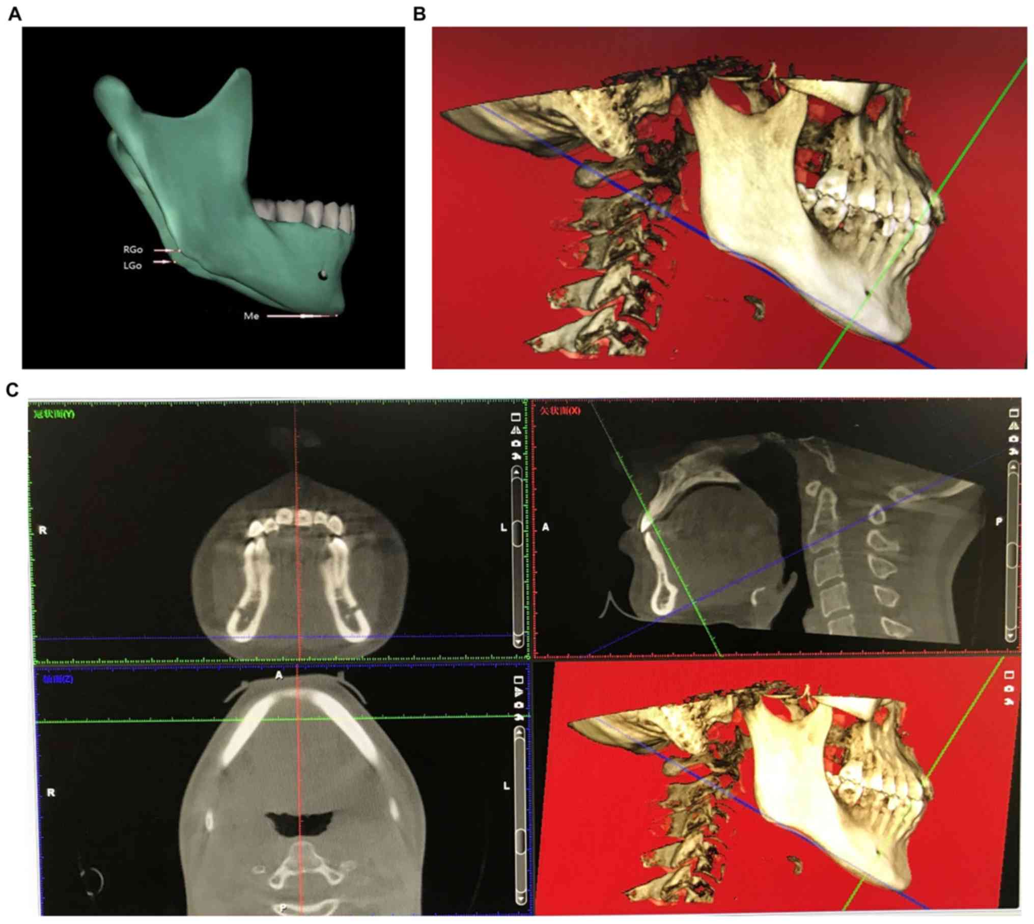
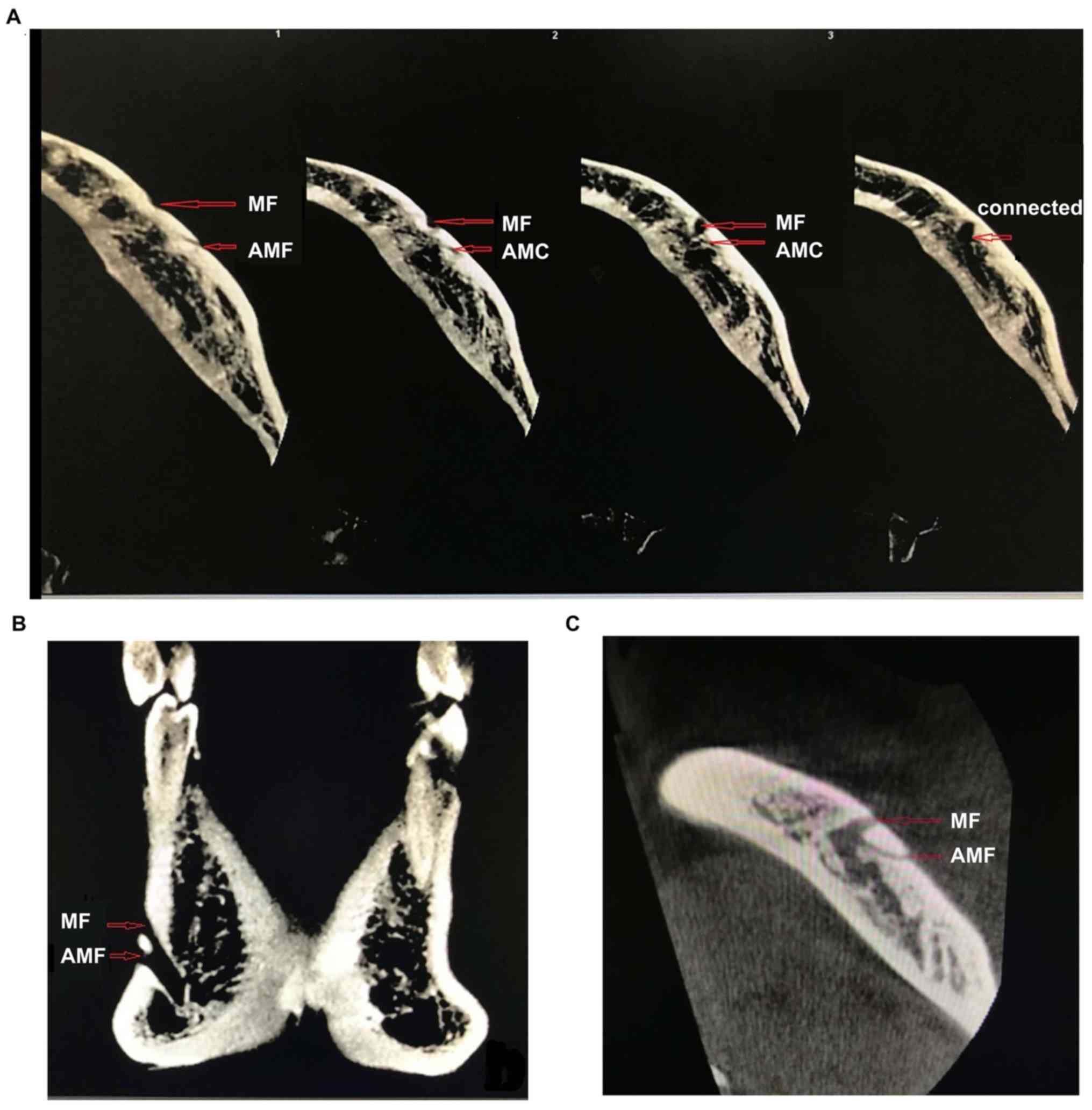
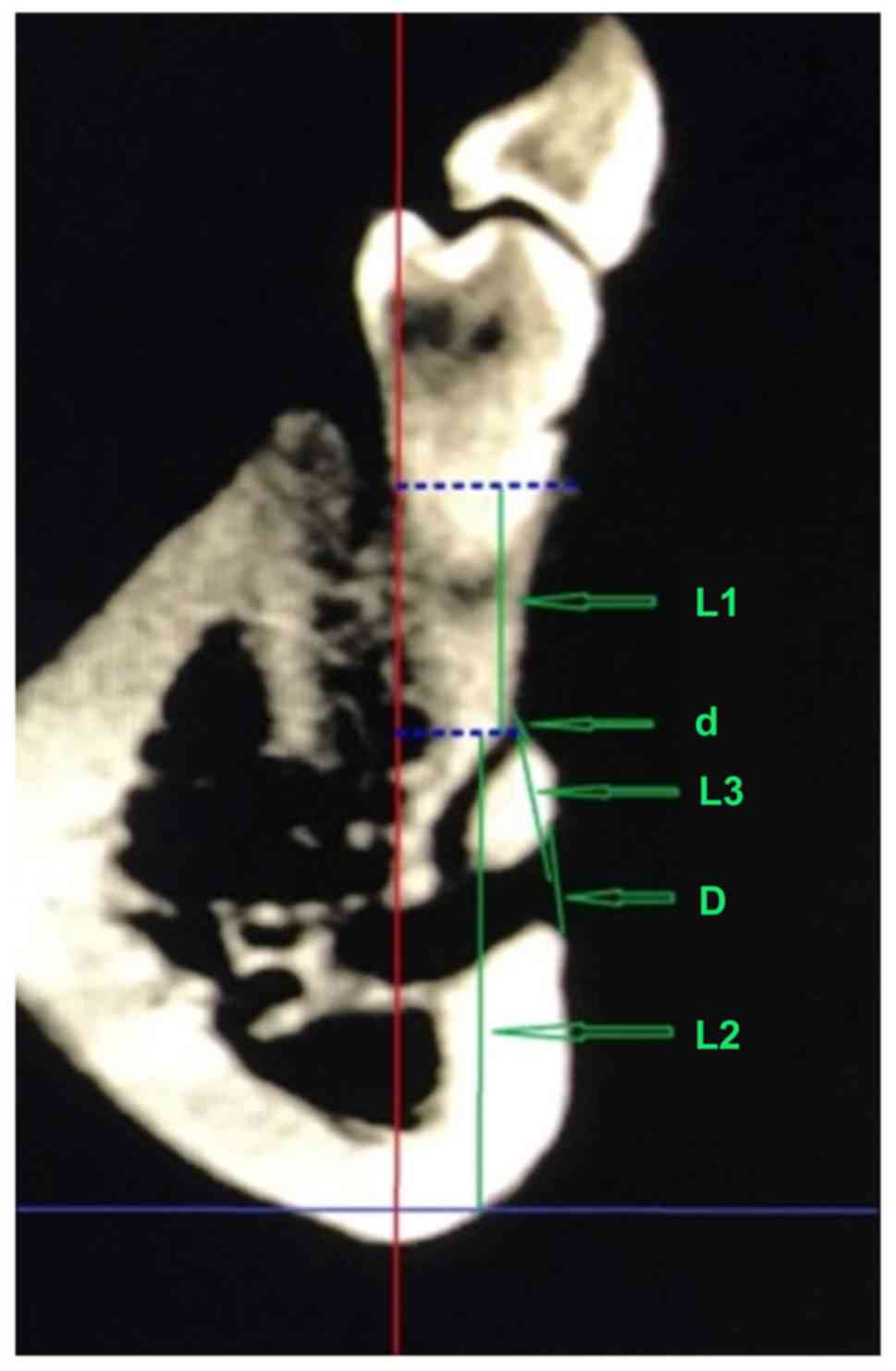
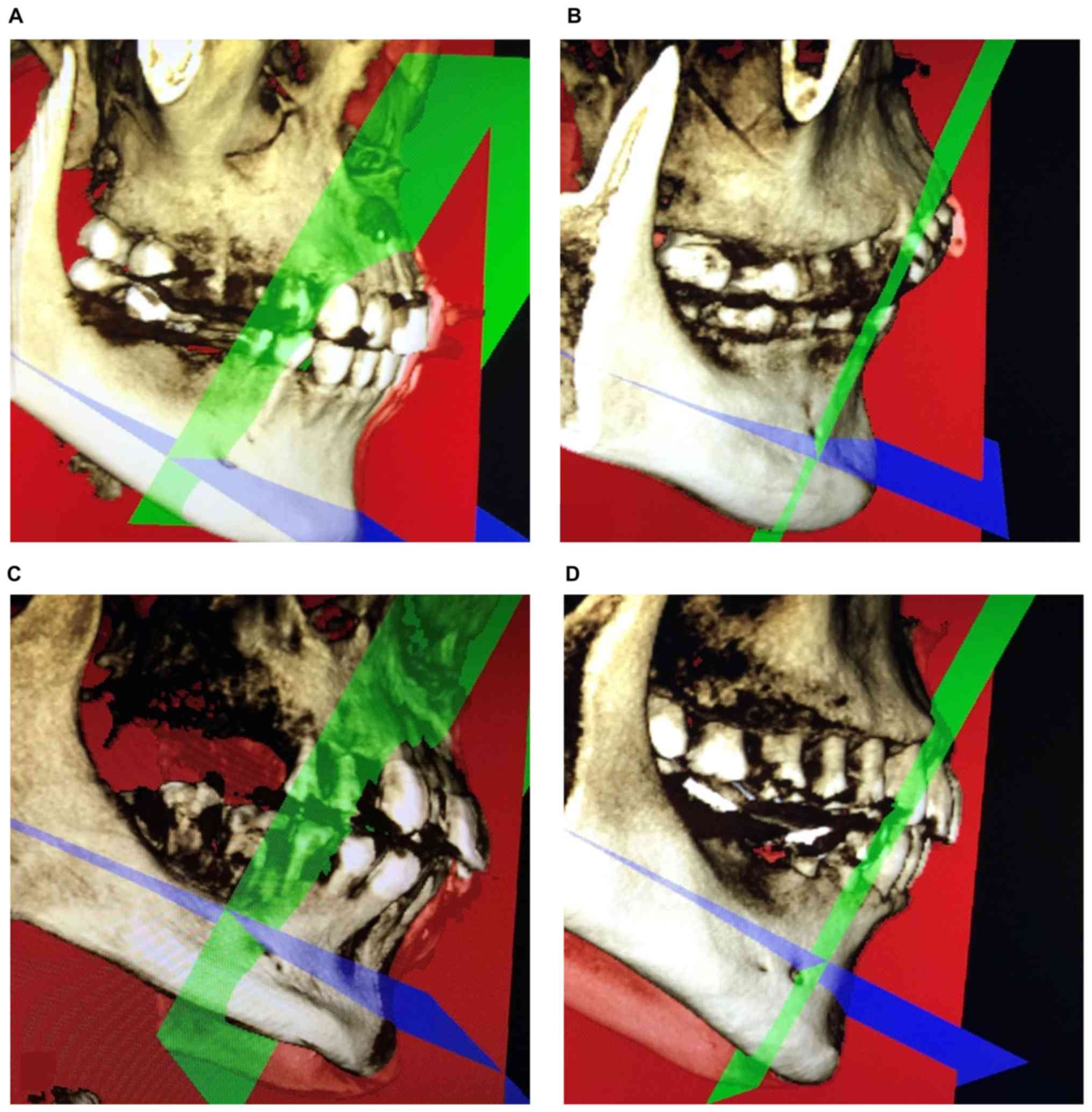
|
1
|
Koutroumpas DC and Koletsi-Kounari H:
Galen on dental anatomy and physiology. J Hist Dent. 60:37–49.
2012.PubMed/NCBI
|
|
2
|
da Silva Ramos Fernandes LM, Capelozza AL
and Rubira-Bullen IR: Absence and hypoplasia of the mental foramen
detected in CBCT images: A case report. Surg Radiol Anat.
33:731–734. 2011.PubMed/NCBI View Article : Google Scholar
|
|
3
|
Katakami K, Mishima A, Shiozaki K, Shimoda
S, Hamada Y and Kobayashi K: Characteristics of accessory mental
foramina observed on limited cone-beam computed tomography images.
J Endod. 34:1441–1445. 2008.PubMed/NCBI View Article : Google Scholar
|
|
4
|
Muinelo-Lorenzo J, Suárez-Quintanilla JA,
Fernández-Alonso A, Varela-Mallou J and Suárez-Cunqueiro MM:
Anatomical characteristics and visibility of mental foramen and
accessory mental foramen: Panoramic radiography vs. cone beam CT.
Med Oral Patol Oral Cir Bucal. 20:e707–e714. 2015.PubMed/NCBI View Article : Google Scholar
|
|
5
|
Torres MG, Valverde LF, Vidal MT and
Crusoé-Rebello IM: Accessory mental foramen: A rare anatomical
variation detected by cone-beam computed tomography. Imaging Sci
Dent. 45:61–65. 2015.PubMed/NCBI View Article : Google Scholar
|
|
6
|
Cantekin K and Şekerci A: Evaluation of
the accessory mental foramen in a pediatric population using
cone-beam computed tomography. J Clin Pediatr Dent. 39:85–89.
2014.PubMed/NCBI View Article : Google Scholar
|
|
7
|
Boronat López A and Peñarrocha Diago M:
Failure of locoregional anesthesia in dental practice. Review of
the literature. Med Oral Patol Oral Cir Bucal. 11:E510–E513.
2006.PubMed/NCBI
|
|
8
|
Zografos J and Mutzuri A: Incidence of
double mental foramen in a sample of Greek population.
Odontostomatol Proodos. 43:521–523. 1989.PubMed/NCBI(In Greek).
|
|
9
|
Cağirankaya LB and Kansu H: An accessory
mental foramen: A case report. J Contemp Dent Pract. 9:98–104.
2008.PubMed/NCBI
|
|
10
|
Kamburoğlu K, Kiliç C, Ozen T and Yüksel
SP: Measurements of mandibular canal region obtained by cone-beam
computed tomography: A cadaveric study. Oral Surg Oral Med Oral
Pathol Oral Radiol Endod. 107:e34–e42. 2009.PubMed/NCBI View Article : Google Scholar
|
|
11
|
Matherne RP, Angelopoulos C, Kulild JC and
Tira D: Use of cone-beam computed tomography to identify root canal
systems in vitro. J Endod. 34:87–89. 2008.PubMed/NCBI View Article : Google Scholar
|
|
12
|
Vujanovic-Eskenazi A, Valero-James JM,
Sánchez-Garcés MA and Gay-Escoda C: A retrospective radiographic
evaluation of the anterior loop of the mental nerve: Comparison
between panoramic radiography and cone beam computerized
tomography. Med Oral Patol Oral Cir Bucal. 20:e239–e245.
2015.PubMed/NCBI View Article : Google Scholar
|
|
13
|
Santana RR, Lozada J, Kleinman A, Al-Ardah
A, Herford A and Chen JW: Accuracy of cone beam computerized
tomography and a three-dimensional stereolithographic model in
identifying the anterior loop of the mental nerve: A study on
cadavers. J Oral Implantol. 38:668–676. 2012.PubMed/NCBI View Article : Google Scholar
|
|
14
|
Imada TS, Fernandes LM, Centurion BS, de
Oliveira-Santos C, Honório HM and Rubira-Bullen IR: Accessory
mental foramina: Prevalence, position and diameter assessed by
cone-beam computed tomography and digital panoramic radiographs.
Clin Oral Implants Res. 25:e94–e99. 2014.PubMed/NCBI View Article : Google Scholar
|
|
15
|
Pancer B, Garaicoa-Pazmiño C and Bashutski
JD: Accessory mandibular foramen during dental implant placement:
Case report and review of literature. Implant Dent. 23:116–124.
2014.PubMed/NCBI View Article : Google Scholar
|
|
16
|
Kalender A, Orhan K and Aksoy U:
Evaluation of the mental foramen and accessory mental foramen in
Turkish patients using cone-beam computed tomography images
reconstructed from a volumetric rendering program. Clin Anat.
25:584–592. 2012.PubMed/NCBI View
Article : Google Scholar
|
|
17
|
Sawyer DR, Kiely ML and Pyle MA: The
frequency of accessory mental foramina in four ethnic groups. Arch
Oral Biol. 43:417–420. 1998.PubMed/NCBI View Article : Google Scholar
|
|
18
|
Hanihara T and Ishida H: Frequency
variations of discrete cranial traits in major human populations
IV. Vessel and nerve related variations. J Anat. 199:273–287.
2001.PubMed/NCBI View Article : Google Scholar
|
|
19
|
Naitoh M, Hiraiwa Y, Aimiya H, Gotoh K and
Ariji E: Accessory mental foramen assessment using cone-beam
computed tomography. Oral Surg Oral Med Oral Pathol Oral Radiol
Endod. 107:289–294. 2009.PubMed/NCBI View Article : Google Scholar
|
|
20
|
Oliveira-Santos C, Souza PH, De Azambuja
Berti-Couto S, Stinkens L, Moyaert K, Van Assche N and Jacobs R:
Characterisation of additional mental foramina through cone beam
computed tomography. J Oral Rehabil. 38:595–600. 2011.PubMed/NCBI View Article : Google Scholar
|
|
21
|
Haktanir A, Ilgaz K and Turhan-Haktanir N:
Evaluation of mental foramina in adult living crania with MDCT.
Surg Radiol Anat. 32:351–356. 2010.PubMed/NCBI View Article : Google Scholar
|
|
22
|
Kieser J, Kuzmanovic D, Payne A, Dennison
J and Herbison P: Patterns of emergence of the human mental nerve.
Arch Oral Biol. 47:743–747. 2002.PubMed/NCBI View Article : Google Scholar
|
|
23
|
Voljevica A, Talović E and Hasanović A:
Morphological and morphometric analysis of the shape, position,
number and size of mental foramen on human mandibles. Acta Med
Acad. 44:31–38. 2015.PubMed/NCBI View Article : Google Scholar
|
|
24
|
Naitoh M, Yoshida K, Nakahara K, Gotoh K
and Ariji E: Demonstration of the accessory mental foramen using
rotational panoramic radiography compared with cone-beam computed
tomography. Clin Oral Implants Res. 22:1415–1419. 2011.PubMed/NCBI View Article : Google Scholar
|
|
25
|
Sisman Y, Sahman H, Sekerci A, Tokmak TT
and Aksu Y and E: Detection and characterization of the mandibular
accessory buccal foramen using CT. Dentomaxillofac Radiol.
41:558–563. 2012.PubMed/NCBI View Article : Google Scholar
|
|
26
|
Al-Mahalawy H, Al-Aithan H, Al-Kari B,
Al-Jandan B and Shujaat S: Determination of the position of mental
foramen and frequency of anterior loop in Saudi population. A
retrospective CBCT study. Saudi Dent J. 29:29–35. 2017.PubMed/NCBI View Article : Google Scholar
|