Introduction
Molecular techniques, such as quantitative
fluorescence PCR (QF-PCR) and the multiplex ligation-dependent
probe amplification (MLPA), are regularly combined with karyotyping
for rapid aneuploidy testing for the detection of common
aneuploidies during invasive prenatal diagnosis (1,2).
Recently, a bead-based multiplex technique, known as Prenatal
BACs-on-Beads™ (PNBoBs™), has been widely used as an alternative
method for the detection of common aneuploidies, as well as
specific microdeletion syndromes, during prenatal diagnosis
(3-20).
Among these methods, the MLPA technique detects the sequence dosage
differences in a semi-quantitative manner (21), whilst QF-PCR combines qualitative
and semi-quantitative approaches for the interpretation of peak
profiles of target chromosomes (22). However, to date, there has been
little agreement on the criteria to be used for the interpretation
of the data obtained from the PNBoBs™ assay. The instruction
method, provided in the kit, denotes a positive result as one where
three or more probes within a given target region exceed the
threshold, set at ±2 trimmed standard deviations (SDs) of the
autosomal probes. Similarly, Gross et al (3) proposed the ‘n-1 or greater probes’
rule as the criterion for denoting a result as positive in the
PNBoBs™ test. In addition to these qualitative methods, Vialard
et al (5) proposed a
semi-quantitative approach to data interpretation, in which the
mean normalized ratio (MNR) of a probe group is used as a metric
for comparison with the threshold value. In practical terms, this
method may be broadly divided into two types: The ‘fixed threshold’
and the ‘trimmed threshold’ rules (8,12),
according to the choice of threshold. However, certain studies have
not explicitly provided specific data interpretation criteria when
using the PNBoBs™ technique for prenatal diagnosis.
In this context, it is difficult to compare the data
across studies. As such, it is essential to evaluate the impact of
different data interpretation methods used for the results obtained
from PNBoBs™ assays in the available studies. In the present study,
retrospective data from >2,200 prenatal samples were utilized to
evaluate the accuracy of existing methods based on different
performance measures with the aim of proposing a versatile data
interpretation approach for use in the PNBoBs™ assay for prenatal
diagnosis.
Subjects and methods
Subjects
A total of 4,496 prenatal diagnoses were performed
at Women and Children's Hospital, School of Medicine, Xiamen
University (Xiamen, China) between January 2018 and June 2019.
Among them, 2,291 cases who applied for karyotyping and PNBoBs™
testing simultaneously were recruited for the present study. Of
these participants, two with failed karyotype analysis were
excluded. Finally, a total of 2,289 cases, who had karyotypes and
raw data of the PNBoBs™ assay were obtained for subsequent
research. Cases interpreted as aneuploidy in targeted chromosomes
by any of the data interpretation methods available for the PNBoBs™
assay were verified by karyotyping. Cases interpreted as
microdeletion or microduplication in targeted regions by any of the
data interpretation methods available for the PNBoBs™ assay were
verified by chromosome microarray analysis (CMA). Ethics approval
was obtained from the Ethical Review Committee of the Women and
Children's Hospital Affiliated to Xiamen University (Xiamen,
China). Each participant provided written informed consent in
compliance with the Declaration of Helsinki prior to being included
in the present study.
CMA
Prenatal diagnostic samples interpreted as copy
number variation in targeted regions by PNBoBs™ assay were verified
by CMA using a CytoScan® 750K Array Suite kit (cat. no.
901859; Affymetrix Inc.; Thermo Fisher Scientific, Inc.), according
to the manufacturer's protocol. Genomic DNA was extracted from the
amniotic fluid cells using the QIAamp DNA Mini Kit (cat. no. 51306;
Qiagen, Inc.). The resulting DNA concentrations and purities were
estimated by NanoDrop One Spectrophotometer (Thermo Fisher
Scientific, Inc.). Data were analyzed using the Chromosome Analysis
Suite 4.0 (r28959) software (Affymetrix Inc.; Thermo Fisher
Scientific, Inc.).
Performance evaluation of data
interpretation methods for the PNBoBs™ assay
A methodological review of the literature was
performed with the search term of ‘Prenatal BoBs™’ or ‘PNBoBs™’
using the PubMed database, with the last publication date for
inclusion being January 15th, 2020. All related literature that
shared the same or similar positive call criteria of the PNBoBs™
assay were grouped. The performance of the data interpretation
methods was evaluated based on the following statistical
indicators: Sensitivity, specificity, false-positive rate (FPR) and
false-negative rate (FNR).
Optimization targets of the new data
interpretation method
The optimization approach was developed based on
statistical results across existing positive call methods, using
the following optimization targets: i) The accurate detection of
fetal aneuploidy of chromosomes X, Y, 13, 18 and 21; ii)
improvement in the detection sensitivity for
microdeletion/microduplication syndrome; and iii) ensuring a
relatively low rate of false positives in the detection of
aneuploidy and microdeletion/microduplication syndrome.
Statistical methods
Statistical analysis was performed with the SPSS
Statistics version 20 (IBM Corp.). For comparison of the MNR of the
Y chromosome between male and female fetuses, the unpaired
Student's t-test was used. One-way ANOVA was performed to
compare the MNR of different copy number groups of non-Y
chromosomes followed by the Student-Newman-Keuls test for post hoc
analysis.
Results
Characteristics of the analyzed
samples
The typical profiles of the true-positive and
false-positive results of the PNBoBs™ assay are presented in
Fig. 1. In total, 224 cases were
denoted as positive by all existing data interpretation methods.
After validation, the PNBoBs™ assay theoretically detected 52 cases
of an unbalanced chromosomal abnormality in the present sample
sets. Supplementary Table SI
provides further details on these positive cases.
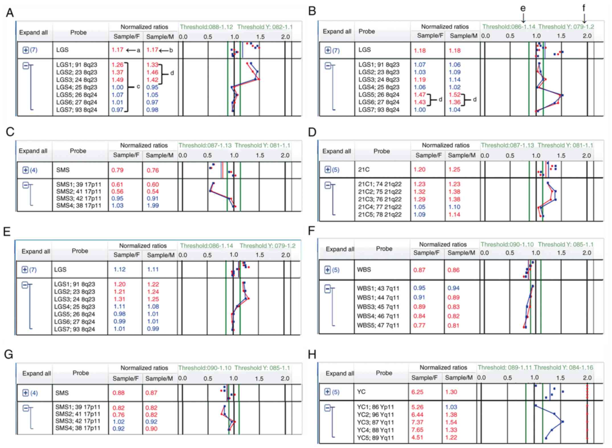 | Figure 1Representative prenatal BACs-on-Beads™
results for true-positive and false-positive cases. (A) The
profiles indicated that both of the MNR of all the probes in the
LGS region exceeded the threshold with three consecutive deviation
probes and this was confirmed by CMA as a true-positive result. (B)
The profiles suggested that both of the MNR of all the probes in
the LGS region exceeded the threshold with two consecutive
deviation probes and this was confirmed by CMA as a true-positive
result. (C) The profiles indicated that both of the MNR of all the
probes in the SMS region exceeded the threshold with two
consecutive deviation probes and this was confirmed by CMA as a
true-positive result. (D) The profiles suggested that both of the
MNR of all the probes in the 21q22 region exceeded the threshold
with three consecutive deviation probes and this was confirmed by
karyotyping as a trisomy 21. (E) The profiles indicated that none
of the MNR of all the probes in the LGS region exceeded the
threshold despite having three consecutive deviation probes and
this was verified by CMA as a false-positive result. (F) The
profiles suggested that both of the MNR of all the probes in the
WBS region exceeded the threshold despite having three and four
consecutive deviation probes and this was verified by CMA as a
false-positive result. (G) The profiles indicated that both of the
MNR of all the probes in the SMS region exceeded the threshold with
two consecutive deviation probes and this was verified by CMA as a
false-positive result. (H) The profiles suggested that the MNR of
the Y chromosome to male reference reached the fixed threshold of
1.3 with four consecutive deviation probes and was verified by
karyotyping as a false-positive result. a, Representative mean
normalized ratio of the sample-to-female reference; b,
Representative mean normalized ratio of the sample-to-male
reference; c, Representative normalized ratio of each probe in the
target region of the sampel-to-reference; d, Representative
normalized ratio of consecutive deviation probes in the target
region of the sample-to-reference; e, Representative threshold
values for the X chromosome and microdeletion syndrome region; and
f, Representative threshold values for the Y chromosome. The
numbers in red indicate that the value exceeds the threshold. The
numbers in blue indicate that the value is within the range of
threshold. The brackets on the right represent the range of the
probes being analyzed. MNR, mean normalized ratio; CMA, chromosome
microarray analysis; LGS, Langer-Giedion syndrome; SMS,
Smith-Magenis syndrome; WBS, Williams-Beuren syndrome; 21C,
Chromosome 21; YC, Y chromosome; M, male reference; F, female
reference. |
A summary of the test results for all samples is
provided in Fig. 2. The mean ± 3SD
of the MNR value for the Y chromosome probe sets in male (n=1,220)
and female samples (n=1,069) is presented in Fig. 2A. A statistical comparison of the
autosomal controls and deviation probes in the selected
true-positive samples (n=36) is provided in Fig. 2B, indicating the presence of a
significant difference (P<0.001) among non-overlapping groups.
The threshold values calculated with 99.7% confidence limits are
listed in Table I.
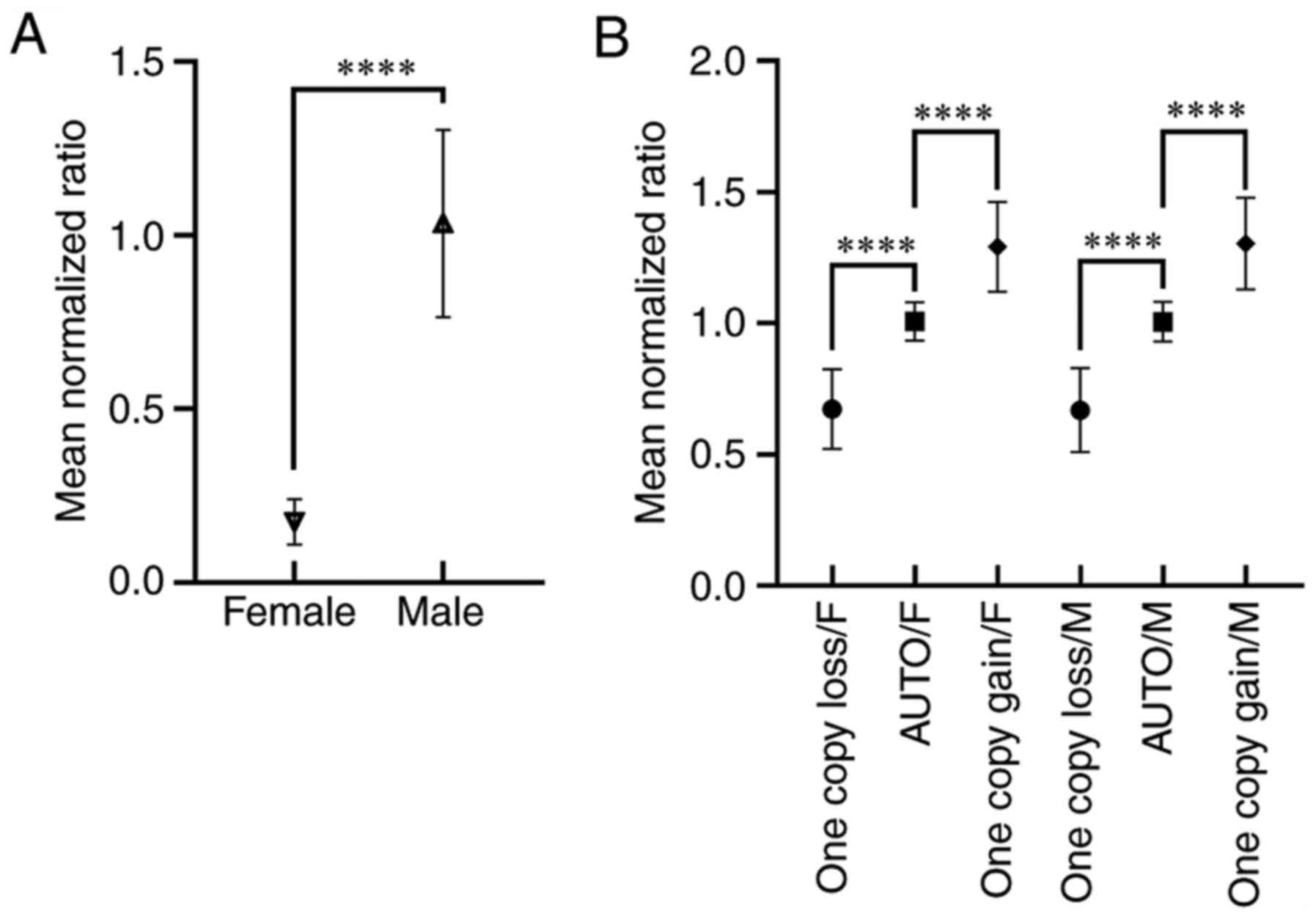 | Figure 2Statistical results of the mean
normalized ratio from probes in the Y chromosome and deviation
targets. (A) Profiles of the MNR ± 3SD of the Y chromosome to the
male reference (female fetuses, n=1,069; male fetuses, n=1,220).
The symbols represent the mean value, while error bars represent
±3SD for each column. (B) The MNR ±3SD profiles of deviation probes
and autosomal control probes of abnormal cases to the female
reference and the male reference. Symbols represent the mean value,
while error bars represent ±3SD for each column.
****P≤0.0001. MNR, mean normalized ratio; SD, standard
deviation; M, male reference; F, female reference; auto, autosomal
control probes. |
 | Table IStatistical results of mean normalized
ratios of copy number variation cases. |
Table I
Statistical results of mean normalized
ratios of copy number variation cases.
| Probe set | n | Mean | SD | Mean ± 3SD range |
|---|
| OCL/Fa | 10 | 0.67 | 0.05 | 0.52-0.82 |
| OCL/Mb | 10 | 0.67 | 0.05 | 0.51-0.83 |
| AUTO/Fa | 36 | 1.00 | 0.02 | 0.93-1.08 |
| AUTO/Mb | 36 | 1.01 | 0.02 | 0.93-1.08 |
| OCG/Fa | 26 | 1.29 | 0.06 | 1.12-1.46 |
| OCG/Mb | 26 | 1.30 | 0.06 | 1.13-1.48 |
| Chromosome
Y/Mb (female
fetuses) | 1,069 | 0.17 | 0.02 | 0.11-0.24 |
| Chromosome
Y/Mb (male
fetuses) | 1,220 | 1.03 | 0.09 | 0.77-1.30 |
Performance of data interpretation
using existing methods
A detailed description of each existing data
interpretation method is given in Table II. Of all 224 positive cases, 143
had three or more probes within a given target region exceeding the
2x trimmed SD threshold. According to the instructions of the
PNBoBs™ assay kit, these samples were interpreted as positive.
After validation, 94 of these (65.7%) were identified as
false-positive results and yielded an FPR of 4.2% (Table III). In addition, two cases with
partial copy number variations in the microdeletion syndrome
regions were classified as ‘missed detection’ due overlooked by the
instruction method due to the presence of < three deviation
probes depite being positive (Fig.
1B and C). The statistical
results indicated that the ‘n-1 or greater probes’ rule had the
highest specificity (99.7%) in all existing methods and the
second-highest FNR (11.5%). Comparison of the results suggested
that the ‘fixed threshold’ rule had the lowest sensitivity (71.2%)
and the second-highest specificity (99.3%). Compared to the other
three approaches, the ‘trimmed SD threshold’ method had the highest
sensitivity (98.1%), with only one overlooked case of low-level
mosaicism. The same FPR (4.2%) was observed between this approach
and the instruction method, although the composition of these two
false-positive groups was markedly different. In the former, the
false-positive results were mainly derived from the microdeletion
syndrome regions (57.4%, 54/94), while in the latter, they were
mostly derived from the Y chromosome (88.3%, 83/94).
| Table IIExisting data interpretation methods
presented for the prenatal BACs-on-Beads™ assay in the
literature. |
Table II
Existing data interpretation methods
presented for the prenatal BACs-on-Beads™ assay in the
literature.
| Study | Specimen type
tested | Positive call
criteria for non-sex chromosome syndromes | Positive call
criteria for sex chromosome syndromes | (Refs.) |
|---|
| Miao et
al | PC | aThree or more probes within a | aThree or more probes within a
given | (20) |
| Fang et
al | PC | given target region
exceeding the ±2x trimmed SD. | target region
exceeding the ±2x trimmed SD for the X chromosome, and ±3x trimmed
SD for the Y chromosome, respectively. | (16) |
| Gross et
al | CL | Ratios of n-1 or
greater probes | Ratios of n-1 or
greater probes within a | (3) |
| Dang et
al | | within a given target
region exceeding the ±2x trimmed SD. to both references | given target region
exceeding the ±2x trimmed SD cutoff for the X chromo some and ±3x
trimmed SD for the Y chromosome, respectively. | (19) |
| Huang et
al | PC | Both of the mean
normalized | Both of the mean
normalized ratios | (17) |
| Choy et
al | PC | ratios within a given
target | within a given target
region exceeding | (10,11) |
| Cheng et
al | CL and PC | region exceeding the
±2x | the ±2x trimmed SD
cutoff for the X | (9) |
| Vialard et
al | PC | trimmed SD. | chromosome and ±3x
trimmed SD for the Y chromosome, respectively. | (5,8) |
| Li et
al | PC | Either of the mean
normalized | Not mentioned. | (18) |
| Garcia-Herrero
et al | PC | ratio within a
given target region equal to or exceeding 1.3 or 0.8 for
duplication and deletion, respectively. | | (12) |
| Rosenfeld et
al | POC | No details. | Not mentioned. | (14) |
| Shaffer et
al | PC | | | (4) |
| Grati et
al | PC | Not Mentioned. | Not mentioned. | (15) |
| Piotrowski et
al | PC | | | (13) |
| Leung et
al | PC | | | (6) |
| Table IIIComparison of different methods on
data interpretation for prenatal BACs-on-Beads™ assay. |
Table III
Comparison of different methods on
data interpretation for prenatal BACs-on-Beads™ assay.
| | Instruction
method | ‘N-1 or greater
probes’ method | ‘Fixed threshold’
method | ‘Trimmed SD
threshold’ method | Optimization
method |
|---|
| Target region | TP | FP | FN | TP | FP | FN | TP | FP | FN | TP | FP | FN | TP | FP | FN |
|---|
| Chr21 | 15 | 10 | 0 | 14 | 0 | 1 | 8 | 0 | 7 | 15 | 2 | 0 | 15 | 0 | 0 |
| Chr18 | 3 | 2 | 0 | 3 | 0 | 0 | 3 | 0 | 0 | 3 | 1 | 0 | 3 | 0 | 0 |
| Chr13 | 3 | 0 | 0 | 3 | 0 | 0 | 1 | 0 | 2 | 3 | 0 | 0 | 3 | 0 | 0 |
| Chr15(PWS) | 2 | 24 | 0 | 2 | 0 | 0 | 2 | 0 | 0 | 2 | 0 | 0 | 2 | 0 | 0 |
| Chr8(LGS) | 1 | 7 | 1 | 0 | 0 | 2 | 2 | 0 | 0 | 2 | 0 | 0 | 2 | 0 | 0 |
| Chr17(MDS) | 1 | 12 | 0 | 1 | 1 | 0 | 1 | 0 | 0 | 1 | 0 | 0 | 1 | 0 | 0 |
| Chr22(DGS) | 4 | 0 | 0 | 4 | 0 | 0 | 3 | 0 | 1 | 4 | 0 | 0 | 4 | 0 | 0 |
| Chr4(WHS) | 2 | 4 | 0 | 2 | 1 | 0 | 2 | 0 | 0 | 2 | 0 | 0 | 2 | 0 | 0 |
| Chr5(CDC) | 1 | 0 | 1 | 1 | 0 | 1 | 1 | 0 | 1 | 1 | 0 | 1 | 1 | 0 | 1 |
| Chr7(WBS) | 0 | 6 | 0 | 0 | 0 | 0 | 0 | 0 | 0 | 0 | 2 | 0 | 0 | 0 | 0 |
| Chr17(SMS) | 0 | 1 | 1 | 0 | 0 | 1 | 1 | 0 | 0 | 1 | 1 | 0 | 1 | 0 | 0 |
| ChrX | 12 | 18 | 0 | 11 | 2 | 1 | 8 | 0 | 4 | 12 | 5 | 0 | 12 | 5 | 0 |
| ChrY | 5 | 10 | 0 | 5 | 2 | 0 | 5 | 16 | 0 | 5 | 83 | 0 | 5 | 3 | 0 |
| Total | 49 | 94 | 3 | 46 | 6 | 6 | 37 | 16 | 15 | 51 | 94 | 1 | 51 | 8 | 1 |
| Statistical
indicators (%) | | | | | | | | | | | | | | | |
|
Sensitivity | 94.2 | 88.5 | 71.2 | 98.1 | 98.1 |
|
Specificity | 95.8 | 99.7 | 99.3 | 95.8 | 99.6 |
|
FPR | 4.2 | 0.3 | 0.7 | 4.2 | 0.4 |
|
FNR | 5.8 | 11.5 | 28.8 | 1.9 | 1.9 |
Performance of the optimization
method
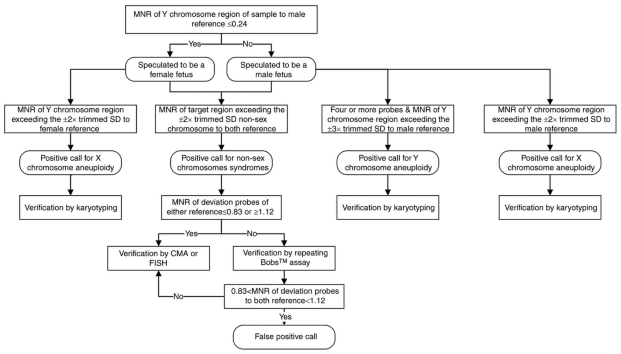
As presented in Fig.
3, a pipeline was developed for the data interpretation of the
PNBoBs™ assay results based on the comparison results described
above. The first step of the analytic process consisted of the
identification of the sex of the fetus by comparing the MNR of the
Y chromosome in a sample to a fixed threshold of 0.24. According to
this criterion, two out of the 1,069 female fetuses were classified
as male fetuses due to the MNR of the Y chromosome slightly
exceeding the cutoff (0.27 and 0.35, respectively). According to
the analysis pipeline applied, the six cases with both MNR present
outside of the 2X trimmed SD threshold in autosomal regions were
classified as inconclusive rather than positive results, since the
corresponding MNRs were at intermediate levels, namely between the
upper limit of the threshold of one probe-set copy and the lower
limit of two probe-set copies (data not shown). In addition, 82 out
of the 83 false-positive results with the Y chromosome, detected by
using the ‘trimmed SD threshold’ method, were excluded in the
combination method due to the presence of fewer than four deviation
probes. Based on this data analysis pipeline, the overall
sensitivity of the present optimization method was 98.1%, with an
FPR of 0.4%.
Discussion
Following the QF-PCR and MLPA techniques, the
PNBoBs™ assay has been widely utilized in recent years due to its
superiority in extending the scope of rapid prenatal diagnosis of
common diseases from fetal aneuploidy to microdeletion syndromes.
In one study from the USA (14),
which included ~2,900 prenatal samples, neither false negatives nor
false positives were observed in the data obtained using the
PNBoBs™ test. Miao et al (20) performed a prospective study on
>4,800 prenatal cases in China and reported false-positive
events; however, no data were provided. A multi-center
retrospective study (15) involving
a cohort of 9,500 pregnancies conducted in EU countries reported an
FPR and FNR of 0.19 and 0.14%, respectively. However, the
preliminary test of the present study indicated recurrent
false-positive results when using the included instruction method
for data interpretation. Therefore, a retrospective comparison of
the literature was performed, revealing one possible reason for
these discrepancies: The criteria for data interpretation described
in the studies were inconsistent.
Gross et al (3), the inventors of the BoBs™ technique,
used the rule of ‘n-1 or greater probes’ beyond the threshold as a
criterion for denoting a positive call. In practical terms, the
researchers specifically indicated that this rule was based on
results obtained from known syndromic cell lines rather than those
from clinical samples. Compared with the instruction method, in
which three deviated probes were sufficient for calling positive
results, using the ‘n-1 or greater probes’ rule theoretically
provided a higher specificity, as most of the desired syndrome
regions (11/14) of the PNBoBs™ kit had at least five probes and at
least four deviated probes are required to call for positive
results under this rule. The results of the present analysis were
consistent with this hypothesis that the ‘n-1 or greater probes’
rule had the highest specificity (99.7%) among the methods
available. At the same time, six true-positive cases were
overlooked under this rigid criterion, which resulted in a higher
FNR (11.5%) compared to the rate (5.8%) obtained using the
instruction method.
In the literature, there were no false results
reported in studies that used the ‘fixed threshold’ rule as the
interpretation method for data obtained using the PNBoBs™ assay
(12,18). However, in the present analysis,
28.8% (15/52) of the positive samples were overlooked when using
this approach. In addition, 16 false-positive cases with a Y
chromosome aneuploidy were called using this method, as the Y
chromosome ratio of the sample reached or exceeded the threshold of
1.3. Given the poor performance in terms of sensitivity observed in
the present study, it is not recommended using the ‘fixed
threshold’ rule alone to interpret data obtained from a PNBoBs™
assay.
Among the previous studies, the ‘trimmed SD
threshold’ rule was the most popular approach for interpreting data
obtained from a PNBoBs™ test (5,8-11,17).
In this approach, a specific sample threshold is used to compare
the MNR of the target region instead of a fixed threshold.
Regarding the performance of this approach, Vialard et al
(8) reported high sensitivity
(97.3%) and specificity (100%) in retrospective samples and
slightly lower sensitivity (95.6%) and specificity (99.7%) in
prospective samples. In a retrospective study performed by Choy
et al (10), the sensitivity
of PNBoBs™ was 96.7%, with a specificity of 100%. In terms of
sensitivity performance, the present result of 98.1% was somewhat
higher than that obtained in earlier studies. The only
false-negative result was from a low-level mosaicism case with an
abnormal cell proportion of 6.57%, which is below the detection
limit of the BoBs™ assay (9). Two
cases with a partial deletion, located in the Langer-Giedion and
the Smith-Magenis syndrome regions, respectively, were classified
as positive results under this rule (i.e. using the trimmed SD
threshold). Vialard et al (8) proposed that at least two consecutive
probes should be observed to be evidently abnormal (ratio <0.8
or >1.3) when calling a partial copy number variation in
microdeletion syndrome regions. These two realistic cases appeared
to be consistent with this criterion. However, given the limited
data, it is difficult to determine which approach is better for
presenting a partial copy number variation at the present time. In
terms of specificity, the results observed in this study were lower
compared to those reported by other studies (5,10). A
total of 94 false-positive results were observed using this rule,
resulting in a high FPR compared with that of the existing methods
(4.2%).
A comparison of the results described above revealed
that none of the existing methods offered optimal levels of
performance for the interpretation of data obtained from the
PNBoBs™ assay, indicating a requirement to optimize these
approaches in order to improve the accuracy of this technique when
used in prenatal diagnosis. To ensure a high degree of sensitivity
and a low rate of false-positives, an analysis workflow was
developed by adopting the advantages and eliminating the
disadvantages of the existing methods. In the analysis pipeline
developed, the first step consists of identifying the sample sex in
order to choose the correct reference sample. Generally, the MNR of
the X chromosome in a normal male sample against the female
reference sample is always less than the 2x trimmed SD threshold;
thus, the sample makes sense only when compared to male reference
DNA. Similarly, the MNR of the X chromosome in a female fetus
should only be compared to the female reference in the present
method. In addition, the upper limit of 99.7% was selected for the
confidence interval of MNR of the Y chromosome rather than the
maximum value that was observed in order to improve the performance
in calling for sex-chromosome mosaicism. Due to its high
sensitivity properties, the ‘trimmed SD threshold’ rule was used as
the framework for the interpretation of data from autosomes. In a
study by Slater et al (2),
using an MLPA for rapid prenatal diagnosis, the threshold values
from a dataset of normal and abnormal samples were used to assign
the test results to different categories. In the present study, a
similar approach was used to further differentiate the cases with
MNR exceeding the 2x trimmed SD. Verification of results obtained
in the present study demonstrated that in cases where the MNR of
the deviation probes in the autosome region was present outside of
the reference intervals, a repeated PNBoBs™ assay was more
consistent with the original intention of using this technique than
with a CMA verification. Compared to the existing methods, one of
the major advantages of the present optimization approach was that
it substantially reduces false positives from the Y chromosome by a
comprehensive analysis of the number of deviation probes and MNR
values, while maintaining a high degree of sensitivity. Re-analysis
of the data using the method optimized herein indicated a high
specificity (99.6%), with a sensitivity that was also at a high
level (98.1%).
In conclusion, a reasonable balance between
sensitivity and specificity was obtained for the interpretation of
data of the PNBoBs™ assay, using all possible methods to reduce the
FPR and providing a basis for reducing the turnaround time and cost
associated with the use of this assay in clinical practice.
However, there are two potential limitations of the present study.
First, the reference intervals established may not be directly used
in another laboratory. Researchers intending to use this method are
encouraged to consider establishing in-house reference thresholds
based on local samples. Furthermore, it should be noted that a
sufficient number of confirmed positive and normal cases should be
included in the reference sample set to meet the desired
statistical requirements. In addition, the clinical implications of
microduplications/microdeletions detected in the present study were
not discussed. Consequently, further studies are required in order
to provide substantial evidence for future guidelines and
recommendations for the PNBoBs™ assay.
Supplementary Material
Details of copy number variation cases
covered by Prenatal BoBs™ assay.
Acknowledgements
The authors would like to thank Dr Qichang Wu,
Department of Obstetrics and Gynecology, Women and Children's
Hospital, School of Medicine, Xiamen University (Xiamen, China) who
contributed the clinical information for the present study.
Funding
The present study was funded by the Young Talent
Support Program of Women and Children's Hospital, School of
Medicine, Xiamen University [grant no. (2018)26) and Xiamen's
Leading Discipline of Medicine [grant no. (2016)601].
Availability of data and materials
Partial output data from the PNBoBs™ assay software
(BoBsoft™) are included in the attached Table SI of this published article. The
datasets used and/or analyzed during the current study are
available from the corresponding author on reasonable request.
Authors' contributions
YJ contributed to writing the manuscript. YJ and JZ
performed the BoBs™ assay. LW helped with the karyotype analysis of
chromosomes. WW and QG performed the statistical analysis. All
authors read and approved the final manuscript.
Ethics approval and consent to
participate
Ethics approval was obtained from the Ethical Review
Committee of the Women and Children's Hospital Affiliated to Xiamen
University (Xiamen, China). Each participant provided written
informed consent in compliance with the Declaration of Helsinki
prior to being included in the present study.
Patient consent for publication
Not applicable.
Competing interests
The authors declare that they have no competing
interests.
References
|
1
|
Adinolfi M and Sherlock J: Prenatal
detection of chromosome disorders by QF-PCR. Lancet. 358:1030–1031.
2001.PubMed/NCBI View Article : Google Scholar
|
|
2
|
Slater HR, Bruno DL, Ren H, Pertile M,
Schouten JP and Choo KH: Rapid, high throughput prenatal detection
of aneuploidy using a novel quantitative method (MLPA). J Med
Genet. 40:907–912. 2003.PubMed/NCBI View Article : Google Scholar
|
|
3
|
Gross SJ, Bajaj K, Garry D, Klugman S,
Karpel BM, Roe AM, Wagner BJ, Zhan J, Apfelroth SD and
Schreiber-Agus N: Rapid and novel prenatal molecular assay for
detecting aneuploidies and microdeletion syndromes. Prenatal Diagn.
31:259–266. 2011.PubMed/NCBI View
Article : Google Scholar
|
|
4
|
Shaffer LG, Coppinger J, Morton SA,
Alliman S, Burleson J, Traylor R, Walker C, Byerly S, Lamb AN,
Schultz R, et al: The development of a rapid assay for prenatal
testing of common aneuploidies and microdeletion syndromes.
Prenatal Diagn. 31:778–787. 2011.PubMed/NCBI View
Article : Google Scholar
|
|
5
|
Vialard F, Simoni G, Aboura A, De Toffol
S, Molina Gomes D, Marcato L, Serero S, Clement P, Bouhanna P,
Rouleau E, et al: Prenatal BACs-on-Beads: A new technology for
rapid detection of aneuploidies and microdeletions in prenatal
diagnosis. Prenatal Diagn. 31:500–508. 2011.PubMed/NCBI View
Article : Google Scholar
|
|
6
|
Leung TY, Wong KM, Wong HK, Leung KO,
Adler K, Lau TK, Wang CC, Schermer M and Choy KW: 725: A
retrospective study of BACs-on-Beads (BoBs) technology for
identification of chromosome abnormalities compared with QF-PCR and
karyotyping in prenatal diagnosis. Am J Obstet Gynecol. 206
(Suppl)(S322)2012.
|
|
7
|
Piotrowski K, Henkelman M and Zajaczek S:
Will the new molecular karyotyping BACs-on-Beads technique replace
the traditional cytogenetic prenatal diagnostics? Preliminary
reports. Ginekol Pol. 83:284–290. 2012.PubMed/NCBI
|
|
8
|
Vialard F, Simoni G, Gomes DM, Abourra A,
De Toffol S, Bru F, Martinez Romero MC, Nitsch L, Bouhanna P,
Marcato L, et al: Prenatal BACs-on-Beads™: The prospective
experience of five prenatal diagnosis laboratories. Prenatal Diagn.
32:329–335. 2012.PubMed/NCBI View
Article : Google Scholar
|
|
9
|
Cheng YK, Wong C, Wong HK, Leung KO, Kwok
YK, Suen A, Wang CC, Leung TY and Choy KW: The detection of
mosaicism by prenatal BoBs™. Prenatal Diagn. 33:42–49.
2013.PubMed/NCBI View
Article : Google Scholar
|
|
10
|
Choy KW, Kwok YK, Cheng YK, Wong KM, Wong
HK, Leung KO, Suen KW, Adler K, Wang CC, Lau TK, et al: Diagnostic
accuracy of the BACs-on-Beads™ assay versus karyotyping for
prenatal detection of chromosomal abnormalities: A retrospective
consecutive case series. BJOG. 121:1245–1252. 2014.PubMed/NCBI View Article : Google Scholar
|
|
11
|
Choy RK, Chen Y, Sun XF, Kwok YK and Leung
TY: BACs-on-beads: A new robust and rapid detection method for
prenatal diagnosis. Expert Rev Mol Diagn. 14:273–280.
2014.PubMed/NCBI View Article : Google Scholar
|
|
12
|
Garcia-Herrero S, Campos-Galindo I,
Martinez-Conejero JA, Serra V, Olmo I, Lara C, Simón C and Rubio C:
BACs-on-Beads technology: A reliable test for rapid detection of
aneuploidies and microdeletions in prenatal diagnosis. Biomed Res
Int. 2014(590298)2014.PubMed/NCBI View Article : Google Scholar
|
|
13
|
Piotrowski K, Halec W, Wegrzynowski J,
Pietrzyk A, Henkelman M and Zajaczek S: Prenatal diagnosis of
Langer-Giedion Syndrome confirmed by BACs-on-Beads technique.
Ginekol Pol. 85:66–69. 2014.PubMed/NCBI View
Article : Google Scholar
|
|
14
|
Rosenfeld JA, Morton SA, Hummel C,
Sulpizio SG, McDaniel LD, Schultz RA, Torchia BS, Ravnan JB,
Ellison JW and Fisher AJ: Experience using a rapid assay for
aneuploidy and microdeletion/microduplication detection in over
2,900 prenatal specimens. Fetal Diagn Ther. 36:231–241.
2014.PubMed/NCBI View Article : Google Scholar
|
|
15
|
Grati FR, Molina Gomes D, Ferreira JC,
Dupont C, Alesi V, Gouas L, Horelli-Kuitunen N, Choy KW,
García-Herrero S, de la Vega AG, et al: Prevalence of recurrent
pathogenic microdeletions and microduplications in over 9500
pregnancies. Prenatal Diagn. 35:801–809. 2015.PubMed/NCBI View
Article : Google Scholar
|
|
16
|
Fang Y, Wang G, Gu L, Wang J, Suo F, Gu M
and Gou L: Application of karyotype analysis combined with
BACs-on-Beads for prenatal diagnosis. Exp Ther Med. 16:2895–2900.
2018.PubMed/NCBI View Article : Google Scholar
|
|
17
|
Huang H, Zhang M, Wang Y, Lin N, He D,
Chen M, Chen L, Lin Y and Xu L: Application of the BACs-on-Beads™
assay for rapid prenatal detection application of BoBs™ for PND of
aneuploidies and microdeletions. Mol Reprod Dev. 85:146–154.
2018.PubMed/NCBI View Article : Google Scholar
|
|
18
|
Li C, Chen B, Zheng J, Cheng L, Song T,
Guo F, Xu H, Yan F, Xu Y, Li Y and Zhang J: Prenatal diagnosis of
BACs-on-beads assay in 3647 cases of amniotic fluid cells. Reprod
Sci. 26:1005–1012. 2019.PubMed/NCBI View Article : Google Scholar
|
|
19
|
Dang Y, Wan S, Zheng Y, Song T, Li C, Li Y
and Zhang J: The prenatal diagnosis of seven fetuses with 7q11.23
microdeletion or microduplication. Fetal Pediatr Pathol.
39:269–276. 2020.PubMed/NCBI View Article : Google Scholar
|
|
20
|
Miao Z, Liu X, Hu F, Zhang M, Yang P and
Wang L: Combined use of bacterial artificial chromosomes-on-beads
with karyotype detection improves prenatal diagnosis. Mol
Cytogenet. 12(9)2019.PubMed/NCBI View Article : Google Scholar
|
|
21
|
Schouten JP, McElgunn CJ, Waaijer R,
Zwijnenburg D, Diepvens F and Pals G: Relative quantification of 40
nucleic acid sequences by multiplex ligation-dependent probe
amplification. Nucleic Acids Res. 30(e57)2002.PubMed/NCBI View Article : Google Scholar
|
|
22
|
Liao C, Yang X, Li FT, Li J and Li DZ: The
detection of aneuploidy and maternal contamination by QF-PCR in
samples undergoing prenatal diagnosis for thalassemia in Southern
China. Eur J Obstet Gynecol Reprod Biol. 144:149–152.
2009.PubMed/NCBI View Article : Google Scholar
|