Introduction
Rheumatoid arthritis (RA), a multi-system autoimmune
inflammatory disease, is accompanied by persistent synovitis and
cartilage destruction, and is characterized by high incidence, high
disability, persistence and recurrence (1). The main clinical manifestations
include redness, swelling, fever, knee pain and severe joint
deformities (2). Currently, the
exact etiology of this disease is still unknown. It has been
reported that the occurrence of RA is associated with the excessive
production of proinflammatory cytokines, such as IL-6,
prostaglandin E2 (PGE2), TNF-α and nitric
oxide (NO), which play crucial roles in the pathogenesis of RA
(3).
Therapy for RA is mainly based on three types of
drugs: Non-steroidal anti-inflammatory drugs, steroidal
anti-inflammatory drugs and immunosuppressive drugs, which are
represented by salicylic acid, glucocorticoid and methotrexate,
respectively (4). However, due to
gastrointestinal ulcers and hemorrhage, hypertension exacerbation,
myelosuppression, neutropenia and other adverse reactions, their
clinical applications are limited (5). Traditional Chinese medicines (TCMs)
have been reported to exhibit anti-inflammatory, immunosuppressive
and other biological activities, and may be used to treat RA in
multi-component, multi-link and multi-target regulatory manners
(6,7). Therefore, there has been increasing
focus on exploring safer, more effective and more widely used
alternative TCMs to achieve optimal therapeutic benefits in RA.
Chloranthus serratus (C. serratus)
Roem. & Schult., a Chloranthus plant in the
Chloranthaceae family, is mainly produced in Anhui, Zhejiang,
Yunnan and other provinces in China. The Dictionary of Traditional
Chinese Medicine records that C. serratus has the ability to
relieve pain and promote blood circulation, and is used in treating
injuries from falls, rheumatic pain and other conditions (8). In addition, Chloranthaceae is
characterized by terpenoids with good anti-inflammatory effects
(9). C. serratus is rich in
terpenoids (such as chlorophenyl lactone C, acetaminone and
shizukanolide C, E and F) and it has good anti-inflammatory effects
in the clinic (10). Our previous
study demonstrated that the water-separated site of C.
serratus exerts anti-inflammatory activity in a dose-dependent
manner in LPS-induced RAW264.7 cells (11). Studies on C. serratus
worldwide have mainly been limited to species identification and
chemical composition analysis (12). Preliminary investigations were made
via visits to the provinces or major medicine markets where C.
serratus is sold that revealed that it induces anti-RA effects
and is widely used as a folk remedy for RA treatment. Among the
root, stem and leaf extracts of C. serratus, the root
extract exerts the most promising effects on adjuvant-induced
arthritis (AA) in rats (13).
However, the anti-RA activity of different isolated sites of the
root extract has not been reported yet.
The complete Freund's adjuvant (CFA)-induced rat
arthritis model has been widely employed in rodents for preclinical
testing due to its pathological similarities to human RA (14). In the present study, the anti-RA
effects on CFA-induced rats were investigated after oral
administration of isolated site extracts. The aim of the present
study was to identify isolated sites with improved anti-RA activity
to lay a foundation for further isolation and screening of active
anti-inflammatory monomer components of C. serratus.
Materials and methods
Reagents
Tripterygium polyglycosides tablets (cat. no.
20171224) were obtained from Shanghai Fudan Fuhua Pharmaceutical
Co., Ltd. CFA (cat. no. SLBC5083) was purchased from
MilliporeSigma. The ELISA kits for IL-6 (cat. no. YC-A-m0258),
IFN-γ (cat. no. YC-A-m0056), immunoglobulin M (IgM; cat. no.
YC-A-m0023), IgG (cat. no. YC-A-m0461), macrophage inhibitor
factor-1 (MIF-1; cat. no. YC-A-m0225), VEGF (YC-A-m0763), TNF-α
(cat. no. YC-A-m0164), IL-1β (cat. no. YC-A-m0157) and
PGE2 (cat. no. YC-A-m0062) were acquired from Shanghai
Youchu Trading Co., Ltd. Superoxide dismutase (SOD; cat. no.
A001-3-2), NO (cat. no. A012-1-2) and malondialdehyde (MDA; cat.
no. A003-1-2) one-step kits were provided by Nanjing Jiancheng
Bioengineering Institute. Rabbit-anti-VEGF antibody (cat. no.
13H26D) and immediate streptavidin-biotin complex (SABC)-POD
(rabbit IgG) kit (cat. no. 12H25C) were purchased from Wuhan Boster
Biological Technology Co., Ltd.
Preparation of plant material
C. serratus was harvested in Yunnan, China.
By referring to the Dictionary of Traditional Chinese Medicine,
C. serratus was positively identified to be genuine by
Professor Jianhua Zhu (Wannan Medical College) (8). A voucher specimen of C.
serratus (Anhui Normal University no. 14096; Xiaoping Zhang)
was deposited in the Herbarium Center, Anhui Normal University
(Anhui, China).
After washing with tap water and drying, the roots
of C. serratus were separated and crushed into coarse
powder. The coarse powder was soaked in 12X 75% ethanol for 0.5 h,
extracted for 1.5 h in a slightly boiling state (78˚C) and then
refluxed with 10X 75% ethanol for 1.5 h; finally, it was extracted
with 8X 75% ethanol for 1 h. The tertiary filtrate was combined and
concentrated under reduced pressure and then successively extracted
with an equal ratio of chloroform, ethyl acetate and n-butanol to
the concentrate. The remaining phase was the water phase. The
solvent was recovered under reduced pressure, and the extracts were
vacuum-dried at 60˚C for 7 days and then filtered through an
80-mesh sieve to obtain four different isolated site extracts,
which were stored at room temperature in a desiccator. Extraction
rate (%)=extract weight (g)/coarse powder weight (g) x100%. The
extraction rates of chloroform, ethyl acetate, n-butanol and water
were 1.28, 1.16, 2.47 and 1.94%, respectively.
Ultraviolet (UV) fingerprint
analysis
The sample solution was obtained by dissolving 0.05
g of chloroform separation site (CF), ethyl acetate separation site
(EA), n-butanol separation site (NB) and water separation site (WA)
with 75% ethanol to a concentration of 1 mg/ml. After blank
correction of a Ultrospec 7000 spectrophotometer (Biochrom, Ltd.)
with 75% ethanol, the solutions were scanned under the following
spectral conditions: Data mode, absorbance; scanning range, 190-500
nm; scanning speed, medium; step, 1.0 nm; bandwidth, 2.0 nm; lamp
mode, pulse (11).
Experimental animals
Male Sprague-Dawley (SD) rats (total, 54; weight,
190-210 g; 6 weeks old) were purchased from Shandong Experimental
Animal Center (cat. no. 2018-0010). Before the experiment, all the
rats were housed in a well-ventilated environment with a relative
humidity of 50-60%, a temperature of 20-25˚C and a 12-h light/dark
cycle, and fed with a standard pellet diet with water available
ad libitum for 1 week. Experiments were conducted with the
approval of The Wannan Medical College Ethics Committee (approval
no. 20180316).
Acute oral toxicity study
An acute oral toxicity study for the CF, EA, NB and
WA was conducted. The test doses (CF, 3.57 mg/kg; EA, 3.36 mg/kg;
NB, 6.93 mg/kg; WA, 5.46 mg/kg) were administered orally to each
rat. If mortality was unclear, two additional animals in each group
were dosed with the CF, EA, NB and WA, respectively. They were
observed for gross morphological changes and mortality up to 48
h.
Animal groups and determination of
administration doses
A total of 42 male SD rats were randomly divided
into the following seven groups (6 rats/group): Control (Con),
model (MD), positive drug (PD), CF, EA, NB and WA groups. According
to the preliminary results and inter-species equivalent dose
conversion table available in the Pharmacological Experimental
Methodology (15), the daily dose
of the rats was determined to be 50X the clinical dose. The daily
dose of the rats (mass of extract, g/kg)=3 g x 0.018 x extraction
rate of isolated site (CF, 1.28%; EA, 1.16%; NB, 2.47%; WA, 1.94%)
x multiple (50) x 5. In the formula, 3 g represents the mass of
C. serratus roots, which is the daily dose for humans based
on the Chinese Medicine Dictionary (8); 0.018 represents the conversion factor;
and the number ‘5’ converts a dose of 200 g/rat to kilogram of body
weight. The calculated doses were 0.17, 0.16, 0.33 and 0.26 g/kg
body weight in the CF, EA, NB and WA groups, respectively.
Experimental induction of AA rats and
different isolated sites of Chloranthus serratus (DISC)
treatment
On the first day, the right hind paw of each rat was
injected subcutaneously with 0.75 ml/kg CFA, with the exception of
the Con group. On day 12, booster immunization was performed with
0.40 ml/kg CFA. The rats in the Con group were injected with the
same volume of physiological saline in the same manner.
On day 15 after modeling, each extract group was
intragastrically administered a suspension of a specified amount of
DISC, according to the aforementioned doses, and 0.5%
carboxymethylcellulose sodium (CMC-Na) solution. Similarly, the
positive control drug tripterygium polyglycoside (16) was dispersed in 0.5% CMC-Na solution
and administered intragastrically at a dosage of 35 mg/kg/day. The
rats in the Con and MD groups were intragastrically administered an
equal volume of 0.5% CMC-Na solution, respectively. All rats
received treatment once a day for 28 days.
General observation of the rats
The rats were weighed on the initial day (day 0)
before CFA injection and at different time points thereafter (day
7, 15, 23, 28 and 29). The body weight on day 0 was called the
initial weight (IW), and the body weights on any other days were
called the final weight (FWx; x=7, 15, 23 and 28). The
changes in the body weights of the rats (g)=FWx-IW. The
states of the rats (fur color, mental state, activity status, diet
and ankle swelling) were observed and recorded daily according to
previous references (13,17).
Calculation of the swelling rate
The circumference of the right hind foot at 0.5 cm
below the ankle joint was measured on day 0 and on days 4, 11, 15,
19 and 28 after CFA injection to assess the degree of paw swelling.
Moreover, the circumference of the left hind foot at 0.5 cm below
the ankle joint was measured by the same method on days 11, 15, 23
and 28 after modeling. Swelling rate (%)=(circumference measured at
each time-circumference before modeling)/circumference before
modeling x100% (18).
Calculation of arthritis index
(AI)
On days 7, 14, 21 and 28 after modeling, the
severity of arthritis in the rats was evaluated and scored
according to previous criteria in terms of swelling (19) and nodules (20). The scoring criteria were shown in
Table I (21). Scores were calculated and expressed
as the sum of scores to obtain a maximum score of eight points.
 | Table IArthritis scoring. |
Table I
Arthritis scoring.
| Score, points | Ears | Nose | Tail | Feet |
|---|
| 0 | No nodules and
swelling | No nodules and
swelling | No nodules and
swelling | No nodules and
swelling |
| 1 | Nodules or swelling
in one ear | Nodules or
swelling | Nodules or
swelling | Nodules or swelling
in one foot |
| 2 | Nodules or swelling
in both ears | NA | NA | Nodules or swelling
in two feet |
| 3 | NA | NA | NA | Nodules or swelling
in three feet |
| 4 | NA | NA | NA | Nodules or swelling
four feet |
Collection of animal samples and
calculation of organ indexes
At the end of the experimental period, blood samples
were collected from the medial retroorbital venous plexus of the
rats with capillary tubes after anesthetization by ether inhalation
(19,22) and the samples were centrifuged at
4˚C and 1006.2 g for 10 min to obtain serum, which was stored at
-80˚C. Ether inhalation was approved for use as an anesthetic in
the present study by The Wannan Medical College Ethics Committee.
After blood collection, the rats still under anesthesia were
euthanized via cervical dislocation. The thymuses, adrenal glands
and spleens of the rats were collected and weighed to calculate
organ indexes. Organ index (%)=organ mass (g)/rat mass (g) x100%.
The ankle joints of the rats were collected and stored at -80˚C for
pathological examination.
Determinations of serum
indicators
The serum was defrosted at room temperature. The
levels of NO, MDA and SOD were detected by one-step method, TBA
method and hydroxylamine method, respectively. TNF-α, IL-1β, IL-6,
PE2, MIF-1, VEGF, IgG, IgM and IFN-γ in serum were
determined using ELISA according to the manufacturer's
protocols.
Histopathological detection
After defrosting the anatomical joints at 25˚C for 1
h, they were fixed with 10% formalin for 2 days at room
temperature. Decalcification was performed with 3% nitric acid
solution at room temperature, and the decalcification solution was
replaced every 48 h for a total of 12 times. Subsequently, the
joint samples were dehydrated (50, 75, 85, 95 and 100% ethanol),
embedded in paraffin, sectioned (thickness, 5 µm) and stained with
hematoxylin (8 min) and eosin (1 min) at room temperature for
pathological examination. Specific pathological changes (synovial
hyperplasia, inflammatory cell infiltration and bone or cartilage
destruction) were observed and images were captured using a CKX3
Olympus light microscope (magnification, x200; six fields; Olympus
Corporation) by two pathologists who were unaware of the animal
treatment groups. The severity of polyarthritis was scored on a
scale of 0-4 (0, normal; 1, minimal; 2, mild; 3, moderate; and 4,
severe) (14).
Immunohistochemical analysis
The slices were dewaxed and rehydrated, and
endogenous enzymes were inactivated with 3%
H2O2 solution at room temperature for 10 min.
After digestion at room temperature for 10 min, the slices were
blocked with 5% BSA solution (cat. no. 201804; Gibco; Thermo Fisher
Scientific, Inc.) at 37˚C for 30 min and then incubated with the
aforementioned primary antibody rabbit-anti-VEGF (1:100) for 2 h at
4˚C and goat anti-rabbit IgG (1:5,000) at 37˚C for 30 min.
Afterward, the slices were incubated with SABC reagent at 37˚C for
30 min, then treated with DAB for 10 min and counterstained with
hematoxylin for 30 sec, both at room temperature. Finally, the
slices were observed and images were captured using a CKX3 Olympus
microscope (magnification, x200; six fields). Images of the cross
section of the ankle joint were captured (six rats in each group)
and the expression of the target protein was quantitatively
analyzed using ImageJ 1.8.0 software (National Institutes of
Health).
Statistical analysis
All results were reported as the mean ± SD and were
statistically evaluated with SPSS 22.0 software (IBM Corp.). The
differences among multiple samples were evaluated using one-way
ANOVA, and then Tukey's post hoc was performed. Kruskal-Wallis H
test and Dunn's post hoc test were used when appropriate. P<0.05
was considered to indicate a statistically significant
difference.
Results
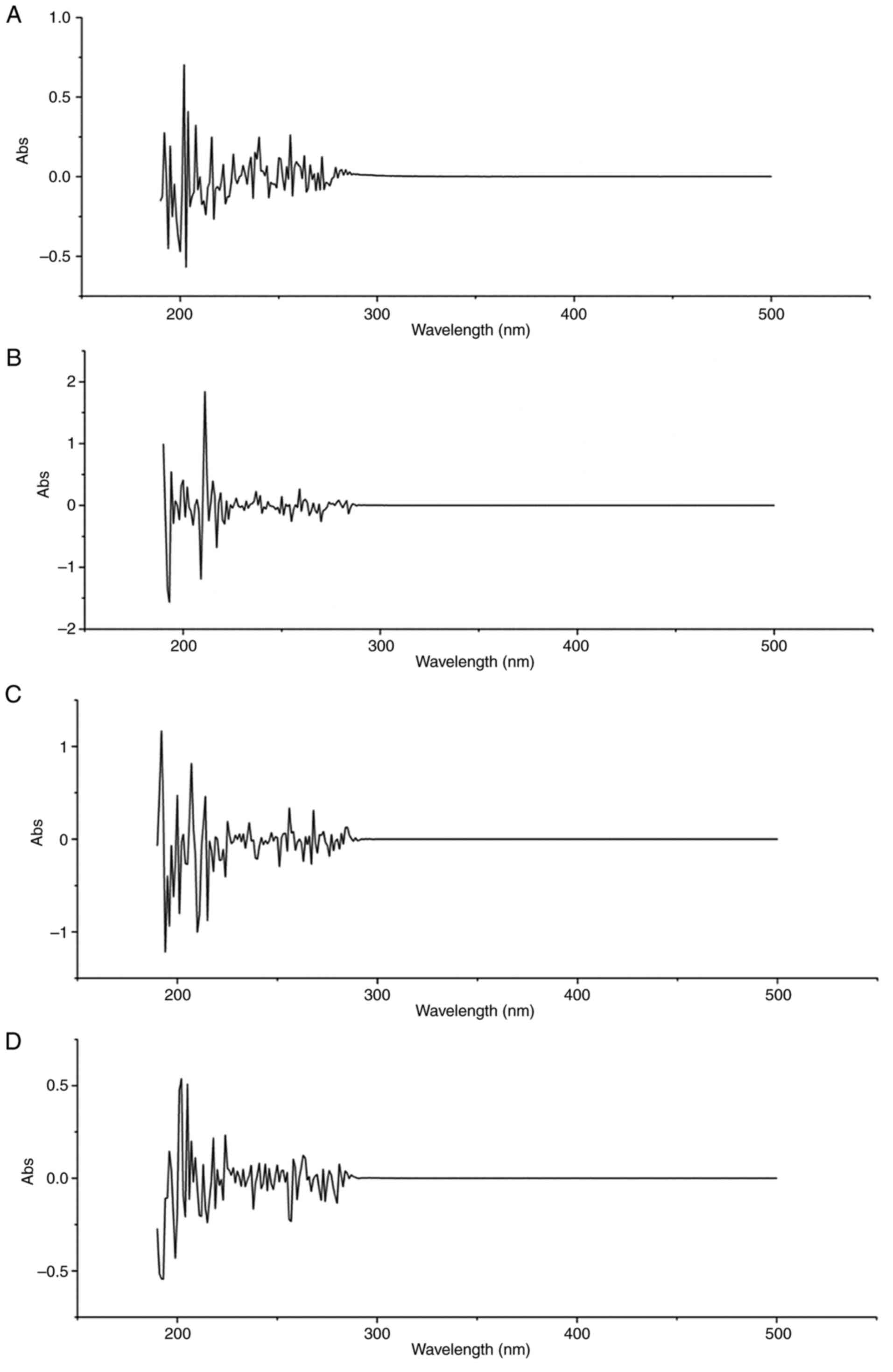
UV fingerprint analysis
As seen from the full-wavelength scanning spectrum
of the samples (Fig. 1), the main
absorption range was 192-256 nm. The CF had absorption peaks at
256, 208, 204, 202 and 192 nm (Fig.
1A); the EA had absorption peaks at 220, 214, 211, 199 and 196
nm (Fig. 1B); the NB had absorption
peaks at 214, 207, 200 and 192 nm (Fig.
1C); and the WA had absorption peaks at 205, 202, 201 and 196
nm (Fig. 1D). These findings
indicated that they had absorption at characteristic wavelengths,
and there was no interference in the range of visible light.
Acute oral toxicity analysis
In order to investigate the safety of the C.
serratus plant, an acute toxicity test was conducted followed
by an anti-inflammatory experiment. Doses were not deliberately
used to cause animal death, but higher doses of the C.
serratus plant were used in the acute toxicity test compared
with the doses used in the anti-inflammatory experiment. If the
acute toxicity test doses of the C. serratus plant had no
effects on the rats, it indicated that the anti-inflammatory test
doses were safe. There were no deaths at oral doses of 3.57, 3.36,
6.93 and 5.46 mg/kg in the CF, EA, NB and WA groups, respectively.
The half-maximal lethal dose values in the CF, EA, NB and WA groups
were found to be >3.57, 3.36, 6.93 and 5.46 mg/kg, respectively.
In addition, the calculated daily doses, 0.17, 0.16, 0.33 and 0.26
mg/kg, of the CF, EA, NB and WA were <3.57, 3.36, 6.93 and 5.46
mg/kg, respectively, which indicated that the anti-inflammatory
test doses were safe. Therefore, 0.17, 0.16, 0.33 and 0.26 mg/kg
were selected as the doses of the CF, EA, NB and WA extracts for
evaluating anti-arthritic activity in CFA-induced arthritic
rats.
Effects on general condition of the
rats
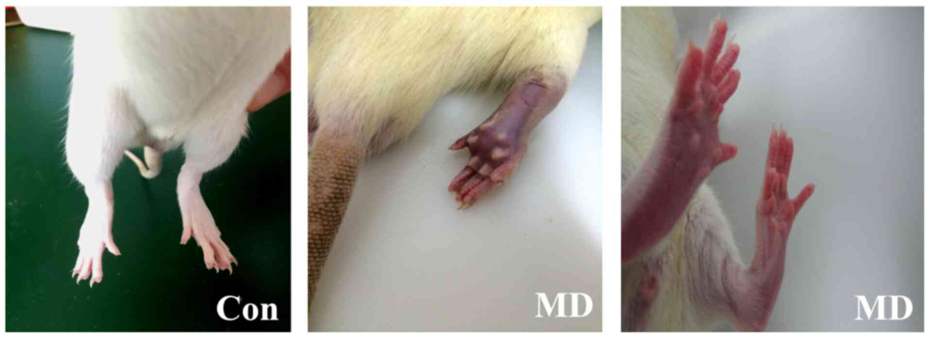
CFA-induced arthritis rats had dull and severely
lost fur, accompanied by reduced food intake and activities,
bleeding nostrils and swollen feet (Fig. 2). Some rats exhibited stiff joints
and tails, impeding their ability to walk. After the DISC or PD
treatment, the rats showed different degrees of decreases in food
intake and activity times in the CF, EA and NB groups, and the fur
color, fur shedding, diet and activities and swollen feet in the WA
and PD groups were improved and similar to those of the Con group
(Table II). It was preliminarily
judged that the WA had obvious anti-inflammatory activity.
| Table IIEffects on general condition of the
rats. |
Table II
Effects on general condition of the
rats.
| Group | Fur color | Bleeding | Feet condition | Food intake | Activity |
|---|
| Con | Normal | None | No swelling | Normal | Normal |
| MD | Dull and severely
lost | Nose | Swollen and
stiff | Greatly
reduced | Greatly
reduced |
| PD | Normal | None | No swelling | Normal | Normal |
| CF | Dull and severely
lost | Slight | Swollen | Greatly
reduced | Greatly
reduced |
| EA | Slightly dull | None | Slightly
swollen | Slightly
reduced | Reduced |
| NB | Slightly dull | None | Slightly
swollen | Normal | Slightly
reduced |
| WA | Normal | None | No swelling | Normal | Normal |
Effects on body weight changes
The weights of CFA-induced arthritic rats are
associated with the severity of inflammation (23). In the present study, compared with
the Con group, the rats injected with CFA exhibited significantly
decreased body weights on days 7-28 (P<0.01). After treatment
with the DISC or PD, the body weight of the rats gradually
recovered, particularly in the WA and PD groups, which approached
the level of the Con group (Table
III), whereas there were no significant changes in the CF group
on days 23 and 28 after modeling compared with the MD group. It
could be judged that the anti-inflammatory effect of the WA was the
best, while the anti-inflammatory effect of the CF was not
obvious.
| Table IIIChanges in body weight of the rats in
each group. |
Table III
Changes in body weight of the rats in
each group.
| Group | Weight on day 7,
g | Weight on day 15,
g | Weight on day 23,
g | Weight on day 38,
g |
|---|
| Con | 34.66±3.51 | 71.70±1.68 | 89.67±2.52 | 100.00±5.29 |
| MD |
26.50±0.96a |
59.23±2.77a |
72.90±4.06a |
76.27±1.94a |
| PD | 26.97±0.74 | 60.93±5.47 |
86.47±2.91c |
92.10±7.48c |
| CF | 26.23±0.60 | 57.43±0.85 | 74.97±1.89 | 81.03±1.50 |
| EA | 27.80±2.63 | 59.57±1.65 |
82.10±1.49c |
89.23±1.60b |
| NB | 26.30±1.65 | 59.20±1.41 |
79.53±1.07b | 83.73±2.65 |
| WA | 27.26±0.60 | 59.53±1.17 |
86.70±1.21c,d |
93.27±2.46c |
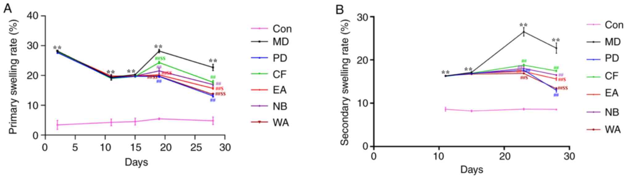
Effects on the primary and secondary
swelling rates
Primary lesions are the proinflammatory edema caused
by the injection of CFA, which can be investigated by calculating
swelling rate (20). Secondary
lesions are immune-mediated changes characterized by inflammation
in the non-injected parts (hind legs, forepaws, ears and nose)
(18). As shown in Fig. 3, the swelling of the left hind foot
(Fig. 3B) was lower compared with
that of the right hind foot (Fig.
3A), and the secondary swelling rate peaked on day 23.
The primary and secondary swelling rates in
CFA-induced arthritis rats were significantly increased (P<0.01)
compared with those of the Con group. From day 19 to day 28 after
modeling, the inflammatory swelling was inhibited by the different
extracts of C. serratus or PD (P<0.01; Fig. 3). Among them, the inhibitory effects
of the EA, WA and PD were more notable compared with that of the
NB. It could be seen that the anti-inflammatory effect of the WA
was better than other extracts.
Effects on AI
The AI value is used to reflect the extent of
inflammation. A Kruskal-Wallis test showed that there were
statistically significant differences in AI between the groups on
days 23 and 28 (χ2=15.51, P=0.02 and
χ2=19.57, both P<0.001; Table IV). On day 23, the mean rank AI was
18.00, 9.67, 14.83 and 7.33 for the CF, EA, NB and WA groups,
respectively; on day 28, the corresponding values were 20.00,
11.00, 16.50 and 7.83. This showed that the WA had obvious
anti-inflammatory activity, followed by the EA.
| Table IVRat AI of each group. |
Table IV
Rat AI of each group.
| | Day 23 | Day 28 |
|---|
| Group | AI | χ2 |
P-valuea | AI | χ2 |
P-valuea |
|---|
| Con | 2.00 | 15.51 | 0.02 | 3.50 | 19.57 | <0.001 |
| MD | 16.17 | | | 14.50 | | |
| PD | 9.00 | | | 5.17 | | |
| CF | 18.00 | | | 20.00 | | |
| EA | 9.67 | | | 11.00 | | |
| NB | 14.83 | | | 16.50 | | |
| WA | 7.33 | | | 7.83 | | |
Effects on organ indexes
The organ indexes can directly reflect the immune
function of the body. As presented in Table V, there was no significant
difference in the adrenal index between the MD and Con groups.
However, after the establishment of the model, the spleen index
increased significantly and the thymus index decreased
significantly (P<0.01). After treatment with the different
extracts or PD, EA, WA and PD significantly reversed these changes
(P<0.01), especially the WA and PD groups. The NB reversed the
spleen index (P<0.05); however, it had no significant effect on
the thymus index. In summary, the DISC only affected part of the
organ indexes.
| Table VEffects on organ indexes of the
rats. |
Table V
Effects on organ indexes of the
rats.
| Group | Spleen, % | Thymus, % | Adrenal gland,
% |
|---|
| Con | 0.18±0.04 | 0.18±0.02 | 0.02±0.01 |
| MD |
0.28±0.02a |
0.11±0.01a | 0.02±0.02 |
| PD |
0.22±0.04c |
0.17±0.02c | 0.02±0.01 |
| CF | 0.24±0.01 | 0.13±0.05 | 0.01±0.03 |
| EA |
0.21±0.02c |
0.17±0.02c | 0.02±0.02 |
| NB |
0.23±0.03b | 0.14±0.01 | 0.02±0.04 |
| WA |
0.20±0.05c |
0.17±0.03c | 0.02±0.05 |
Effects on the serum levels of NO, MDA
and SOD
The application of CFA induced an increase in the
levels of NO and MDA, and a decrease in the level of SOD in the MD
group (P<0.01). It is noteworthy that the WA and PD not only
significantly reduced the NO and MDA levels (P<0.05 or
P<0.01), but also raised the SOD level close to that of the Con
group. By contrast, the CF group showed a significant increase in
NO and MDA contents compared with the MD group (P<0.01), whereas
the changes in the EA and NB groups were not significant (Fig. 4). These results indicated that WA
alleviated arthritis damage by mediating oxidative stress.
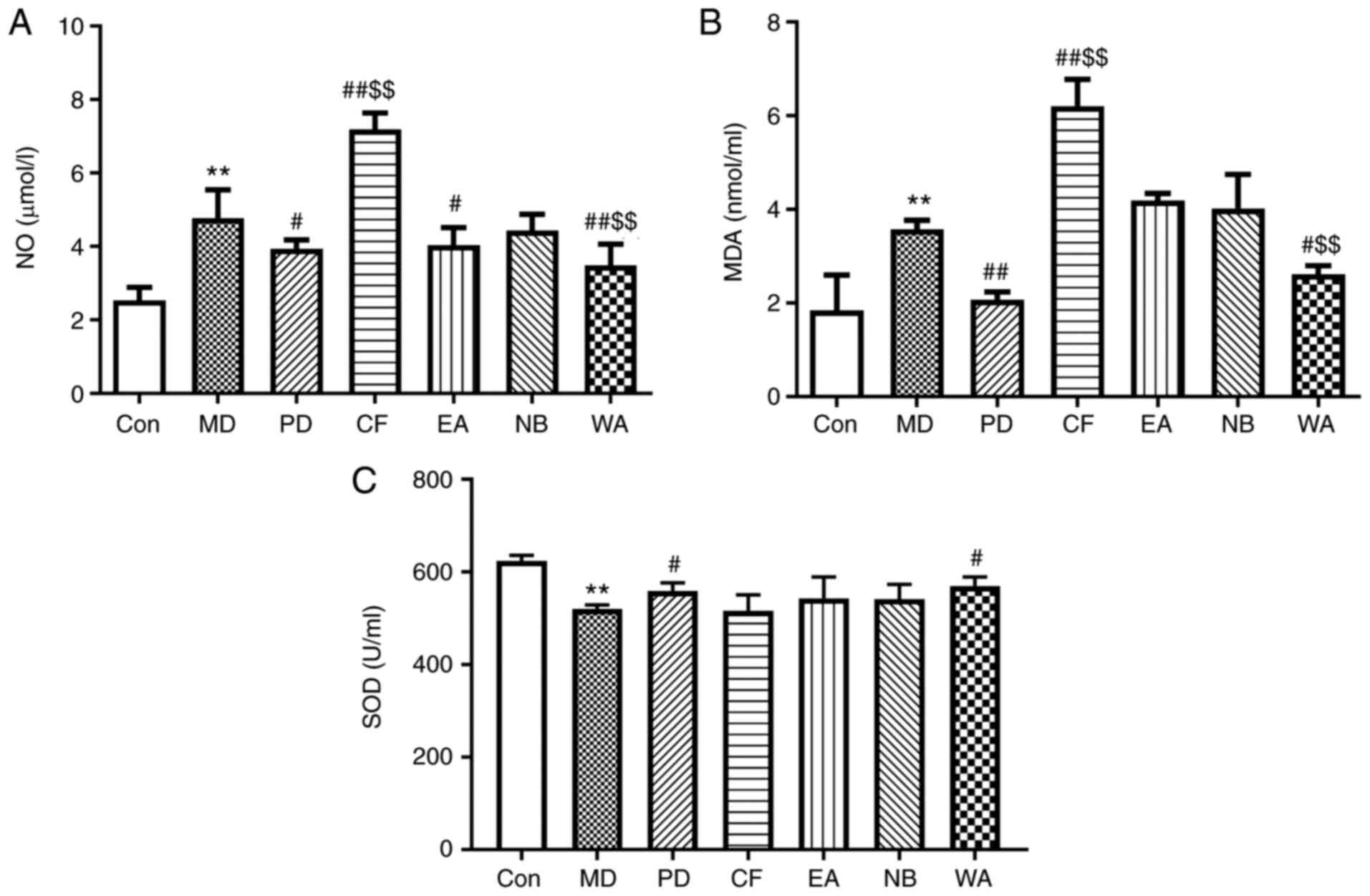 | Figure 4Effects on the serum inflammatory
factors and oxidative stress indicators. (A) NO, (B) MDA and (C)
SOD. n=6. **P<0.01 vs. Con; #P<0.05,
##P<0.01 vs. MD; $$P<0.01 vs. NB. Con,
control; MD, model; PD, positive drug; CF, chloroform separation
site; EA, ethyl acetate separation site; NB, n-butanol separation
site; WA, water separation site; NO, nitric oxide; MDA,
malondialdehyde; SOD, superoxide dismutase. |
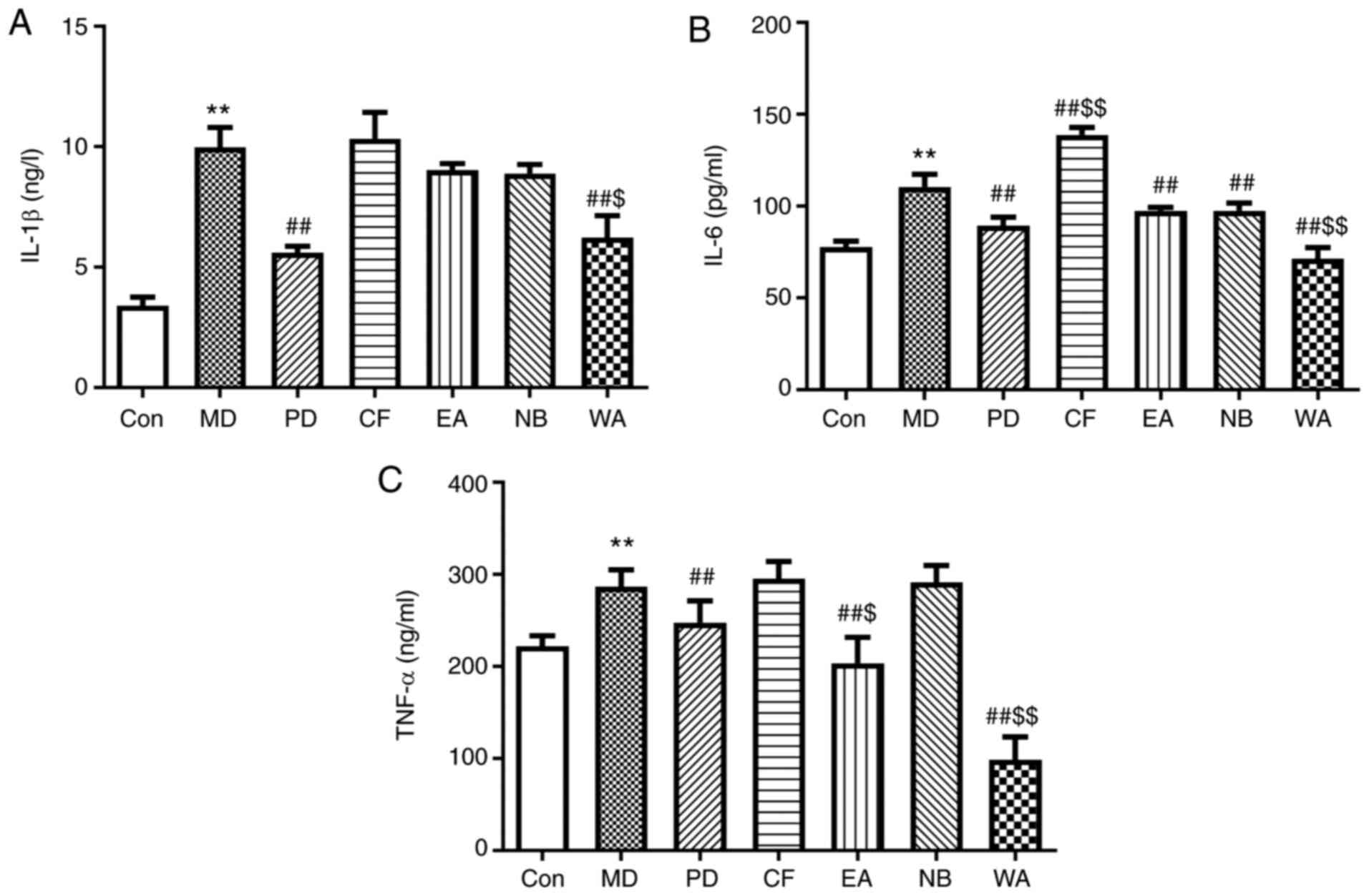
Effects on the serum levels of IL-1β,
IL-6 and TNF-α
A significant elevation in the serum levels of
IL-1β, TNF-α and IL-6 was observed in CFA-induced rats compared
with the rats in the Con group (all P<0.01). After exposure to
the DISC or PD, compared with the MD group, the levels of IL-1β,
IL-6 and TNF-α in the PD and WA groups decreased significantly
(P<0.01), the contents of IL-6 and TNF-α in the EA and NB groups
decreased significantly (P<0.01) and IL-6 showed an significant
decrease in the NB group (P<0.01). However, the level of IL-6
was significantly increased in the CF group (P<0.01), although
the levels of IL-1β and TNF-α were not significantly impacted, as
presented in Fig. 5. The results
showed that the DISC had a certain anti-inflammatory effect, and
the anti-inflammatory activity of the WA group was more
obvious.
Effects on the levels of MIF-1,
PGE2 and VEGF
Injection with CFA resulted in significant increases
in MIF-1, PGE2 and VEGF levels (P<0.05 or P<0.01).
Treatment with the CF had no significant impact on the three
indicators. The three indicators were decreased in the EA and NB
groups but this was not a significant change (P>0.05; Fig. 6). The three indicators were
significantly decreased in the WA and PD groups compared with those
in the arthritic group (P<0.05 or P<0.01). This showed that
the WA had the best anti-inflammatory activity, followed by the EA
and NB, and the CF was the least obvious.
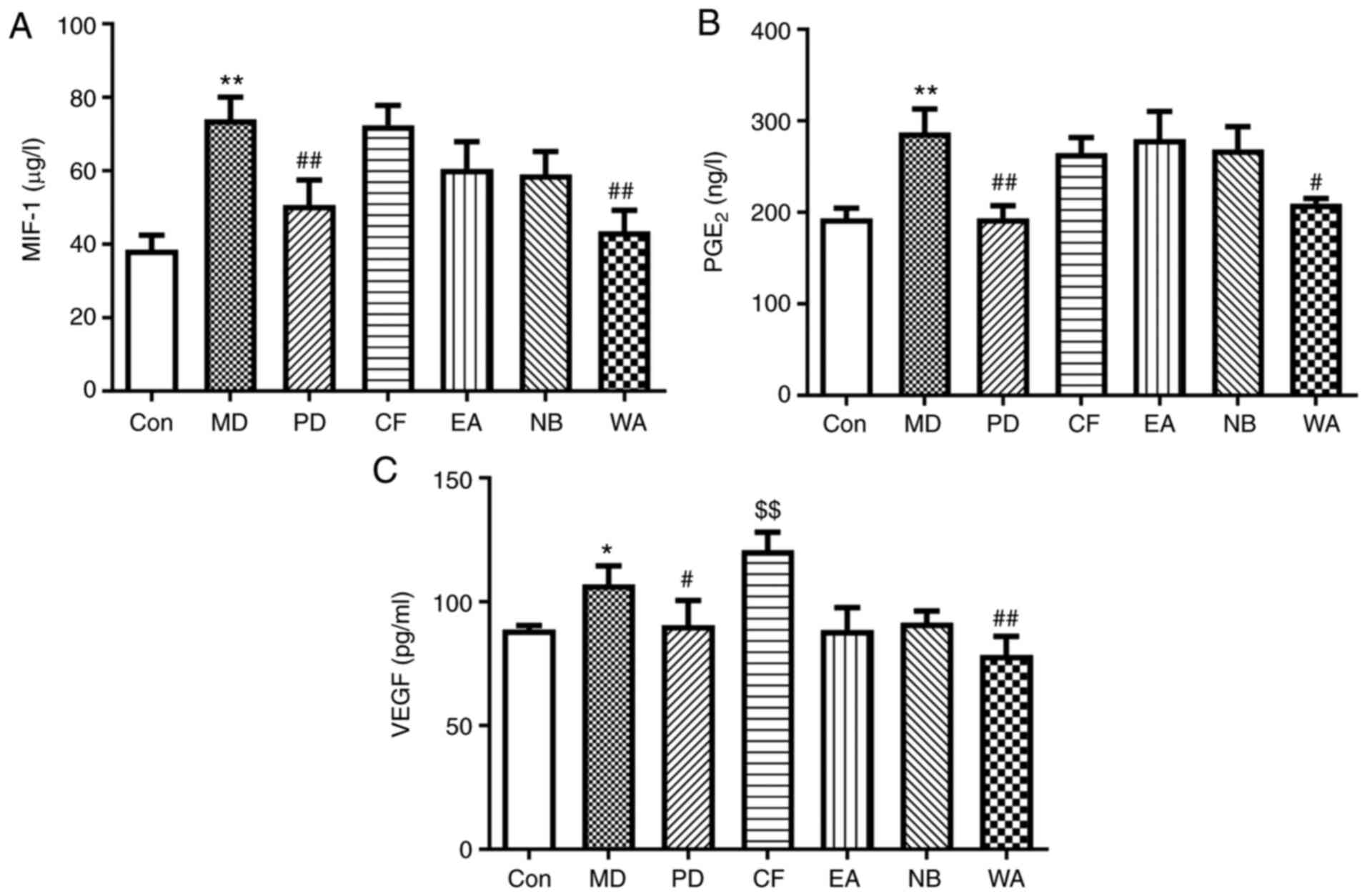 | Figure 6Effects on the serum inflammatory
indicators. (A) MIF-1, (B) PGE2 and (C) VEGF. n=6.
*P<0.05, **P<0.01 vs. Con;
#P<0.05, ##P<0.01 vs. MD;
$$P<0.01 vs. NB. Con, control; MD, model; PD,
positive drug; CF, chloroform separation site; EA, ethyl acetate
separation site; NB, n-butanol separation site; WA, water
separation site; MIF-1, macrophage inhibitor factor-1;
PGE2, prostaglandin E2. |
Effects on serum levels of IgG, IgM
and IFN-γ
The levels of IgG and IgM were significantly
increased, and the levels of IFN-γ were decreased after intradermal
injection of CFA compared with the Con (all P<0.01). When
treated with the DISC or PD, there were no significant changes in
the three indicators in the CF group (P>0.05). Compared with the
MD group, there were significant changes in the three indicators in
the PD and WA groups (P<0.05 or P<0.01) and the levels of IgG
and IFN-γ decreased significantly in the EA group (P<0.01). The
content of IgG decreased significantly in the NB group compared
with the MD group, while there were no significant changes in IFN-γ
in the NB group (P>0.05); these changes were significant in the
WA and PD groups (P<0.01; Fig.
7). These results indicated that the alleviation of arthritis
damage of the DISC was related to participating in the immune
response.
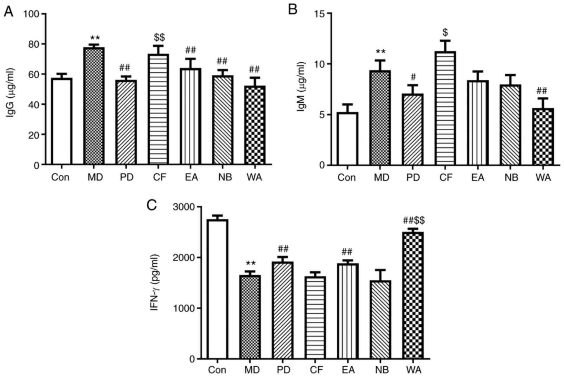 | Figure 7Effects on the serum immune
indicators. (A) IgG, (B) IgM and (C) IFN-γ. **P<0.01
vs. Con; #P<0.05, ##P<0.01 vs. MD;
$P<0.05, $$P<0.01 vs. NB. Con, control;
MD, model; PD, positive drug; CF, chloroform separation site; EA,
ethyl acetate separation site; NB, n-butanol separation site; WA,
water separation site; Ig, immunoglobulin. |
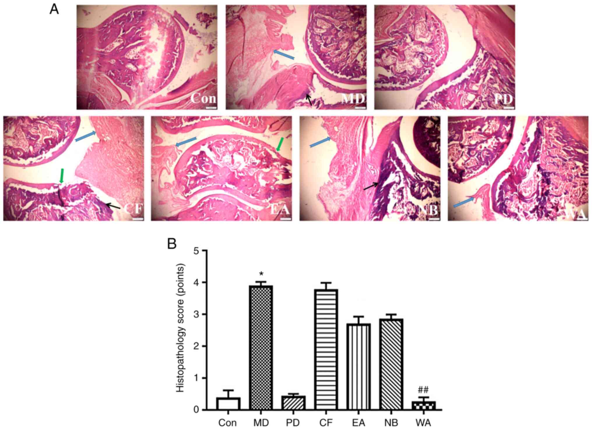
Effects on joint histopathology
The anti-inflammatory effects of the DISC were
confirmed via histopathological examination. As shown in Fig. 8, there was no notable synovial
hyperplasia or inflammatory cell infiltration in the Con group. In
contrast, the articular endometrium of the AA rats was thickened
with a large amount of inflammatory cell infiltration, tissue
granulation, damaged articular cartilage tissues and pannus
formation in the synovium. However, the DISC or PD treatment
reduced the symptoms of synovial hyperplasia, inflammatory cell
infiltration and cartilage destruction. The recovered
histopathological characteristics in the WA and PD groups appeared
to be similar to those of the Con group. A Kruskal-Wallis test
showed that there were statistically significant differences in
histopathology scores across the groups (χ2=24.25,
P<0.001), with a mean rank histopathology score of 24.00, 15.75,
17.25 and 4.38 for the CF, EA, NB and WA groups, respectively
(Table VI). There was a
significant difference between the Con and MD groups (P<0.05).
There was significant difference between the WA and MD groups
(P<0.01). The pathological scores of other DISC groups were
lower compared with that of the MD group, but there were no
significant differences. The above showed that the DISC could
alleviate the pathological damage of joints, of which the WA was
the most obvious. The EA and NB showed no obvious changes compared
with the MD group.
| Table VIHistopathology score of
representative images of hematoxylin and eosin staining. |
Table VI
Histopathology score of
representative images of hematoxylin and eosin staining.
| Group | Histopathology
score, points | χ2 |
P-valuec |
|---|
| Con | 7.25 | 24.25 | <0.001 |
| MD | 25.00a | | |
| PD | 7.88 | | |
| CF | 24.00 | | |
| EA | 15.75 | | |
| NB | 17.25 | | |
| WA | 4.38b | | |
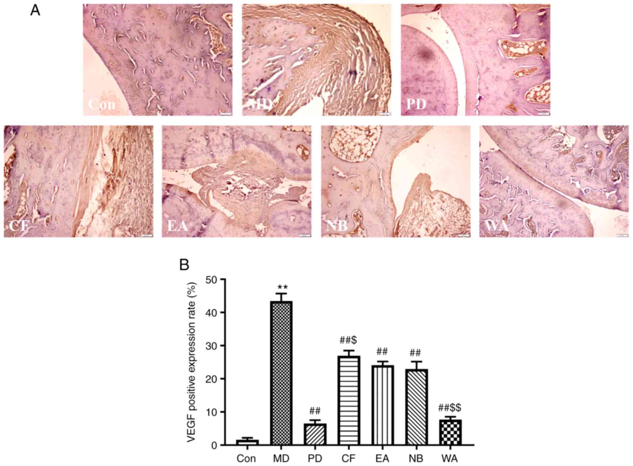
Immunohistochemical analysis of
VEGF
After immunohistochemical staining of the synovial
tissues of the arthritis rats, the positive expression of VEGF was
mainly manifested as clear brownish-yellow particles in the
cytoplasm of the cells (Fig. 9A)
(24). As shown in Fig. 9B, increased VEGF expression occurred
in the arthritic rats compared with the Con group (P<0.01). The
positive expression of VEGF in the extract and PD groups was
significantly decreased compared with that of the MD group
(P<0.01). By comparing each extract group, it was revealed that
the positive expression rate of VEGF in the WA group was
significantly lower compared with that in the NB group (P<0.01),
while the EA group was not significantly different; by contrast,
the positive expression rate of VEGF in the CF group was
significantly increased compared with that in the NB group
(P<0.05). It was indicated that among the extract groups, the WA
group had the most significant decrease, followed by the EA and NB
groups. In summary, it was shown that the DISC had a certain
anti-inflammatory effect, and the WA had the best anti-inflammatory
activity.
Discussion
In the pre-experiment of the anti-inflammatory
experiment, 12.5, 25 and 50-fold values of the clinical dose were
used for optimization. Some indicators such as general condition of
the rats, body weight change, primary and secondary swelling rates,
AI and organ indexes were examined. There were no clear
inflammatory effects at 12.5X of the dose; 25X dose showed that
DISC had anti-inflammatory effects (data not shown) and there were
significant differences among the four DISCs at 50X dose.
Overall, 50X of the clinical dose after gradient
extraction did not cause toxicity and the anti-inflammatory effects
were good. Therefore, 50X the clinical dose per extract (0.17,
0.16, 0.33 and 0.26 mg/kg for the CF, EA, NB and WA, respectively)
was used to investigate the anti-inflammatory activity of the four
different separation sites in CFA-induced arthritic rats.
RA is a type of chronic inflammation mediated by
cytokines and cross-reactive antigens. As a result of various
imbalances between proinflammatory and anti-inflammatory cytokine
activities, the clinical features of RA involve systemic
inflammation, leading to a variety of systemic immune complications
(25). Currently, the drugs used to
treat RA only temporarily relieve joint pain and discomfort rather
than providing fundamental treatment (26). The study of effective drugs for the
treatment of RA has attracted increasing attention of experts and
scholars (27).
In the current study, the AA rat model was used to
evaluate the anti-arthritic effects of the DISC on various
arthritic phenomena, including inflammatory cell infiltration. It
is a convenient and ideal model for screening anti-RA drugs and
studying immune inflammatory diseases. Based on the principles of
RA and the processes of immune diseases, CFA-induced arthritis has
numerous similarities to human RA in terms of histopathology and
serum biochemical indicators (28).
In RA, complicated cytokine networks regulate chronic inflammation
and joint destruction (29).
Changes in the levels of these proinflammatory mediators (IL-6,
PGE2, TNF-α and NO) reflect the severity of inflammation
and joint damage in AA rats (30).
Among these inflammatory factors, IL-6 is highly
expressed in the synovium and serum of patients with RA, leading to
inflammatory cell infiltration and synovial hyperplasia (31). Excessive levels of the long-lasting
inflammatory cytokine IL-1β contribute to the development of RA
(32). PGE2 regulates
hyperalgesia; its overexpression in RA promotes vasodilation in
synovial tissues, and PGE2 synergizes with other
inflammatory cytokines, such as IL-1β, to accelerate the process of
inflammation and induce chronic inflammation (32,33).
During the development of RA, MIF-1 induces the release of
proinflammatory cytokines, including IL-1β, IL-6, PGE2
and TNF-α (34). Decreasing the
level of MIF-1 can control inflammation and cartilage destruction
(35). TNF-α induces the production
of other inflammatory cytokines, particularly in the occurrence and
development of RA (36). In the
present study, the WA had significant inhibitory effects on the
levels of IL-1β, IL-6, TNF-α, MIF-1 and PGE2, whereas
there were no inhibitory effects on the levels of IL-1β and
PGE2 in the CF, EA and NB groups, indicating that the WA
induced the most potent anti-RA effects.
In the AA environment, increases in the levels of
TNF-α, IL-6 and PGE2 induce the production of VEGF; in
turn, VEGF promotes the production and release of these
inflammatory mediators (24). VEGF
increases vascular permeability, provides adequate blood flow and
nutrition for the proliferation and migration of synovial cells,
and induces the development of synovitis, resulting in joint
swelling and dysfunction (37). In
the current study, the WA significantly inhibited the expression of
VEGF in synovial tissues, which is consistent with the level of
VEGF in serum.
IgM, IgG and other immunoconjugates formed with
antibodies accumulate in the joints and synovium of patients with
RA, which induce the production of various inflammatory factors,
such as TNF-α, IL-6 and VEGF, causing articular cartilage damage
(38). IFN-γ is an important immune
regulator and macrophage-stimulating factor, and is synthesized
during the cellular immune response (39). It has two-way immunomodulatory
ability, plays a notable role in the prevention and treatment of
immune diseases, and has potential therapeutic effects on RA
(40). In the present study, the WA
significantly reversed CFA-induced increases of IgG and IgM levels,
and decreases in the IFN-γ level, with similar but weaker effects
observed in the EA and NB groups, which indicated that the WA
exerted the most potent anti-inflammatory effects, followed by the
EA and NB.
A significant increase in the NO level in serum
leads to increased oxidative stress, causing damage to cells and
tissues, and accelerating the process of joint damage (41). SOD scavenges oxygen free radicals,
regulates the balance of oxidants and antioxidants in vivo,
and reduces lipid peroxidation to protect cells and tissues from
oxidative stress-induced damage (42). MDA, a free radical, reflects the
degree of lipid peroxidation and has been widely used as a marker
of lipid peroxidation products (43). In the present study, the WA
significantly reversed the changes in the levels of NO, MDA and SOD
induced by CFA, illustrating that the anti-inflammatory activity of
the WA was the strongest, with lesser effects induced by the EA and
NB. These results suggested that oxidative stress was involved in
the anti-RA mechanisms of DISC.
Furthermore, after the DISC treatment, CFA-induced
proliferation of synovial cells and a large amount of inflammatory
cell infiltration were alleviated to varying degrees. The
recoveries in the WA group were the most increased, which is
consistent with the results of the serum indicators; thus, these
findings indicated that the WA exerted the strongest
anti-inflammatory activity.
The spleen and thymus are important lymphoid organs,
and the adrenal gland is an important endocrine organ. The organ
indexes can roughly estimate the strength of immune function
(44). Inflammation stimulates the
immune function of the body and damages the endocrine organs, so
the more increased the anti-inflammatory activity is, the more
effectively it can reduce the immune responses and damage to the
adrenal gland in inflammatory rats (45). In the present study, except for the
CF, all the isolated sites could significantly reduce the spleen
indexes; however, only the EA and WA increased the thymus indexes.
Moreover, all the isolated sites could restore weight gain to
varying degrees in AA rats on days 23 and 28 after modeling,
particularly the EA and WA. Although a variety of confounding
factors affected organ indexes (46) and body weight changes (47), the four separated site groups were
tested under identical conditions. Therefore, these two indicators
are comparable among groups. All these indicated that the EA and WA
exhibited anti-inflammatory activity. Among them, the WA appeared
to possess the most potent anti-inflammatory activity.
In addition, changes in the primary and secondary
swelling rates are associated with the accumulation of granulocytes
and monocytes in the joint tissues (48). In the present study, the four
isolated sites significantly inhibited the swelling rates in
CFA-induced arthritis rats, and the inhibition of the WA was the
most significant. Except for the CF, all of the isolated sites
could reduce AI, particularly the WA, indicating that the three
isolated sites exhibited anti-inflammatory activity.
In conclusion, the WA of C. serratus
exhibited the greatest effect on anti-RA activity, followed by the
EA and NB, which was associated with oxidative stress and decreased
inflammatory factor release. These results suggested that C.
serratus may serve as a potential candidate for the treatment
of RA, providing a scientific basis for the exploration and
utilization of C. serratus, as well as providing a reference
for further development of drugs against RA. The anti-inflammatory
mechanisms of C. serratus will be explored further in
subsequent studies, and this study lays a foundation for further
separation and purification of monomer anti-inflammatory
components.
Acknowledgements
The authors would like to acknowledge Professor
Jianhua Zhu of Wannan Medical College for the plant identification,
Associate Professor Yinhua Liu of Wannan Medical College for the
pathological staining of the ankle joints and Professor Xiaoping
Zhang of Wannan Medical College for the voucher.
Funding
Funding: The present study was supported by The Key Research and
Development Projects (grant no. 1804h08020271) from The Anhui
Provincial Department of Science and Technology.
Availability of data and materials
The datasets used and/or analyzed during the current
study are available from the corresponding author on reasonable
request.
Authors' contributions
All the authors participated in the experimental
processes, either in whole or in part. SS designed the experiments.
SS, YD and SL performed the experiments. SS, YD, SL, BG, RX, WC, CZ
and EZ analyzed the data and drafted the initial part of the
manuscript. SS and YD confirmed the authenticity of all the raw
data. All authors read and approved the final manuscript.
Ethics approval and consent to
participate
Experiments were carried out with the approval of
The Wannan Medical College Ethics Committee (approval no.
20180316).
Patient consent for publication
Not applicable.
Competing interests
The authors declare that they have no competing
interests.
References
|
1
|
Wei ST, Sun YH, Zong SH and Xiang YB:
Serum levels of IL-6 and TNF-α may correlate with activity and
severity of rheumatoid arthritis. Med Sci Monit. 21:4030–4038.
2015.PubMed/NCBI View Article : Google Scholar
|
|
2
|
Zhang Q, Yu Y, Li J, Guan Y, Huang J, Wang
Z, Zhang Z, Zhang W, Guo J, Li J, et al: Anti-arthritic activities
of ethanol extracts of circaea mollis Sieb.& Zucc. (whole
plant) in rodents. J Ethnopharmacol. 225:359–366. 2018.PubMed/NCBI View Article : Google Scholar
|
|
3
|
Abdel El-Gaphar OAM, Abo-Youssef AM and
Abo-Saif AA: Effect of losartan in complete Freund's
adjuvant-induced arthritis in rats. Iran J Pharm Res. 17:1420–1430.
2018.PubMed/NCBI
|
|
4
|
Kocyigit A, Guler EM and Kaleli S:
Anti-inflammatory and antioxidative properties of honey bee venom
on Freund's complete adjuvant-induced arthritis model in rats.
Toxicon. 161:4–11. 2019.PubMed/NCBI View Article : Google Scholar
|
|
5
|
Ingawale DK and Patel SS: Hecogenin
exhibits anti-arthritic activity in rats through suppression of
pro-inflammatory cytokines in complete Freund's adjuvant-induced
arthritis. Immunopharmacol Immunotoxicol. 40:59–71. 2018.PubMed/NCBI View Article : Google Scholar
|
|
6
|
da Rocha ML, Oliveira LE, Patrício Santos
CC, de Sousa DP, de Almeida RN and Araújo DA: Antinociceptive and
anti-inflammatory effects of the monoterpene α,β-epoxy-carvone in
mice. J Nat Med. 67:743–749. 2013.PubMed/NCBI View Article : Google Scholar
|
|
7
|
Qiao J, Xu LH, He J, Ouyang DY and He XH:
Cucurbitacin E exhibits anti-inflammatory effect in RAW 264.7 cells
via suppression of NF-κB nuclear translocation. Inflamm Res.
62:461–469. 2013.PubMed/NCBI View Article : Google Scholar
|
|
8
|
Nanjing University of Traditional Chinese
Medicine: Dictionary of Traditional Chinese Medicine. Vol 1. 2nd
edition. Shanghai Science and Technology Press, Shanghai,
pp310-311, 2006.
|
|
9
|
Tang L, Zhu H, Yang X, Xie F, Peng J,
Jiang D, Xie J, Qi M and Yu L: Shizukaol D, a dimeric sesquiterpene
isolated from Chloranthus serratus, represses the growth of
human liver cancer cells by modulating wnt signalling pathway. PLoS
One. 11(e0152012)2016.PubMed/NCBI View Article : Google Scholar
|
|
10
|
Zhang M, Wang J, Luo J, Wang P, Guo C and
Kong L: Labdane diterpenes from Chloranthus serratus.
Fitoterapia. 91:95–99. 2013.PubMed/NCBI View Article : Google Scholar
|
|
11
|
Sun SP, Du YY, Yin CL, Suo XG, Wang R, Xia
RP and Zhang XP: Water-separated part of Chloranthus
serratus alleviates lipopolysaccharide-induced RAW264.7 cell
injury mainly by regulating the MAPK and Nrf2/HO-1 inflammatory
pathways. BMC Complement Altern Med. 19:343–355. 2019.PubMed/NCBI View Article : Google Scholar
|
|
12
|
Yuan T, Zhu RX, Yang SP, Zhang H, Zhang CR
and Yue JM: Serratustones A and B representing a new dimerization
pattern of two types of sesquiterpenoids from Chloranthus
serratus. Org Lett. 14:3198–3201. 2012.PubMed/NCBI View Article : Google Scholar
|
|
13
|
Sun S, Li S, Du Y, Wu C, Zhang M, Li J and
Zhang X: Anti-inflammatory effects of the root, stem and leaf
extracts of Chloranthus serratus on adjuvant-induced
arthritis in rats. Pharm Biol. 58:528–537. 2020.PubMed/NCBI View Article : Google Scholar
|
|
14
|
Saleem A, Saleem M, Akhtar MF, Shahzad M
and Jahan S: Moringa rivae leaf extracts attenuate complete
Freund's adjuvant-induced arthritis in Wistar rats via modulation
of inflammatory and oxidative stress biomarkers.
Inflammopharmacology. 28:139–151. 2020.PubMed/NCBI View Article : Google Scholar
|
|
15
|
Wei W, Wu XM and Li YJ: Pharmacological
experimental methodology. In: Appendix. 4th edition. Beijing:
People's Medical Publishing House, pp1698, 2010.
|
|
16
|
Yao YM, Fang X, Li J, Zhang JQ, Ruan KF
and Liang S: Study on anti-inflammatory effects of diterpenoids and
different polarity fractions from Rhododendron molle G. Don in
vivo and in vitro. J Tradit Chin Med Sci. 33:84–88.
2019.
|
|
17
|
Yang X, Yang J and Zou H: Baicalin
inhibits IL-17-mediated joint inflammation in murine
adjuvant-induced arthritis. Clin Dev Immunol.
2013(268065)2013.PubMed/NCBI View Article : Google Scholar
|
|
18
|
Lu SW, Su H, Yu FM, Wang QS, Guo YY, LI GY
and Kuang HX: Therapeutic effect and mechanism of caulophyllum
robustum maxim extract on adjuvant arthritis rats. Med Plant.
9:50–55. 2018.
|
|
19
|
Yamagishi Y, Igarashi M, Suzuki A, Suguro
S, Hirano SI and Nagaoka I: Evaluation of the effect of methionine
and glucosamine on adjuvant arthritis in rats. Exp Ther Med.
4:640–644. 2012.PubMed/NCBI View Article : Google Scholar
|
|
20
|
Gohil P, Patel V, Deshpande S, Chorawala M
and Shah G: Anti-arthritic activity of cell wall content of
Lactobacillus plantarum in Freund's adjuvant-induced arthritic
rats: Involvement of cellular inflammatory mediators and other
biomarkers. Inflammopharmacology. 26:171–181. 2018.PubMed/NCBI View Article : Google Scholar
|
|
21
|
Miao CG: The role of SFRP4 in the
pathological change of adjuvant arthritis in rats and its
epigenetic modifications. Anhui Med Univ. 3(22)2014.
|
|
22
|
Tu Q, Li Y, Jin J, Jiang X, Ren Y and He
Q: Curcumin alleviates diabetic nephropathy via inhibiting podocyte
mesenchymal transdifferentiation and inducing autophagy in rats and
MPC5 cells. Pharm Biol. 57:778–786. 2019.PubMed/NCBI View Article : Google Scholar
|
|
23
|
Syed Zameer Ahmed K, Ahmed SSZ,
Thangakumar A and Krishnaveni R: Therapeutic effect of parmotrema
tinctorum against complete Freund's adjuvant-induced arthritis in
rats and identification of novel isophthalic ester derivative.
Biomed Pharmacother. 112(108646)2019.PubMed/NCBI View Article : Google Scholar
|
|
24
|
Kim HR, Kim KW, Kim BM, Cho ML and Lee SH:
The effect of vascular endothelial growth factor on
osteoclastogenesis in rheumatoid arthritis. PLoS One.
10(e0124909)2015.PubMed/NCBI View Article : Google Scholar
|
|
25
|
Pretorius E, Akeredolu OO, Soma P and Kell
DB: Major involvement of bacterial components in rheumatoid
arthritis and its accompanying oxidative stress, systemic
inflammation and hypercoagulability. Exp Biol Med (Maywood).
242:355–373. 2017.PubMed/NCBI View Article : Google Scholar
|
|
26
|
Mateen S, Zafar A, Moin S, Khan AQ and
Zubair S: Understanding the role of cytokines in the pathogenesis
of rheumatoid arthritis. Clin Chim Acta. 455:161–171.
2016.PubMed/NCBI View Article : Google Scholar
|
|
27
|
Perera PK, Cheng P, Xue L, Yun-Man L,
Wei-Rong F and Cai-Feng H: Effects of Yishen Juanbi (YJB) pill on
experimental rheumatoid arthritis. Chin J Nat Med. 8:57–61.
2010.PubMed/NCBI View Article : Google Scholar
|
|
28
|
Miao C, Chang J, Zhong G, Yu H, Zhou L,
Zhou G and Zhao C: CUL4B promotes the pathology of adjuvant-induced
arthritis in rats through the canonical Wnt signaling. J Mol Med
(Berl). 96:495–511. 2018.PubMed/NCBI View Article : Google Scholar
|
|
29
|
Zhang C, Zhang W, Shi R, Tang B and Xie S:
Coix lachryma-jobi extract ameliorates inflammation and oxidative
stress in a complete Freund's adjuvant-induced rheumatoid arthritis
model. Pharm Biol. 57:792–798. 2019.PubMed/NCBI View Article : Google Scholar
|
|
30
|
Fernandes J and Gupta GL: N-acetylcysteine
attenuates neuroinflammation associated depressive behavior induced
by chronic unpredictable mild stress in rat. Behav Brain Res.
364:356–365. 2019.PubMed/NCBI View Article : Google Scholar
|
|
31
|
Kamel KM, Gad AM, Mansour SM, Safar MM and
Fawzy HM: Novel anti-arthritic mechanisms of polydatin in complete
Freund's adjuvant-induced arthritis in rats: Involvement of IL-6,
STAT-3, IL-17, and NF-кB. Inflammation. 41:1974–1986.
2018.PubMed/NCBI View Article : Google Scholar
|
|
32
|
Chen X, Lu J, An M, Ma Z, Zong H and Yang
J: Anti-inflammatory effect of resveratrol on adjuvant arthritis
rats with abnormal immunological function via the reduction of
cyclooxygenase-2 and prostaglandin E2. Mol Med Rep.
9:2592–2598. 2014.PubMed/NCBI View Article : Google Scholar
|
|
33
|
Zhang P and Gan YH: Prostaglandin
E2 upregulated trigeminal ganglionic sodium channel 1.7
involving temporomandibular joint inflammatory pain in rats.
Inflammation. 40:1102–1109. 2017.PubMed/NCBI View Article : Google Scholar
|
|
34
|
Kim KW and Kim HR: Macrophage migration
inhibitory factor: A potential therapeutic target for rheumatoid
arthritis. Korean J Intern Med. 31:634–642. 2016.PubMed/NCBI View Article : Google Scholar
|
|
35
|
Li S, Zhang R, Li P, Yi W, Zhang Z, Chen
S, Su S, Zhao L and Hu C: Development of a novel method to measure
macrophage migration inhibitory factor (MIF) in sera of patients
with rheumatoid arthritis by combined electrochemical immunosensor.
Int Immunopharmacol. 8:859–865. 2008.PubMed/NCBI View Article : Google Scholar
|
|
36
|
Yoshino S and Ohsawa M: The role of
lipopolysaccharide injected systemically in the reactivation of
collagen-induced arthritis in mice. Brit J Pharmacol.
129:1309–1314. 2000.PubMed/NCBI View Article : Google Scholar
|
|
37
|
Boissier MC: Cell and cytokine imbalances
in rheumatoid synovitis. Joint Bone Spine. 78:230–234.
2011.PubMed/NCBI View Article : Google Scholar
|
|
38
|
Boissier MC, Semerano L, Challal S,
Saidenberg-Kermanac'h N and Falgarone G: Rheumatoid arthritis: from
autoimmunity to synovitis and joint destruction. J Autoimmun.
39:222–228. 2012.PubMed/NCBI View Article : Google Scholar
|
|
39
|
Schurgers E, Mertens F, Vanoirbeek JA, Put
S, Mitera T, De Langhe E, Billiau A, Hoet PH, Nemery B, Verbeken E
and Matthys P: Pulmonary inflammation in mice with collagen-induced
arthritis is conditioned by complete Freund's adjuvant and
regulated by endogenous IFN-r. Eur J Immunol. 42:3223–3234.
2012.PubMed/NCBI View Article : Google Scholar
|
|
40
|
Bi D, Bi D, Zhong M, Zhang H, Jin S, Ma S
and Luo H: Effects of leukotriene B4 on interleukin-32,
interferon-γ and chemokines in rats with rheumatoid arthritis. Exp
Ther Med. 14:2925–2930. 2017.PubMed/NCBI View Article : Google Scholar
|
|
41
|
Cicala C, Ianaro A, Fiorucci S, Calignano
A, Bucci M, Gerli R, Santucci L, Wallace JL and Cirino G:
NO-naproxen modulates inflammation, nociception and downregulates T
cell response in rat Freund's adjuvant arthritis. Brit J Pharmacol.
130:1399–1405. 2000.PubMed/NCBI View Article : Google Scholar
|
|
42
|
Lee J, Homma T, Kobayashi S, Ishii N and
Fujii J: Unveiling systemic organ disorders associated with
impaired lipid catabolism in fasted SOD1-deficient mice. Arch
Biochem Biophys. 654:163–171. 2018.PubMed/NCBI View Article : Google Scholar
|
|
43
|
Aryaeian N, Djalali M, Shahram F, Jazayeri
SH, Chamari M and Nazari S: Beta-carotene, vitamin E, MDA,
glutathione reductase and arylesterase activity levels in patients
with active rheumatoid arthritis. Iran J Public Health. 40:102–109.
2011.PubMed/NCBI
|
|
44
|
Wan L, Liu J, Huang CB, Zhang XJ, Wang YL,
Zhang PH, Sun Y and Liu L: Effects of triptolide on the autophagy
in synovial, spleen and thymus of rats with adjuvant arthritis.
Sichuan Da Xue Xue Bao Yi Xue Ban. 48:520–525. 2017.PubMed/NCBI(In Chinese).
|
|
45
|
Sattler J, Tu J, Stoner S, Li J,
Buttgereit F, Seibel MJ, Zhou H and Copper MS: Role of 11β-HSD type
1 in abnormal HPA axis activity during immune-mediated arthritis.
Endocr Connect. 7:385–394. 2018.PubMed/NCBI View Article : Google Scholar
|
|
46
|
LI X, Wu Z, He B and Zhong W: Etrandrine
alleviates symptoms of rheumatoid arthritis in rats by regulating
the expression of cyclooxygenase-2 and inflammatory factors. Exp
Ther Med. 16:2670–2676. 2018.PubMed/NCBI View Article : Google Scholar
|
|
47
|
Tu Y, Wang K, Liang Y, Jia X, Wang L, Wan
JB, Han J and He C: Glycine tabacina ethanol extract ameliorates
collagen-induced arthritis in rats via inhibiting pro-inflammatory
cytokines and oxidation. J Ethnopharmacol. 237:20–27.
2019.PubMed/NCBI View Article : Google Scholar
|
|
48
|
Holmdahl R, Malmström V and Burkhardt H:
Autoimmune priming, tissue attack and chronic inflammation-the
three stages of rheumatoid arthritis. Eur J Immunol. 44:1593–1599.
2014.PubMed/NCBI View Article : Google Scholar
|