|
1
|
Eurostat: Your key to European statistics:
https://ec.europa.eu/eurostat/statistics-xplained/index.php/Causes_of_death_statistics_-people_over_65
https://ec.europa.eu/eurostat/statistics-explained/index.php/Cardiovascular_diseases_statistics.
|
|
2
|
Singh RB, Mengi SA, Xu YJ, Arneja AS and
Dhalla NS: Pathogenesis of atherosclerosis: A multifactorial
process. Exp Clin Cardiol. 7:40–53. 2002.PubMed/NCBI
|
|
3
|
Albanese I, Khan K, Barratt B, Al-Kindi H
and Schwertani A: Atherosclerotic calcification: Wnt is the hint. J
Am Heart Assoc. 7(e007356)2018.PubMed/NCBI View Article : Google Scholar
|
|
4
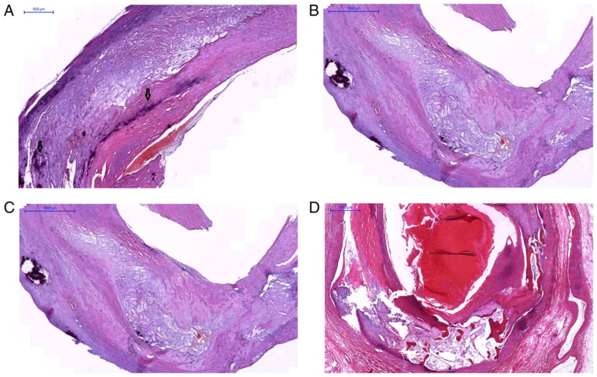
|
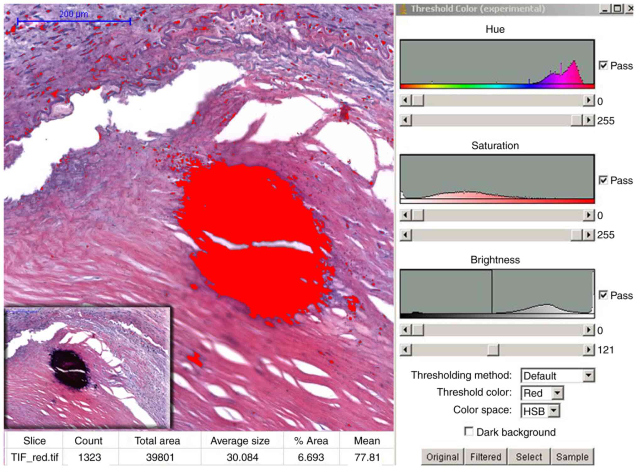
Peace A, van Mil A, Jones H and Thijssen
DHJ: Similarities and differences between carotid artery and
coronary artery function. Curr Cardiol Rev. 14:254–263.
2018.PubMed/NCBI View Article : Google Scholar
|
|
5
|
Schiano V, Sirico G, Giugliano G,
Laurenzano E, Brevetti L, Perrino C, Brevetti G and Esposito G:
Femoral plaque echogenicity and cardiovascular risk in claudicants.
JACC Cardiovasc Imaging. 5:348–357. 2012.PubMed/NCBI View Article : Google Scholar
|
|
6
|
Golomb BA, Dang TT and Criqui MH:
Peripheral arterial disease: Morbidity and mortality implications.
Circulation. 114:688–699. 2006.PubMed/NCBI View Article : Google Scholar
|
|
7
|
Maleckis K, Anttila E, Aylward P, Poulson
W, Desyatova A, MacTaggart J and Kamenskiy A: Nitinol Stents in the
femoropopliteal artery: A mechanical perspective on material,
design, and performance. Ann Biomed Eng. 46:684–704.
2018.PubMed/NCBI View Article : Google Scholar
|
|
8
|
Kwee RM: Systematic review on the
association between calcification in carotid plaques and clinical
ischemic symptoms. J Vasc Surg. 51:1015–1025. 2010.PubMed/NCBI View Article : Google Scholar
|
|
9
|
Shioi A and Ikari Y: Plaque Calcification
during atherosclerosis progression and regression. J Atheroscler
Thromb. 25:294–303. 2018.PubMed/NCBI View Article : Google Scholar
|
|
10
|
Yang J, Pan X, Zhang B, Yan Y, Huang Y,
Woolf AK, Gillard JH, Teng Z and Hui P: Superficial and multiple
calcifications and ulceration associate with intraplaque hemorrhage
in the carotid atherosclerotic plaque. Eur Radiol. 28:4968–4977.
2018.PubMed/NCBI View Article : Google Scholar
|
|
11
|
Shi X, Gao J, Lv Q, Cai H, Wang F, Ye R
and Liu X: Calcification in atherosclerotic plaque vulnerability:
Friend or Foe? Front Physiol. 11(56)2020.PubMed/NCBI View Article : Google Scholar
|
|
12
|
Tang L, Cui QW, Liu DP and Fu YY: The
number of stents was an independent risk of stent restenosis in
patients undergoing percutaneous coronary intervention. Medicine
(Baltimore). 98(e18312)2019.PubMed/NCBI View Article : Google Scholar
|
|
13
|
Laird JR and Yeo KK: The treatment of
femoropopliteal in-stent restenosis: Back to the future. J Am Coll
Cardiol. 59:24–25. 2012.PubMed/NCBI View Article : Google Scholar
|
|
14
|
Gerardi D, Alfani A, Tesorio T, Cioppa A,
Esposito G and Stabile E: Drug-coated balloon in superficial
femoral artery in-stent restenosis. Postepy Kardiol Interwencyjnej.
14:9–14. 2018.PubMed/NCBI View Article : Google Scholar
|
|
15
|
Gaudry M, Bartoli JM, Bal L, Giorgi R, De
Masi M, Magnan PE and Piquet P: Anatomical and technical factors
influence the rate of in-stent restenosis following carotid artery
stenting for the treatment of post-carotid endarterectomy stenosis.
PLoS One. 11(e0161716)2016.PubMed/NCBI View Article : Google Scholar
|
|
16
|
Katano H, Nishikawa Y, Yamada H and Mase
M: Calcification in original plaque and restenosis following
carotid artery stenting. Surg Neurol Int. 8(279)2017.PubMed/NCBI View Article : Google Scholar
|
|
17
|
Katano H, Mase M, Nishikawa Y, Yamada H
and Yamada K: Analysis of recurrent stenosis after carotid
endarterectomy featuring primary plaque calcification.
Neurosurgery. 80:863–70. 2017.PubMed/NCBI View Article : Google Scholar
|
|
18
|
Herisson F, Heymann MF, Chetiveaux M,
Charrier C, Battaglia S, Pilet P, Rouillon T, Krempf M, Lemarchand
P, Heymann D and Gouëffic Y: Carotid and femoral atherosclerotic
plaques show different morphology. Atherosclerosis. 216:348–354.
2011.PubMed/NCBI View Article : Google Scholar
|
|
19
|
Kelly-Arnold A, Maldonado N, Laudier D,
Aikawa E, Cardoso L and Weinbaum S: Revised microcalcification
hypothesis for fibrous cap rupture in human coronary arteries.
PNAS. 110:10741–1046. 2013.PubMed/NCBI View Article : Google Scholar
|
|
20
|
Jinnouchi H, Sato Y, Sakamoto A,
Cornelissen A, Mori M, Kawakami R, Gadhoke NV, Kolodgie FD, Virmani
R and Finn AV: Calcium deposition within coronary atherosclerotic
lesion: Implications for plaque stability. Atherosclerosis.
306:85–95. 2020.PubMed/NCBI View Article : Google Scholar
|
|
21
|
Han RI, Wheeler TM, Lumsden AB, Reardon
MJ, Lawrie GM, Grande-Allen KJ, Morrisett JD and Brunner G:
Morphometric analysis of calcification and fibrous layer thickness
in carotid endarterectomy tissues. Comput Biol Med. 70:210–219.
2016.PubMed/NCBI View Article : Google Scholar
|
|
22
|
Tavakoli S and Sadeghi MM:
18F-NaF PET and plaque calcification: How complicated
can it be? Circ Cardiovasc Imaging. 12(e008712)2019.
|
|
23
|
Stary HC: Natural history and histological
classification of atherosclerotic lesion: An update. Arterioscler
Thromb Vasc Biol. 20:1177–1178. 2000.PubMed/NCBI View Article : Google Scholar
|
|
24
|
Ferreira T and Rasband W: ImageJ User
Guide. Image Processing and Analysis in Java. National Institutes
of Health, 2012. http://rsb.info.nih.gov/ij.
|
|
25
|
Redgrave JNE, Lovett JK, Gallagher PJ and
Rothwell P: Histological assessment of 526 symptomatic carotid
plaques in relation to the nature and timing of ischemic symptoms:
The Oxford plaque study. Circulation. 113:2320–2328.
2006.PubMed/NCBI View Article : Google Scholar
|
|
26
|
Zhu G, Hom J, Li Y, Jiang B, Rodriguez F,
Fleischmann D, Saloner D, Porcu M, Zhang Y, Saba L and Wintermark
M: Carotid plaque imaging and the risk of atherosclerotic
cardiovascular disease. Cardiovasc Diagn Ther. 10:1048–1067.
2020.PubMed/NCBI View Article : Google Scholar
|
|
27
|
Reneman RS, Arts T and Hoeks AP: Wall
shear stress-an important determinant of endothelial cell function
and structure-in the arterial system in vivo discrepancies with
theory. J Vasc Res. 43:251–269. 2006.PubMed/NCBI View Article : Google Scholar
|
|
28
|
VanderLaan PA, Reardon CA and Getz GS:
Site specificity of atherosclerosis: Site selective responses to
atherosclerotic modulators. Arterioscler Thromb Vasc Biol.
24:12–22. 2004.PubMed/NCBI View Article : Google Scholar
|
|
29
|
Otsuka F, Sakakura K, Yahagi K, Joner M
and Virmani R: Has our understanding of calcification in human
coronary atherosclerosis progressed? Arterioscler Thromb Vasc Biol.
34:724–736. 2014.PubMed/NCBI View Article : Google Scholar
|
|
30
|
Chistiakov DA, Myasoedova VA, Melnichenko
AA, Grechko AV and Orekhov AN: Calcifying matrix vesicles and
atherosclerosis. Biomed Res Int. 2017(7463590)2017.PubMed/NCBI View Article : Google Scholar
|
|
31
|
Janzen J: The microscopic transitional
zone between elastic and muscular arteries. Arch Mal Coeur Vaiss.
97:909–914. 2004.PubMed/NCBI
|
|
32
|
Amann K: Media calcification and intima
calcification are distinct entities in chronic kidney disease. Clin
J Am Soc Nephrol. 3:1599–1605. 2008.PubMed/NCBI View Article : Google Scholar
|
|
33
|
Allison MA, His S, Wassel CL, Morgan C, Ix
JH, Wright CM and Criqui MH: Calcified atherosclerosis in different
vascular beds and the risk of mortality. Arterioscler Thromb Vasc
Biol. 32:140–146. 2012.PubMed/NCBI View Article : Google Scholar
|
|
34
|
Zettervall SL, Marshall AP, Fleser P and
Guzman RJ: Association of arterial calcification with chronic limb
ischemia in patients with peripheral artery disease. J Vasc Surg.
67:507–513. 2018.PubMed/NCBI View Article : Google Scholar
|
|
35
|
Huang CL, Wu IH, Wu YW, Hwang JJ, Wang SS,
Chen WJ, Lee WJ and Yang WS: Association of lower extremity
arterial calcification with amputation and mortality in patients
with symptomatic peripheral artery disease. PLoS One.
9(e90201)2014.PubMed/NCBI View Article : Google Scholar
|
|
36
|
Blacher J, Guerin AP, Pannier B, Marchais
SJ and London GM: Arterial calcifications, arterial stiffness, and
cardiovascular risk in end-stage renal disease. Hypertension.
38:938–942. 2001.PubMed/NCBI View Article : Google Scholar
|
|
37
|
Guzman RJ, Brinkley DM, Schumacher PM,
Donahue RMJ, Beavers H and Qin X: Tibial artery calcification as a
marker of amputation risk in patients with peripheral arterial
disease. J Am Coll Cardiol. 51:1967–1974. 2008.PubMed/NCBI View Article : Google Scholar
|
|
38
|
Rambhia SH, Liang X, Xenos M, Alemu Y,
Maldonado N, Kelly A, Chakraborti S, Weinbaum S, Cardoso L, Einav S
and Bluestein D: Microcalcifications increase coronary vulnerable
plaque rupturepotential: A patient-based micro-CT fluid-structure
interaction study. Ann Biomed Eng. 40:1443–1454. 2012.PubMed/NCBI View Article : Google Scholar
|
|
39
|
Lee RT, Grodzinsky AJ, Frank EH, Kamm RD
and Schoen FJ: Structure-dependent dynamic mechanical behavior of
fibrous caps from human atherosclerotic plaques. Circulation.
83:1764–1770. 1993.PubMed/NCBI View Article : Google Scholar
|
|
40
|
Wang Y, Osborne MT, Tung B, Li M and Li Y:
Imaging cardiovascular calcification. J Am Heart Assoc.
7(e008564)2018.PubMed/NCBI View Article : Google Scholar
|
|
41
|
Dweck MR, Aikawa E, Newby DE, Tarkin JM,
Rudd JH, Narula J and Fayad ZA: Noninvasive molecular imaging of
disease activity in atherosclerosis. Circ Res. 119:330–340.
2016.PubMed/NCBI View Article : Google Scholar
|