Introduction
Ovarian cancer exhibits the highest mortality of all
gynecological malignancies, due to the non-specific symptoms and
insufficient diagnostic approaches. The majority of patients do not
know they have the disease until they receive a diagnosis at an
advanced stage. Cytoreductive surgery plus post-operative
adjunctive chemotherapy is currently considered the standard
protocol for treatment. Due to the undesirable efforts of the major
administration on late-stage ovarian cancer, feasible chemotherapy
after surgery is of high importance (1,2). As a
cell cycle non-specific agent, cisplatin suppresses the DNA
duplication and transcription by binding intracellular DNA into a
cisplatin-DNA adduct or complexing with cytoplasmic proteins and
nucleoproteins. Accordingly, cisplatin has been adopted in
chemotherapy for numerous types of cancer (e.g., ovarian cancer,
gastric cancer and bladder cancer) (3). Paclitaxel refers to one type of
antimicrotubular agent, intracellularly binding to microvascular
proteins with the effect of facilitating polymerization, and
thwarts normal microvascular depolymerization. Thus, the agent can
regulate microvascular homeostasis in the body, thwart the mitosis
and proliferation of cancer cells, and predominate in the treatment
of breast cancer and ovarian cancer (4). According to several years of clinical
practice, the strategy of paclitaxel plus cisplatin has enhanced
the clinical efficacy of ovarian cancer. Despite enormous efforts
to improve surgical treatment and combination chemotherapy for the
disease, the 5-year survival rate remains to be 25-35% (5).
As the studies on the pathogenesis of ovarian cancer
have been deepening, molecular targeted therapy for cancer
continues to make novel achievements. Targeted drugs exert specific
killing effects on tumor cells based on single drug, combined
chemotherapeutic agents or combined targeted drugs; they have been
adopted as a novel strategy for ovarian cancer therapy. Poly
ADP-ribose polymerase (PARP), a DNA repair enzyme, exists in most
eukaryotic cells, and it causes cleavage of caspase, a core member
in apoptosis. By recognizing the impaired structure of DNA pieces,
PARP can be activated to mitigate DNA damage signaling pathways. It
has been commonly considered a sensor of DNA damage and to be
critical in DNA damage repair (6,7).
Recently, studies have suggested that the most acceptable
mechanisms underlying DNA damage repair include base excision
repair (BER), nucleotide excision repair (NER), mismatch repair
(MMR), as well as homologous recombination (HR). To be specific,
PARP largely predominates the repair of single-strand breaks (SSBs)
of BER (8). When PARP is devoid, or
its inhibitory activity occurs, i.e., functional defects in BER,
the resulting irreparable SSBs will form double-strand breaks
(DSBs) due to the decreased replication forks. Nevertheless, the
continuously accumulated DSBs become irreparable by HR, and the
consequent cytotoxicity leads to synthetic lethality, killing
targeted tumor cells and exerting anti-tumor effects. The PARP
family consists of 17 members with PARP-1/2 as the existing
research emphasis (9). Poly
ADP-ribosylation (PARylation), catalyzed by the two enzymes
participating in DNA damage repair, is vital to the pathogenesis
and development of multiple types of tumor (10,11).
BER is critical to restore SSBs by PARP-1/2. However, it is the
restoration that develops radiotherapy or chemotherapy resistance
(e.g., alkylating agents) (12,13).
Numerous studies have suggested that the overexpression of PARP in
tumor cells may induce therapy resistance. Since the suppression of
PARP can decrease anti-tumor drug resistance, PARP has been
highlighted as a novel target for cancer therapy (14).
PARP inhibitor as a novel targeted agent for ovarian
cancer is a potential option for targeted therapy, by selectively
inhibiting PARP. Numerous studies have revealed that PARP
inhibitors have favorable anti-tumor effects on recurrent ovarian
cancer with a good drug tolerance, significantly prolonging the
progression-free survival (PFS) of patients with the BRCA gene,
i.e., a susceptibility gene of breast cancer (15). Olaparib is a newly approved peroral
PARP inhibitor, having been prioritized in a review from European
Medicines Agency (EMA) and Food and Drug Administration (FDA) in
sequence (16). It has been
approved in Europe and the USA on December 18 and 19, 2014,
respectively (17). Another study
demonstrated that olaparib, a PARP inhibitor, has anti-tumor
effects on high-grade-serous ovarian cancer, and is closely
associated with platinum sensitivity (18). The combination of olaparib and other
chemotherapeutic agents also exhibits significant anti-tumor
effects, with basic drug tolerance (19,20). A
recent study reported that treatment with olaparib improves the PFS
of patients with epithelial ovarian cancer that possess a
non-mutated BRCA gene (21). These
results indicated that the patients with homologous recombination
deficits also potentially respond to PARP inhibitors, except for
those that possess a mutated BRCA gene. The present study aimed to
determine the effects of single or pairwise combinations of
olaparib, cisplatin and paclitaxel on ovarian cancer cell lines,
with the hope of providing a novel strategy for its treatment in
the clinical setting.
Materials and methods
Cell lines and culture conditions
The A2780 and OVCAR-3 human epithelial ovarian
cancer cell lines were obtained from the American Type Culture
Collection. A2780 cells were cultured in a humidified 5%
CO2 incubator at 37˚C with RPMI-1640 medium supplemented
with 10% fetal bovine serum (Gibco, Invitrogen) and 1%
penicillin/streptomycin solution. OVCAR-3 cells were cultured in a
humidified 5% CO2 incubator at 37˚C with RPMI-1640
medium (Gibco; Invitrogen) supplemented with 20% fetal bovine serum
and 1% penicillin/streptomycin solution. The medium was replaced
every other day. The cells were detached with 0.25% tyrosinase and
passaged when the cells reached 75-85% confluency.
Drug treatments
Olaparib, cisplatin and paclitaxel were purchased
from Selleck Chemicals and prepared in 10 mM stocks. The initial
concentrations of olaparib, cisplatin and paclitaxel were 160 µM,
800 µM and 160 nM, respectively, for the half maximal inhibitory
concentration (IC50) determinations. Agents were diluted
to 7 concentrations with 4-fold serial dilutions. After the
IC50 values were determined, concentrations of three
agents were set to 0.0625x, 0.125x, 0.25x, 0.5x, 1.0x and 2.0x
IC50 in the subsequent drug combination experiments.
Proliferation inhibitory rate via cell
counting kit-8 (CCK-8) assay
The CCK-8 assay (Dojindo Molecular Technologies,
Inc.) was utilized for the detection of proliferation inhibitory
rate. A2780 and OVCAR-3 cells were treated by diluted agents
(Gibco, Invitrogen) in 96-well culture plates at 5x10³
cells per well for 48 h. After 2 h incubation with the CCK-8
reagent (10 µl/well) at 37˚C, the OD450 values of plate
wells were recorded with a microplate reader. The proliferation
inhibitory rates of A2780 and OVCAR-3 cells were ascertained using
the following equation: Proliferation inhibitory
rate=[(1-(ODtreatment group-ODintact
group)/(ODcontrol group-ODintact
group)] x100%. Each group had 3 readings, and the experiment
was performed in triplicate.
Crystal violet staining
Cells were inoculated in 60 mm cell culture dishes
at a density of 1x10⁵ cells per dish in triplicate. The
concentration of cisplatin and olaparib were administered as 0.25x
IC50 for both single and combination use. The cells were
cultured for 48 h before being stained with crystal violet at room
temperature. Images of cells were captured with a fluorescence
microscope at x100 magnification, and the area fraction of the cell
staining was analyzed by ImageJ 2.0 software.
Flow cytometry detection
A2780 and OVCAR-3 cells were inoculated into 6-well
plates. When the cell confluency reached 60-70%, cisplatin,
olaparib, cisplatin + paclitaxel, paclitaxel + olaparib, cisplatin
+ olaparib, and the medium set as the control group were added at a
density of 1x10⁵ cells per well in triplicate.
Cisplatin, paclitaxel and olaparib were administered at 0.25x
IC50 for both single and combination use. After 48 h
incubation, cells in all groups were collected and then centrifuged
at 1,000 rpm for 3 min at 4˚C; next, the supernatant was removed,
and 1 ml PBS was added for resuspension, and the cells were
recentrifuged at 1,000 rpm for 3 min at 4˚C. The selected cells
were stained using an Annexin V-FITC apoptosis kit (Dojindo), and
cell apoptosis was determined via flow cytometry. FITC and PI
fluorescence were detected by 525 and 620 nm bandpass filters
excited at the wavelength of 488 nm, and fluorescence signals of
20,000 cells were collected in each sample.
Compusyn software analysis
Compusyn software (ComboSyn, Inc.) analysis was used
to assess the synergistic, additive and antagonistic effects of the
anti-cancer agents. The Chou-Talalay method (22) for drug combination was employed to
quantitatively determine the interactions between two agents. The
combination index (CI) value was based on the multiple drug-effect
equation and it could be calculated by
CI=(D1/DX1) + (D2/DX2).
D1 and D2 denote the doses of agents 1 and 2
respectively, as the combination reached a certain proliferation
inhibitory rate, while DX1 and DX2 denote the
doses of single agents 1 and 2 under that proliferation inhibitory
rate. CI<1 is considered to indicate synergistic effects of the
two combined agents on inhibition of tumor cell proliferation,
while CI>1 was considered an antagonistic effect.
Statistical analysis
Each experiment was performed in triplicate. All
data were analyzed using SPSS software (version 20.0). Student's
t-test was used for comparisons between two groups. The differences
among multiple groups were analyzed via one-way ANOVA with Tukey's
post hoc test. P<0.05 was considered to indicate a statistically
significant difference.
Results
Proliferation inhibitory effects of
single and combined anti-cancer agents on A2780 and OVCAR-3
cells
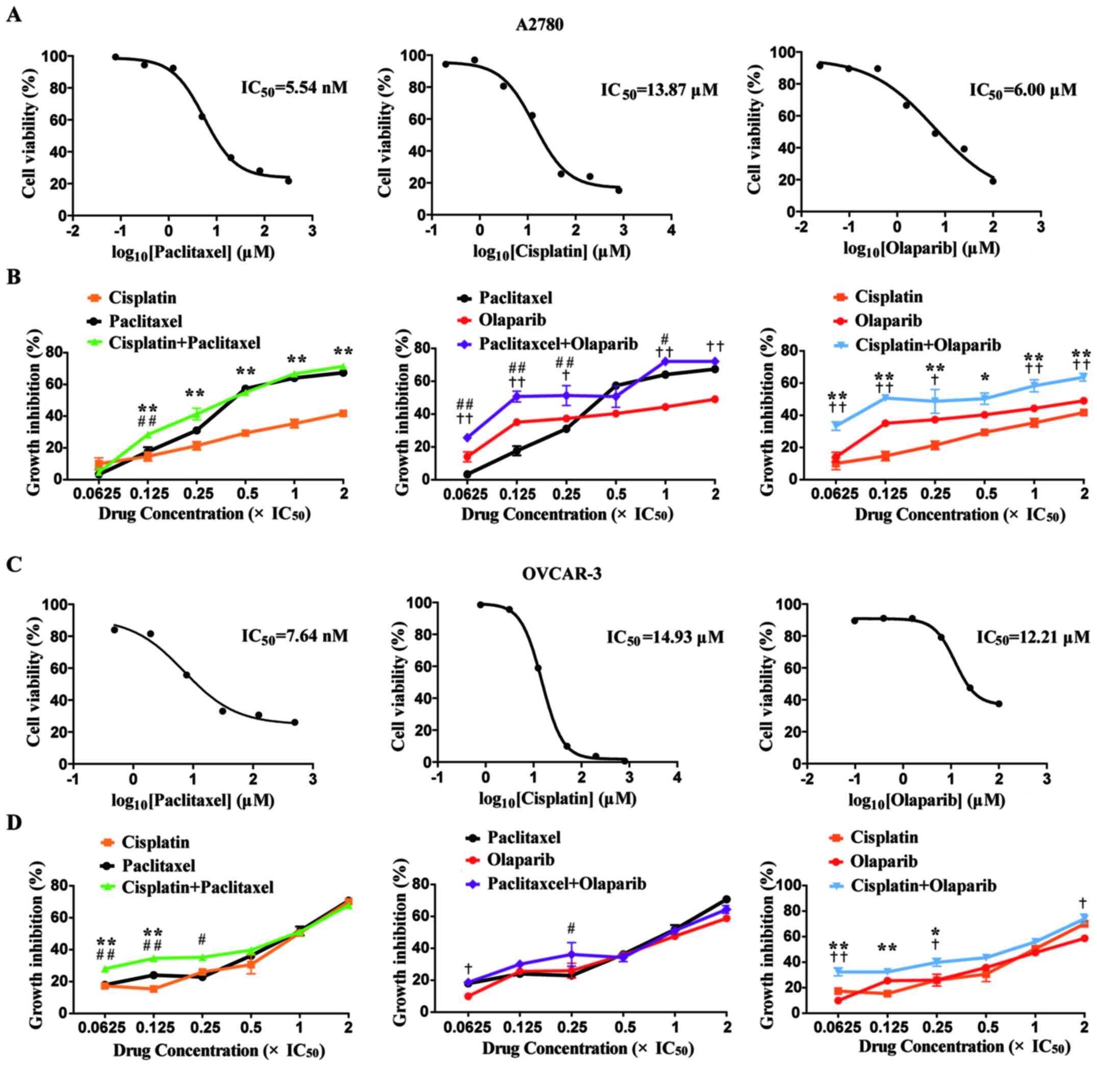
In the present study, the IC50 values of
cisplatin, paclitaxel and olaparib were determined via a CCK-8
assay. For the A2780 cell line, the IC50 values of
cisplatin, paclitaxel and olaparib cell lines were 13.87±0.08,
5.54±0.21 and 6.00±0.35 (SEM) µM, respectively. For the OVCAR-3
cell line, the IC50 values of cisplatin, paclitaxel and
olaparib were 14.93±0.07, 7.64±0.14 and 12.21±0.10 (SEM) µM,
respectively (Fig. 1A and C).
To assess the inhibitory effects of the single or
combined use of the agents on the proliferation of A2780 and
OVCAR-3 cells, three groups were set as two single agent groups and
one combination agent group. The proliferation inhibitory rates of
the A2780 and OVCAR-3 cells at 6 concentrations (0.0625x, 0.125x,
0.25x, 0.5x, 1.0x and 2.0x IC50) were ascertained via a
CCK-8 assay. With the rise in drug concentrations, the
proliferation inhibitory rates of A2780 cells reached 49.0, 41.7
and 67.3% with single olaparib, cisplatin and paclitaxel from 14.0,
10.0 and 3.3%, respectively, in a concentration-dependent manner.
The proliferation inhibitory rates of OVCAR-3 cells reached 58.7,
70.1 and 70.7% with single olaparib, cisplatin and paclitaxel from
9.9, 17.4 and 18.0%, respectively, in a concentration-dependent
manner. It was noted that the inhibitory rates of two types of
ovarian cell lines in the cisplatin + olaparib group were higher
than those in any of the single group at all concentrations
(Fig. 1B and D).
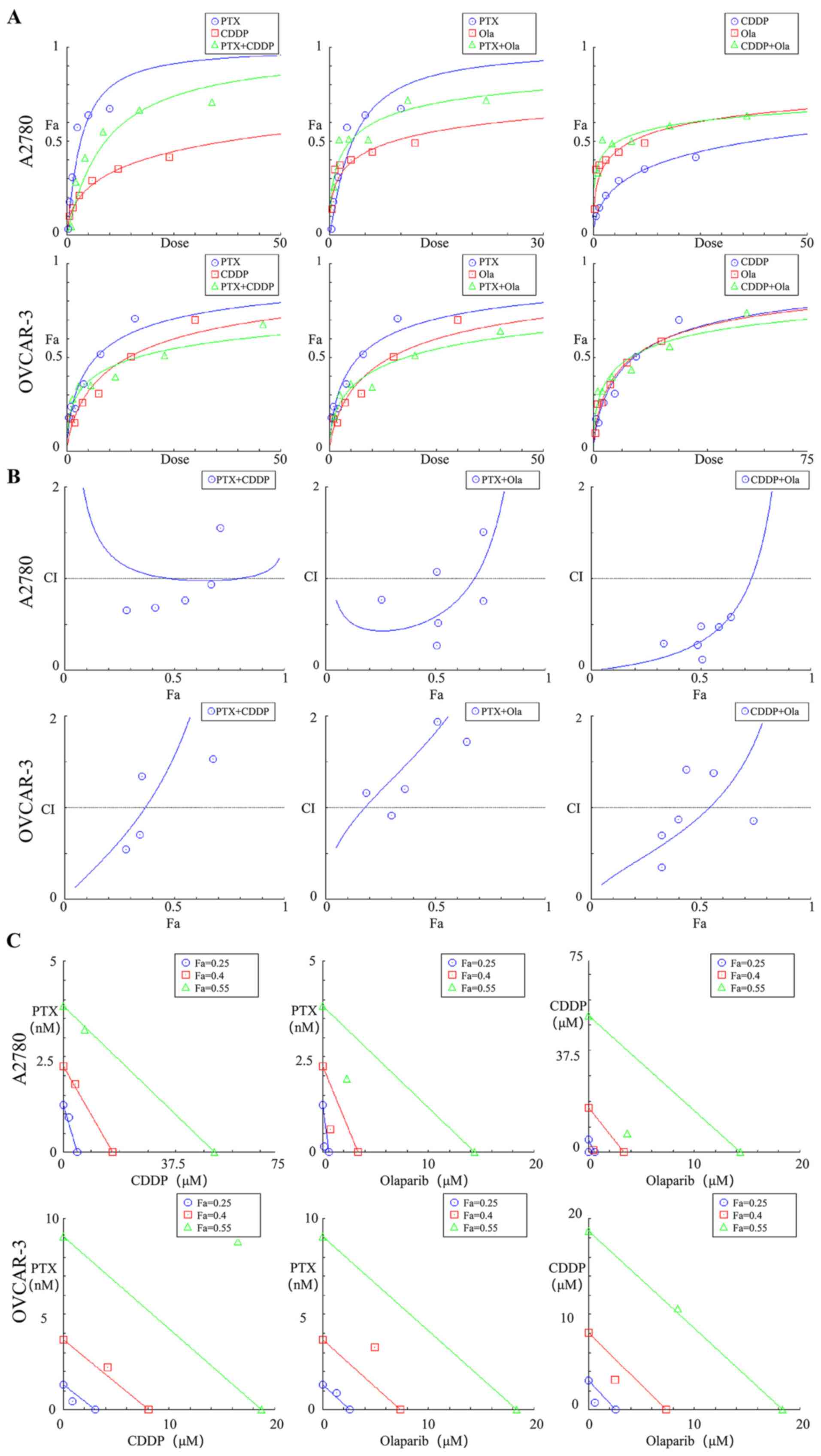
Synergistic, additive and antagonistic
effects of combined agents on the proliferation inhibitory rate of
A2780 and OVCAR-3 cells
The data for the proliferation inhibitory rates of
the three agents in single or combination use at 6 concentrations
(0.0625x, 0.125x, 0.25x, 0.5x, 1.0x and 2.0x IC50) were
substituted into Compusyn software, and the Compusyn provided
graphic representations. The association between dose and effect
was ascertained in accordance with the median-effect principle
(Fig. 2A) (23). The synergistic, additive or
antagonistic effects on proliferation inhibitory rates of the cells
were dependent of the CI values. As a result, for the two ovarian
cell lines, the cisplatin + olaparib group exhibited lower CI
values than the other two combination groups at all concentrations.
For A2780 cells, the CI value of cisplatin + olaparib group was
upregulated to 1.14 from 0.32 across EC50 to
EC75 (Table I).
Cisplatin + olaparib with low concentrations (0.0625x, 0.125x and
0.25x IC50) exerted a strong synergistic effect, with CI
values ranging from 0.1-0.3 for the fraction affected by the dose
(Fa) from 33.3-50.7%. Cisplatin + olaparib at high concentrations
(0.5x, 1.0x and 2.0 IC50) exhibited a synergistic effect
with CI values from 0.3-0.7, and Fa values from 50.3-63.7%. For
OVCAR-3 cells, cisplatin + olaparib at low concentrations (0.0625x,
0.125x and 0.25x IC50) exerted a synergistic effect,
with CI values ranging from 0.35-0.87, and Fa values from
32.4-39.9% (Fig. 2B). As the Fa
values were 0.25, 0.40 and 0.55, the cisplatin + olaparib
combination demonstrated a stronger synergistic effect than the
other two combinations. For OVCAR-3 cells, the Fa values were 0.25
and 0.40, and thus the cisplatin + olaparib combination
demonstrated stronger synergistic effects than the other two
combinations (Fig. 2C).
 | Table ICI values of growth inhibition by
paclitaxel/cisplatin/olaparib in combination for A2780 and OVCAR-3
cell lines. |
Table I
CI values of growth inhibition by
paclitaxel/cisplatin/olaparib in combination for A2780 and OVCAR-3
cell lines.
| | CI | |
|---|
| Cell line | Combination |
0.0625xIC50 |
0.125xIC50 |
0.25xIC50 |
0.5xIC50 |
1xIC50 |
2xIC50 | CI
ED50 | CI
ED75 | CI
ED90 | CI Ave
ED50-90 |
|---|
| A2780 |
Paclitaxel+Cisplatin | 6.43 | 0.66 | 0.68 | 0.77 | 0.94 | 1.55 | 1.00 | 0.99 | 1.06 | 1.02 |
| |
Paclitaxel+Olaparib | 0.77 | 0.27 | 0.52 | 1.07 | 0.76 | 1.51 | 0.59 | 1.38 | 4.02 | 2.00 |
| |
Cisplatin+Olaparib | 0.29 | 0.11 | 0.28 | 0.48 | 0.48 | 0.58 | 0.32 | 1.14 | 0.68 | 0.71 |
| OVCAR-3 |
Paclitaxel+Cisplatin | 0.55 | 0.71 | 1.35 | 2.05 | 2.08 | 1.54 | 1.57 | 3.89 | 9.65 | 17.93 |
| |
Paclitaxel+Olaparib | 1.16 | 0.92 | 1.21 | 2.75 | 1.94 | 1.72 | 1.82 | 2.80 | 4.31 | 5.77 |
| |
Cisplatin+Olaparib | 0.35 | 0.70 | 0.87 | 1.41 | 1.38 | 0.86 | 0.92 | 1.75 | 3.35 | 5.22 |
Furthermore, for the two types of ovarian cell
lines, the cisplatin + olaparib group at all concentrations
(0.0625x, 0.125x, 0.25x, 0.5x, 1.0x and 2.0x IC50)
exhibit a higher dose-reduction index >1 than the other two
combinations. For A2780 cells, single cisplatin and olaparib
displayed 26.11- and 12.73-fold dose reductions at 0.125x
IC50 (Table II). It was
suggested that, in the A2780 cell line, the CI values between the
low concentrations (0.0625x, 0.125x and 0.25x IC50) and
high concentrations (0.5x, 1.0x and 2.0x IC50) of
cisplatin + olaparib were significantly different (P<0.05;
Fig. 3A). In the OVCAR-3 cell line,
the CI values of low concentrations (0.0625x, 0.125x and 0.25x
IC50) presented synergistic effects (Fig. 3B).
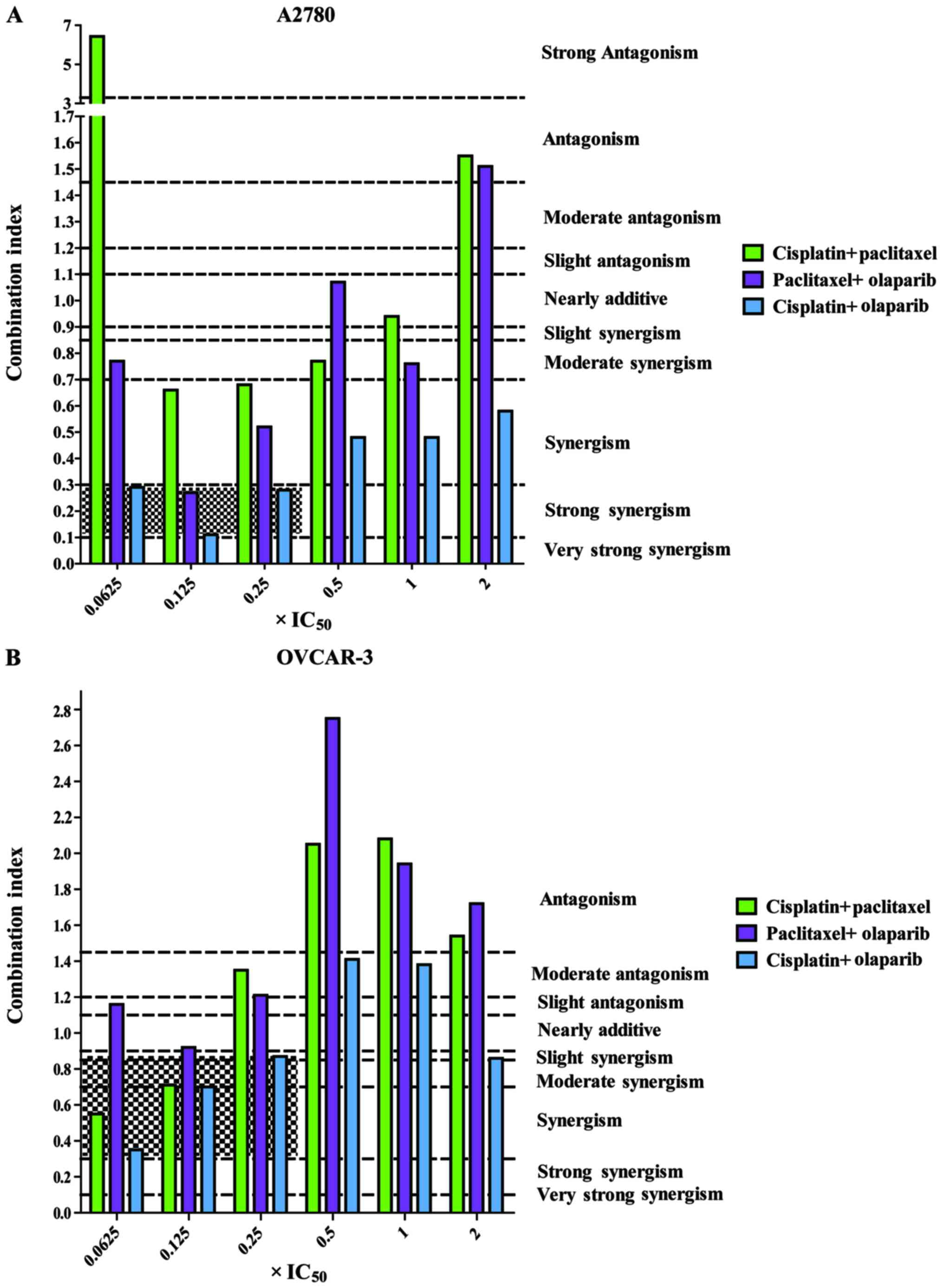 | Figure 3CI values of the combination of three
agents in pairs at six concentrations (0.0625, 0.125, 0.25, 0.5, 1
and 2x IC50). (A) CI values of the combinations in A2780
cells. (B) CI values of the combinations in OVCAR-3 cells. Range of
CI: <0.1, very strong synergism; 0.1-0.3, strong synergism;
0.3-0.7, synergism; 0.7-0.85, moderate synergism; 0.85-0.90, slight
synergism; 0.90-1.10, nearly additive; 1.10-1.20, slight
antagonism; 1.20-1.45, moderate antagonism; 1.45-3.3, antagonism;
3.3-10, strong antagonism; >10, very strong antagonism. CI,
combination index. |
| Table IIDRI values for growth inhibition by
paclitaxel/cisplatin/olaparib in combination for A2780 and OVCAR-3
cell lines. |
Table II
DRI values for growth inhibition by
paclitaxel/cisplatin/olaparib in combination for A2780 and OVCAR-3
cell lines.
| | DRI |
|---|
| Cell line | Combination | Component |
0.0625xIC50 |
0.125xIC50 |
0.25xIC50 |
0.5xIC50 |
1xIC50 |
2xIC50 |
|---|
| A2780 |
Paclitaxel+Cisplatin | Paclitaxel | 0.75 | 2.29 | 1.89 | 1.53 | 1.17 | 0.70 |
| | | Cisplatin | 0.20 | 4.49 | 6.50 | 8.97 | 11.12 | 8.04 |
| |
Paclitaxel+Olaparib | Paclitaxel | 4.09 | 5.26 | 2.69 | 1.32 | 1.46 | 0.73 |
| | | Olaparib | 1.89 | 12.73 | 6.74 | 3.18 | 14.15 | 7.07 |
| |
Cisplatin+Olaparib | Cisplatin | 13.83 | 26.11 | 11.27 | 6.34 | 5.74 | 4.36 |
| | | Olaparib | 4.54 | 12.73 | 5.26 | 3.06 | 3.31 | 2.84 |
| OVCAR-3 |
Paclitaxel+Cisplatin | Paclitaxel | 3.30 | 2.60 | 1.37 | 0.90 | 0.91 | 1.27 |
| | | Cisplatin | 4.07 | 3.11 | 1.63 | 1.06 | 1.02 | 1.33 |
| |
Paclitaxel+Olaparib | Paclitaxel | 1.51 | 1.91 | 1.45 | 0.64 | 0.90 | 1.01 |
| | | Olaparib | 2.00 | 2.54 | 1.94 | 0.85 | 1.21 | 1.37 |
| |
Cisplatin+Olaparib | Cisplatin | 5.45 | 2.72 | 2.14 | 1.32 | 1.32 | 2.03 |
| | | Olaparib | 6.02 | 3.01 | 2.45 | 1.53 | 1.62 | 2.72 |
Combinational interactions of
cisplatin and olaparib against the proliferation of A2780 and
OVCAR-3 cells at low doses
The experiment was divided into four groups,
cisplatin, olaparib, cisplatin + olaparib, and control group. The
proliferation inhibitory effect of A2780 and OVCAR-3 cells was
visually observed following crystal violet solution staining.
Compared with the control group, the experimental groups
(cisplatin, olaparib, cisplatin + olaparib) at 0.25x
IC50 all exhibited different levels of proliferation
inhibition (Fig. 4A and C). The combination group exhibited the
minimum crystal violet and the lowest fraction area of stained
cells, as analyzed by ImageJ software, in each dish, revealing that
the proliferation inhibitory effect was the strongest among four
groups (P<0.01; Fig. 4B and
D). In addition, the potent
synergistic effects of cisplatin + olaparib were further revealed
through cell apoptosis and SP cell via flow cytometry in A2780 and
OVCAR-3 (Fig. S1). When the
concentrations of cisplatin and olaparib were at 0.25x
IC50, the apoptotic rate in A2780 cells in the control,
cisplatin, olaparib, cisplatin + paclitaxel, paclitaxel + olaparib,
cisplatin + olaparib groups reached 2.08, 3.95, 6.40, 7.28, 8.49
and 21.94%, respectively. The apoptotic rate in OVCAR-3 cells in
the control, cisplatin, olaparib, cisplatin + paclitaxel,
paclitaxel + olaparib, cisplatin + olaparib groups reached 3.01,
5.24, 8.74, 9.78, 12.25 and 19.42%, respectively (Fig. 4E and G). It was therefore demonstrated that
combination group of cisplatin and olaparib at low doses can
significantly induce the apoptosis of A2780 and OVCAR-3 cells
compared with each single group and control group (P<0.01;
Fig. 4F and H). Besides, through sorting the
side-population cells in A2780 and OVCAR-3, the percentage of SP
cells showed quite different in the six groups. Following
cisplatin, olaparib, cisplatin + paclitaxel, paclitaxel + olaparib,
cisplatin + olaparib treatment, SP proportion in A2780 was
respectively 8.11, 8.02, 7.47, 5.91 and 2.42%, SP proportion in
OVCAR-3 was respectively 8.05, 7.93, 6.21, 5.99 and 2.38%.
Cisplatin + olaparib can obviously decrease the proportion of SP
cells (Fig. S1).
Discussion
Overall, combination chemotherapeutic agents exhibit
improved efficacy than single administration for the majority of
malignancies. The additive or synergistic effects of multiple
agents usually lead to noticeably higher clinical benefit of the
agents. Researchers have achieved breakthroughs in high-throughput
sequencing and molecular targeted drugs, patients with ovarian
cancer greatly benefit from these advanced technologies.
Molecular-targeted drugs indicate that small molecule drugs
primarily target critical sites to interfere with the pathogenesis
and development of malignancies in a pathophysiological manner. The
targeted drugs can not only be utilized in combination with
chemotherapy, but can also act as maintenance therapy to promote
patient survival time. At present, olaparib has exhibited
encouraging therapeutic effects for the treatment of ovarian
cancer. Olaparib combined with chemotherapy has been proven to
enhance the efficacy of chemotherapy in patients with
platinum-sensitive ROC. From the experimental results of the
present study, it has been revealed that cisplatin + olaparib was
the only group with a CI<0.7 under all Fa values, revealing that
the combination exerts synergistic or strong synergistic effects.
Furthermore, the CI value was smaller than those of the other two
combinations for each experimental concentration, which
demonstrated its synergistic effect was superior.
Conventional anti-tumor therapies (radiotherapy and
chemotherapy) exhibit favorable short-term efficacy largely by
decimating tumor cells, resulting in fast shrinking of tumor
lesions. Nevertheless, these methods are always accompanied by
metastatic or recurrent tumors, which frustrate former efforts of
the conventional strategies. Therefore, the combined use of PARP
inhibitors can enhance the efficacy of radiotherapy, alkylation
agents and platinum drug chemotherapy by inhibiting DNA damage
repair of tumor cells and accelerating apoptosis of tumor cells
(24). Adverse reactions can also
be inhibited by decreasing chemoradiotherapy or radiation doses. It
has been proved that low-dose chemotherapy drugs do not affect
prognosis, and they can also significantly decrease the incidence
of postoperative adverse reactions (25). As reported by Garcia and Singh
(26) bevacizumab, in combination
with low-dose chemotherapeutic agents, decreases the recurrent
ovarian tumor recurrence and inhibits the growth of the lesion. The
graphic representations and quantification outcomes in the present
study demonstrate that the combination of cisplatin and olaparib
exerts superior synergistic effects on the inhibition of A2780 and
OVCAR-3 cell lines proliferation, particularly at low doses. The
low-dose group (0.0625x, 0.125x and 0.25x IC50) of
cisplatin + olaparib even could reach the same proliferation
inhibitory rate as high-dose group (0.5x, 1.0x and 2.0x
IC50) in the A2780 cells (P>0.05).
Chemotherapy-induced cell cycle arrest is commonly considered a
result of DNA damage. When DNA damage repair induced by
chemotherapy cannot be achieved, cell aging or even apoptosis will
be immediately initiated (27). In
the present study, cisplatin + olaparib at low concentrations
successfully induced the apoptosis of A2780 and OVCAR-3 ovarian
cancer cell lines, exhibiting higher apoptosis rates than either
single application. The combination significantly inhibited cell
proliferation following apoptosis.
Furthermore, many studies have confirmed that
cisplatin combined with olaparib has a synergistic effect on tumors
in vivo. Minami et al (28) researched the effectiveness of the
cisplatin with olaparib in a PTEN-deficient lung cancer xenograft
model, they found that cisplatin plus olaparib could inhibit tumor
growth than other treatment groups in PC-9PTEN-xenograft
model. Yasukawa et al (29)
examined the effects of PARP inhibitor (AZD2281) with cisplatin on
oral cancer xenografted model. Results showed that combination
treatment with AZD2281 and cisplatin significantly inhibited
xenografted tumor growth compared with control and single
treatment. de Groot et al (30) demonstrated that combined cisplatin
and PARP1 inhibition could successfully attenuated tumor onset in a
mouse model of BRCA1-associated breast cancer. Nevertheless,
further studies are needed to elucidate the mechanisms behind the
synergistic effect of olaparib combine with cisplatin on ovarian
cancer cells in vivo and in vitro.
Supplementary Material
Cisplatin combined with olaparib
decreases the percentage of the SP cells. (A) Flow cytometric
patterns of SP cells in A2780 cells after cisplatin, paclitaxel,
olaparib, cisplatin+paclitaxel, paclitaxel+olaparib and
cisplatin+olaparib treatment. (B) Flow cytometric patterns of SP
cells in OVCAR-3 cells after cisplatin, paclitaxel, olaparib,
cisplatin+paclitaxel, paclitaxel+olaparib and cisplatin+olaparib
treatment. The percentages of SP cells are indicated. SP,
side-population.
Acknowledgements
Not applicable.
Funding
Funding: The present study was supported by funding from
National Key R&D Program of China (grant no. 2016YFC1303100)
and National Natural Science Foundation of China (grant nos.
31570803, 81773090, 81272879 and 81402151).
Availability of data and materials
All data generated or analyzed during this study are
included in this published article.
Authors' contributions
JG analyzed the growth inhibitory effects of single
and combined anticancer agents on A2780 and OVCAR-3 cells and
interpreted the CompuSyn report. ZW performed the experiments in
cellular function and was a major contributor in writing the
manuscript. JF and JA performed data acquisition. YO was involved
in the conceptualization and investigation. CX conceived the study
and contributed to review/editing of the manuscript. All authors
read and approved the final manuscript.
Ethics approval and consent to
participate
Not applicable.
Patient consent for publication
Not applicable.
Competing interests
The authors declare that they have no competing
interests.
References
|
1
|
Siegel R, Ma J, Zou Z and Jemal A: Cancer
statistics, 2014. CA Cancer J Clin. 64:9–29. 2014.PubMed/NCBI View Article : Google Scholar
|
|
2
|
Bacalbasa N, Balescu I, Dima S, Brasoveanu
V and Popescu I: Hematogenous splenic metastases as an independent
negative prognosis factor at the moment of primary cytoreduction in
advanced stage epithelial ovarian cancer-a single center
experience. Anticancer Res. 35:5649–5654. 2015.PubMed/NCBI
|
|
3
|
Rosell R, Lord RV, Taron M and Reguart N:
DNA repair and cisplatin resistance in non-small-cell lung cancer.
Lung Cancer. 38:217–227. 2002.PubMed/NCBI View Article : Google Scholar
|
|
4
|
Michailidou M, Brown HK, Lefley DV, Evans
A, Cross SS, Coleman RE, Brown NJ and Holen I: Microvascular
endothelial cell responses in vitro and in vivo: Modulation by
zoledronic acid and paclitaxel? J Vasc Res. 47:481–493.
2010.PubMed/NCBI View Article : Google Scholar
|
|
5
|
Scholz HS, Tasdemir H, Hunlich T, Turnwald
W, Both A and Egger H: Multivisceral cytoreductive surgery in FIGO
stages IIIC and IV epithelial ovarian cancer: Results and 5-year
follow-up. Gynecol Oncol. 106:591–595. 2007.PubMed/NCBI View Article : Google Scholar
|
|
6
|
Vida A, Márton J, Mikó E and Bai P:
Metabolic roles of poly(ADP-ribose) polymerases. Semin Cell Dev
Bio. 65:135–143. 2017.PubMed/NCBI View Article : Google Scholar
|
|
7
|
Schreiber V, Dantzer F, Ame JC and de
Murcia G: Poly(ADP-ribose): Novel functions for an old molecule.
Nat Rev Mo Cell Bio. 7:517–528. 2006.PubMed/NCBI View
Article : Google Scholar
|
|
8
|
Walsh CS: Two decades beyond BRCA1/2:
Homologous recombination, hereditary cancer risk and a target for
ovarian cancer therapy. Gynecol Oncol. 137:343–350. 2015.PubMed/NCBI View Article : Google Scholar
|
|
9
|
He JX, Wang M, Huan XJ, Chen CH, Song SS,
Wang YQ, Liao XM, Tan C, He Q, Tong LJ, et al: Novel PARP1/2
inhibitor mefuparib hydrochloride elicits potent in vitro and in
vivo anticancer activity, characteristic of high tissue
distribution. Oncotarget. 8:4156–4168. 2017.PubMed/NCBI View Article : Google Scholar
|
|
10
|
Phulwani NK and Kielian T: Poly
(ADP-ribose) polymerases (PARPs) 1-3 regulate astrocyte activation.
J Neurochem. 106:578–590. 2008.PubMed/NCBI View Article : Google Scholar
|
|
11
|
Kummar S, Chen A, Parchment RE, Kinders
RJ, Ji J, Tomaszewski JE and Doroshow JH: Advances in using PARP
inhibitors to treat cancer. BMC Med. 10(25)2012.PubMed/NCBI View Article : Google Scholar
|
|
12
|
Ricks TK, Chiu HJ, Ison G, Kim G, McKee
AE, Kluetz P and Pazdur R: Successes and challenges of PARP
inhibitors in cancer therapy. Front Oncol. 5(222)2015.PubMed/NCBI View Article : Google Scholar
|
|
13
|
Löser DA, Shibata A, Shibata AK, Woodbine
LJ, Jeggo PA and Chalmers AJ: Sensitization to radiation and
alkylating agents by inhibitors of poly(ADP-ribose) polymerase is
enhanced in cells deficient in DNA double-strand break repair. Mol
Cancer Ther. 9:1775–1787. 2010.PubMed/NCBI View Article : Google Scholar
|
|
14
|
Dedes KJ, Wilkerson PM, Wetterskog D,
Weigelt B, Ashworth A and Reis-Filho JS: Synthetic lethality of
PARP inhibition in cancers lacking BRCA1 and BRCA2 mutations. Cell
Cycle. 10:1192–1199. 2011.PubMed/NCBI View Article : Google Scholar
|
|
15
|
Ledermann J, Harter P, Gourley C,
Friedlander M, Vergote I, Rustin G, Scott CL, Meier W,
Shapira-Frommer R, Safra T, et al: Olaparib maintenance therapy in
patients with platinum-sensitive relapsed serous ovarian cancer: A
preplanned retrospective analysis of outcomes by BRCA status in a
randomised phase 2 trial. Lancet Oncol. 15:852–861. 2014.PubMed/NCBI View Article : Google Scholar
|
|
16
|
Parkes EE and Kennedy RD: Clinical
application of Poly(ADP-ribose) polymerase inhibitors in high-grade
serous ovarian cancer. Oncologist. 21:568–593. 2016.PubMed/NCBI View Article : Google Scholar
|
|
17
|
Qin C, Zhang C, Zhu F, Xu F, Chen SY,
Zhang P, Li YH, Yang SY, Wei YQ, Tao L and Chen YZ: Therapeutic
target database update 2014: A resource for targeted therapeutics.
Nucleic Acids Res. 42 (Database Issue):D1118–D1123. 2014.PubMed/NCBI View Article : Google Scholar
|
|
18
|
Fong PC, Yap TA, Boss DS, Carden CP,
Mergui-Roelvink M, Gourley C, De Greve J, Lubinski J, Shanley S,
Messiou C, et al: Poly(ADP)-ribose polymerase inhibition: Frequent
durable responses in BRCA carrier ovarian cancer correlating with
platinum-free interval. J Clin Oncol. 28:2512–2519. 2010.PubMed/NCBI View Article : Google Scholar
|
|
19
|
Lee JM, Hays JL, Annunziata CM, Noonan AM,
Minasian L, Zujewski JA, Yu M, Gordon N, Ji J, Sissung TM, et al:
Phase I/Ib study of olaparib and carboplatin in BRCA1 or BRCA2
mutation-associated breast or ovarian cancer with biomarker
analyses. J Natl Cancer Inst. 106(dju089)2014.PubMed/NCBI View Article : Google Scholar
|
|
20
|
Del Conte G, Sessa C, von Moos R, Viganò
L, Digena T, Locatelli A, Gallerani E, Fasolo A, Tessari A,
Cathomas R and Gianni L: Phase I study of olaparib in combination
with liposomal doxorubicin in patients with advanced solid tumours.
Br J Cancer. 111:651–659. 2014.PubMed/NCBI View Article : Google Scholar
|
|
21
|
Konstantinopoulos PA, Ceccaldi R, Shapiro
GI and D'Andrea AD: Homologous recombination deficiency: Exploiting
the fundamental vulnerability of ovarian cancer. Cancer Discov.
5:1137–1154. 2015.PubMed/NCBI View Article : Google Scholar
|
|
22
|
Chou TC and Martin N: CompuSyn for Drug
Combinations: PC Software and User's Guide: A computer program for
quantitation of synergism and antagonism in drug combinations, and
the determination of IC50 and ED50 and
LD50 values. ComboSyn Inc., Paramus NJ, 2005.
|
|
23
|
Ashton JC: Drug combination studies and
their synergy quantification using the Chou-Talalay method-letter.
Cancer Res. 75(2400)2015.PubMed/NCBI View Article : Google Scholar
|
|
24
|
Oza AM, Cibula D, Benzaquen AO, Poole C,
Mathijssen RH, Sonke GS, Colombo N, Špaček J, Vuylsteke P, Hirte H,
et al: Olaparib combined with chemotherapy for recurrent
platinum-sensitive ovarian cancer: A randomised phase 2 trial.
Lancet Oncol. 16:87–97. 2015.PubMed/NCBI View Article : Google Scholar
|
|
25
|
Kamada K, Nakanishi T, Kitamoto M, Aikata
H, Kawakami Y, Ito K, Asahara T and Kajiyama G: Long-term prognosis
of patients undergoing transcatheter arterial chemoembolization for
unresectable hepatocellular carcinoma: Comparison of cisplatin
lipiodol suspension and doxorubicin hydrochloride emulsion. J Vasc
Interv Radiol. 12:847–854. 2001.PubMed/NCBI View Article : Google Scholar
|
|
26
|
Garcia A and Singh H: Bevacizumab and
ovarian cancer. Ther Adv Med Oncol. 5:133–141. 2013.PubMed/NCBI View Article : Google Scholar
|
|
27
|
Salminen A, Ojala J and Kaarniranta K:
Apoptosis and aging: Increased resistance to apoptosis enhances the
aging process. Cell Mol Life Sci. 68:1021–1031. 2011.PubMed/NCBI View Article : Google Scholar
|
|
28
|
Minami D, Takigawa N, Takeda H, Takata M,
Ochi N, Ichihara E, Hisamoto A, Hotta K, Tanimoto M and Kiura K:
Synergistic effect of olaparib with combination of cisplatin on
PTEN-deficient lung cancer cells. Mol Cancer Res. 11:140–148.
2013.PubMed/NCBI View Article : Google Scholar
|
|
29
|
Yasukawa M, Fujihara H, Fujimori H,
Kawaguchi K, Yamada H, Nakayama R, Yamamoto N, Kishi Y, Hamada Y
and Masutani M: Synergetic effects of PARP inhibitor AZD2281 and
cisplatin in oral squamous cell carcinoma in vitro and in vivo. Int
J Mol Sci. 17(272)2016.PubMed/NCBI View Article : Google Scholar
|
|
30
|
de Groot JS, van Diest PJ, van Amersfoort
M, Vlug EJ, Pan X, Ter Hoeve ND, Rosing H, Beijnen JH, Youssef SA,
de Bruin A, et al: Intraductal cisplatin treatment in a
BRCA-associated breast cancer mouse model attenuates tumor
development but leads to systemic tumors in aged female mice.
Oncotarget. 8:60750–60763. 2017.PubMed/NCBI View Article : Google Scholar
|