Introduction
Inferior pole fracture of the patella is an
extra-articular injury that accounts for 5% of all patellar
fractures and usually requires operative treatment if displaced or
associated with complete disruption of the extensor mechanism
(1). However, displaced fracture
fragments are typically small and comminuted and it is difficult to
fix and maintain anatomical reduction (2). The inferior patellar pole is
continuous with the patellar tendon and the key to reconstructing
the fracture fragment surgically is to reestablish the extensor
mechanism while simultaneously restoring articular congruency. The
traditional treatment for displaced comminuted inferior pole
fractures is partial patellectomy followed by repair of the
patellar tendon. However, this treatment may potentially lead to a
shorter long axis of the patella, which affects the functioning of
the patellofemoral joint. Comparatively better outcomes have been
achieved from fixation of displaced fragments (3,4).
Most surgical methods use metallic fixation for
inferior patellar pole fractures, such as tension band wiring
(1), separate vertical wiring
(5,6), cannulated lag screws (7) and basket plates (8). Although these surgeries may ultimately
achieve satisfactory outcomes, patients frequently require
reoperation for implant removal or have soft-tissue irritation
secondary to prominent hardware. Studies have reported on suture
fixation to treat inferior patellar pole fractures, including
transosseous tunnel suture (TTS) repair (9,10) and
anchor suture (AS) fixation (11,12).
Compared with using metal implants, suture fixation has similar
functional outcomes but fewer hardware-related complications and
lower implant removal rates. Sutures such as #5 Ethibond and
Fiberwire have been proven to be similar in strength to 18-gauge
stainless steel wires (13,14). Compared with transosseous
techniques, AS offers the potential for minimal surgical
dissection, no drilling and possibly reduced tissue trauma.
However, to the best of our knowledge, no studies have compared the
clinical outcomes of TTS with those of AS for inferior patellar
pole fractures.
The purpose of the present study was to evaluate and
compare the functional outcomes and complication rates in inferior
patellar pole fractures treated with TTS and AS. It was
hypothesized that patients would experience similar outcomes
regardless of the fixation method.
Patients and methods
Patients
The present study provided a retrospective analysis
of patients with sustained patellar inferior pole fracture treated
at the Department of Orthopedics of the Second Affiliated Hospital
of Nanchang University (Nanchang, China) between June 2014 and
April 2018. The inclusion criteria were the use of TTS or AS for
fracture fixation, age of >18 years and a minimum follow-up of
at least 12 months. The exclusion criteria were open fractures,
concomitant knee fractures other than those of the patella,
multiple traumata, acute infection, osteoarthritis, rheumatoid
arthritis or musculoskeletal disorders.
The present study was approved by the Ethics
Committee of the Second Affiliated Hospital of Nanchang University
(Nanchang, China) and all patients or their relatives provided
written informed consent prior to inclusion. A total of 14 patients
(9 males and 5 females; mean age, 47.6 years; age range, 32-70
years) with patellar inferior pole fracture who underwent TTS and
21 patients (12 males and 9 females; mean age, 45.6 years; age
range, 25-68 years) who underwent AS were enrolled in the present
study. The 2 groups were comparable in terms of age, sex and
operative side. The condition of the soft tissue was carefully
assessed prior to surgery to understand the optimal timing of the
operation. The group treated with TTS underwent surgery at an
average of 3.6 days after trauma (range, 3-6 days) and the AS group
underwent surgery at an average of 4.0 days (range, 2-8 days) after
trauma.
Surgical techniques
After general anesthesia or lumbar anesthesia, the
patient was placed in a supine position on the operating table with
tourniquet inflation. All patients received antibiotics
(first-generation cephalosporin) 30 min prior to surgery. A total
of three surgeons from the same group at our department performed
all surgeries together. A midline longitudinal skin incision on the
knee was made. The distal pole patella fragment was identified, and
hematoma and soft tissue from the fracture site were removed. The
proximal fracture edges were irrigated with saline and debrided
with a curette and rongeur to promote fracture healing. With regard
to the TTS technique, the surgery was performed similar to the
example of Swensen et al (10). A total of three transpatellar
tunnels were drilled inferior to superior using a 2.5-mm drill bit
or pin at the central, medial and lateral proximal patellar sites.
Next, #5 Ethibond (Ethicon) braided polyester sutures were used in
Krackow locking fashion up and down the medial and lateral edges of
the patellar tendon. The suture limbs were passed through the
central, medial and lateral tunnels using a suture passer. The
fracture was next reduced with bone reduction forceps and C-arm
fluoroscopy indicated good fracture reduction. The sutures were
then tied at the superior pole with the knee in extension. For the
AS group, two holes were drilled into the medial and lateral
proximal fracture edges and suture anchors (4.5 mm Healix Advance
anchor; DePuy Mitek, Inc.) with two Orthocord sutures were placed
into each of the holes. After reduction with bone reduction forceps
and verification with C-arm fluoroscopy, the sutures from each anchor
were passed through the medial and lateral sides of the patellar
tendon in a horizontal Krackow locking fashion. In addition, tears
observed in the retinaculum were repaired and after subcutaneous
layer closure, the skin layer was closed with running subcuticular
sutures. The operation time, incision length and total cost were
determined and recorded for comparison between groups.
Rehabilitation protocol
In all patients, the knee was immobilized at 0°
postoperatively with an orthesis or plaster for 4 weeks to allow
for healing of the retinacular tears and patellar ligament to the
bone. The patients were allowed to begin partial weight-bearing at
50% of their weight at 2 weeks postoperatively, with full weight by
3-4 weeks. Both active and passive range of motion (ROM) exercises
were started at the fourth week postoperatively with the goal of
90˚ flexion for the next 2 weeks and full ROM was allowed from the
sixth week postoperatively.
Clinical assessment
The patients were evaluated both radiologically and
clinically at 3, 6 and 12 months after the surgery and then until
the last follow-up. Antero-posterior and lateral radiographs were
obtained to assess reduction quality and bone union. The degree of
pain was assessed using the visual analog scale (VAS) (15) ranging from 0-10. Knee functional
outcomes were evaluated using the Bostman et al (16) and Lysholm and Gillquist (17) scoring systems. The final knee-joint
ROM was measured. In addition, postoperative complications,
including nonunion, implant failure and infection, were
recorded.
Statistical analysis
Statistical analyses were performed using SPSS
statistical software 23.0 (IBM Corp.). Continuous variables are
presented as the mean ± standard deviation and categorical
variables were expressed as n (%). The Kolmogorov-Smirnov test was
used to check the data for normality of distribution. The t-test
was employed to compare variables with a normal distribution. The
nonparametric Mann-Whitney U-test was used for variables without a
normal distribution, such as the ROM and VAS score. Differences in
categorical variables between the two groups were assessed with
Fisher's exact test. P<0.05 was considered to indicate
statistical significance.
Results
General data
No significant differences in age, sex, operative
side or time to surgery were present between the two groups. 14
patients in the TTS group and 21 in the AS group attained their
final follow-ups. The mean follow-up duration was 22.6±9.7 months
in the TTS group and 18.7±5.9 months in the AS group (P=0.191;
Table I).
 | Table ICharacteristics of the two groups. |
Table I
Characteristics of the two groups.
| Variable | TTS group (n=14) | AS group (n=21) | P-value |
|---|
| Age (years) | 47.6±10.6 | 45.6±11.5 | 0.602a |
| Sex
(male/female) | 9(64)/5(36) | 12(57)/9(43) | 0.737b |
| Side
(left/right) | 6(43)/8(57) | 7(33)/14(67) | 0.724b |
| Time to surgery
(days) | 3.6±0.9 | 4.0±1.5 | 0.606c |
| Follow-up
(months) | 22.6±9.7 | 18.7±5.9 |
0.191a |
| Operation time
(min) | 62.3±6.4 | 41.1±4.2 | <0.01a |
| Incision length
(cm) | 12.4±1.2 | 8.3±1.1 | <0.01c |
| Total cost (¥) | 20,778±489 | 30,856±859 | <0.01a |
Surgical conditions
The average operation time was 62.3±6.4 min in the
TTS group and 41.1±4.2 min in the AS group (P<0.01). The mean
incision length was significantly smaller in the AS group than in
the TTS group (8.3±1.1 vs. 12.4±1.2 cm, P<0.01). However, the AS
group incurred higher hospital costs than the TTS group
(¥30,856±859 vs. 20,778±489, P<0.01; Table I).
Functional outcomes
To evaluate functional outcomes in the present
study, the Bostman and Lysholm scoring systems were utilized. At
the last follow-up, the mean Bostman score was 28.7±1.3 (excellent)
in the group treated with TTS and 27.8±1.4 (good) in the group who
received AS. Furthermore, the mean Lysholm score at the final
follow-up was 94.4±3.9 (good) in the TTS group and 91.4±5.1 (good)
in the AS group. Differences in the Bostman and Lysholm scores were
not statistically significant between the groups (P=0.071 and
P=0.118, respectively). Pain evaluation using the VAS score also
indicated no significant differences between the TTS group and the
AS group (1.6±0.9 vs. 1.4±0.9, P=0.654). At the time of the final
follow-up, the mean knee joint ROM was 133.6±6.3˚ in the TTS group
and 129.8±6.2˚ in the AS group, with no significant differences in
ROM between the two groups (P=0.089; Table II).
| Table IIInter-group comparison of functional
outcomes. |
Table II
Inter-group comparison of functional
outcomes.
| Variable | TTS group | AS group | P-value |
|---|
| ROM, ˚
(extension/flexion) | 0/133.6±6.3 | 0/129.8±6.2 | 0.089 |
| VAS score | 1.6±0.9 | 1.4±0.9 | 0.654 |
| Bostman score | 28.7±1.3 | 27.8±1.4 | 0.071 |
| Lysholm score | 94.4±3.9 | 91.4±5.1 | 0.118 |
| Complications | None | 1 infection | - |
Radiographic results and
Complications
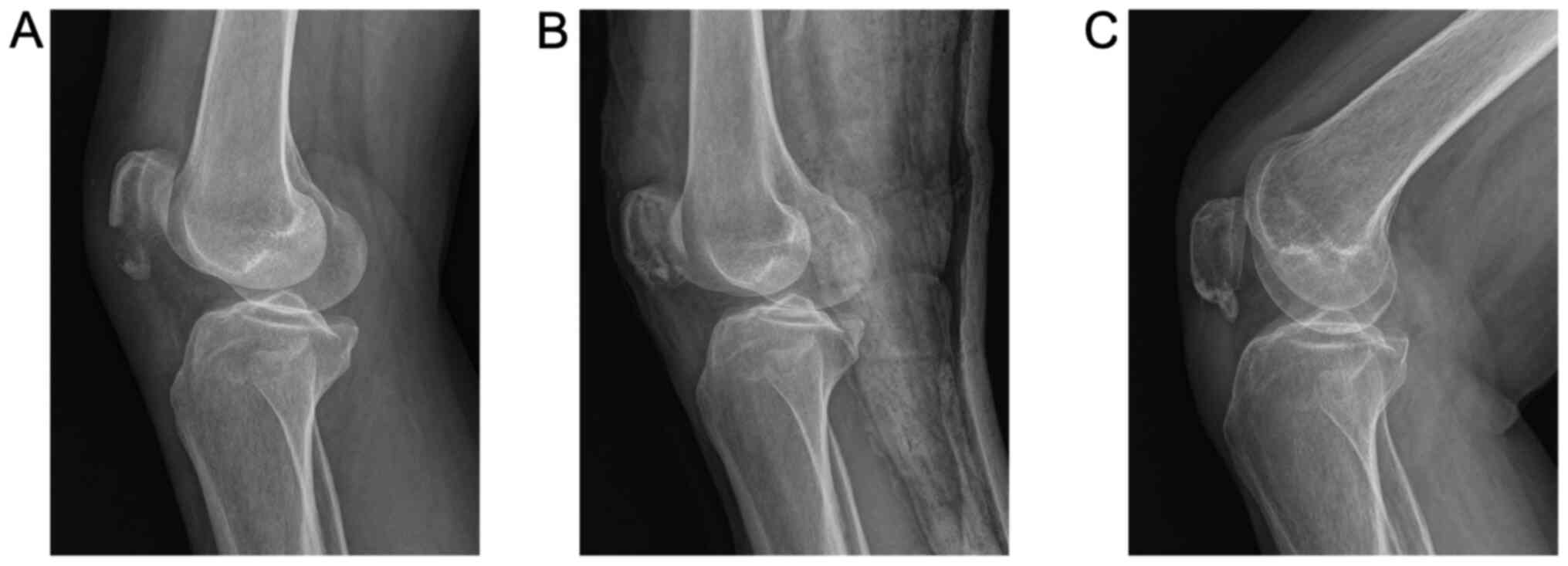
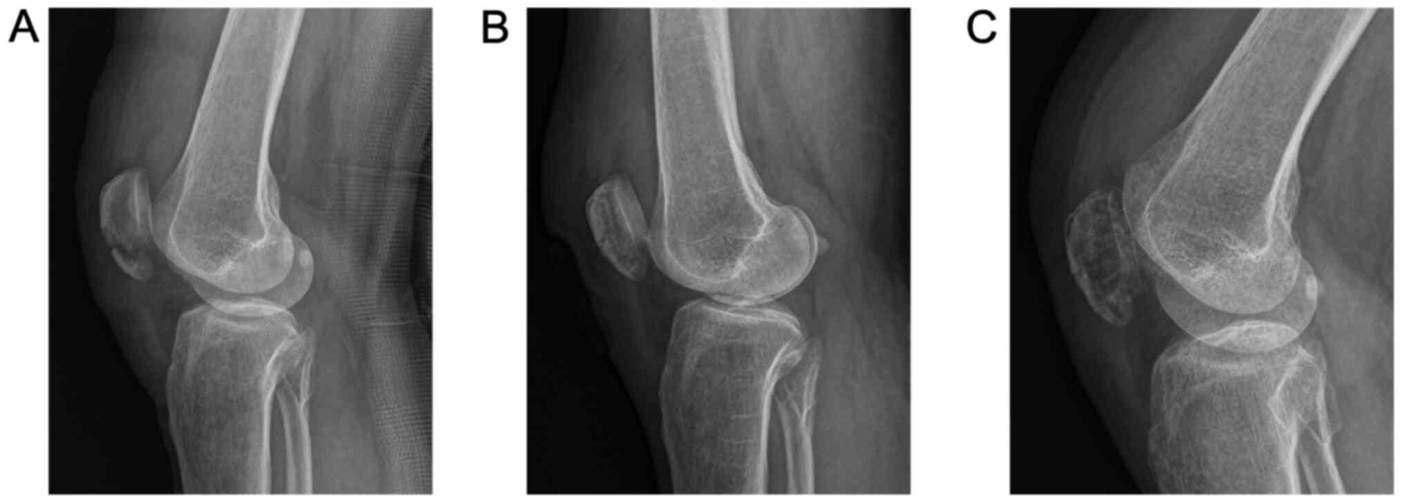
Radiographic signs of bone union were achieved after
approximately 3 months in both groups (Figs. 1 and 2). No postoperative complications, such as
infection, nonunion or implant failure, were observed in the TTS
group. However, one patient in the AS group had a superficial minor
wound infection, which was successfully treated by daily dressing
changes and adapted antibiotics. No cases of deep infection
occurred. Furthermore, none of the patients in either group had any
skin irritation or reduction loss (Table II).
Discussion
Fracture of the inferior patellar pole is
characterized by avulsion of the patellar tendon (18); thus, repairing the extensor
mechanism and stable fixation are vital for osteosynthesis of the
inferior pole. The tension-band-wiring technique has been the most
commonly used method for displaced patellar fracture, but it is
difficult to implement for inferior pole fixation due to the
inherent weakness of small comminuted distal fragments. Other
surgical techniques, such as separate vertical wiring or basket
plate and their modifications, may provide stable fixation and
satisfactory results (2,5,8,19,20).
However, this specific plate is not available in numerous
institutions and the prominent wire knots or the relative bulk of
the plate covered with thin soft tissue may cause skin irritation
in the flexed knee position, possibly necessitating metal implant
removal. Various studies have reported on the use of nonmetallic
implants for fixation of inferior pole fractures, such as
nonabsorbable braided sutures (9,10,21,22)
and suture anchors (11,12).
Egol et al (9) demonstrated that suture repair had
fewer hardware-related postoperative complications, with only
one-quarter of the reoperation rate of standard tension band
fixation. Recently, a retrospective study of surgically treated
patellar fractures was performed in which suture fixation resulted
in a lower number and rate of soft-tissue irritation and
reoperation rate compared with hybrid and metal fixation, even
though these advantages were negated with the addition of a metal
tension band wire (22).
Furthermore, a prior study reported a hardware removal rate of ~52%
following the use of metallic implants (23). In the present study, both TTS and AS
techniques resulted in positive clinical outcomes with low
implant-related complication rates. Only one of 21 patients in the
AS group had a superficial minor wound infection and none of the
patients complained of any symptomatic hardware or required implant
removal.
The major concern in suture replacement of stainless
steel wire for the treatment of patellar fracture is suture
fixation strength. To address this, previous biomechanical studies
have verified the fixation properties of sutures in the tension band
technique (13,14,24).
Patel et al (13) reported
that the #5 Ethibond braided polyester suture is an alternative to
stainless steel wire for fixation of patellar fractures. In
addition, a study evaluated the stiffness and failure strength of
Fiberwire, a braided polyblend suture, using a three-point-bend
model, and indicated that double-strand Fiberwire has a
significantly higher failure load than stainless steel wire
(14). This was also confirmed in
clinical studies; 4 studies comprising 29 patients demonstrated a
convincing clinical outcome using Ethibond or Fiberwire sutures for
the treatment of inferior pole patella fractures. In the present
study, #5 Ethibond was used for the TTS technique and acceptable
clinical outcomes were achieved (9,10,21,22).
Suture anchors have been used extensively for
numerous tendon and ligament repairs (25-27),
and the technique using two suture anchors for the treatment of
patellar inferior pole fractures was recently introduced (11,12).
Compared with the TTS technique for patellar inferior pole
fracture, the AS technique is a more convenient surgical procedure,
given the requirement to expose only the distal pole of the patella
without transosseous drilling. Minimal surgical dissection may be
associated with a shorter operative time, less soft-tissue trauma
and a lower risk of iatrogenic patellar articular cartilage.
Although the fixation strength of AS in the treatment of patellar
fractures has not been proven experimentally, a recent study
evaluated the use of suture anchor fixation for patellar tendon
ruptures and reported that the suture anchor yields biomechanical
effects similar to those of transosseous suture repair (28). It is recommended that bone anchors
are held through intact cortex bone in tendon repairs. However,
with inferior pole fragment avulsion, bone anchors must be inserted
into cancellous bone without using the rim of the cortex, which may
constitute a weak point leading to implant failure. Kadar et
al (12) reported 2 cases
(7.4%) of implant failure in 27 patients with distal pole patellar
fracture who underwent the AS technique. In both cases, the anchor
was pulled out of the main patellar fragment. Regardless, such
failure did not occur in the study by Anand et al (11) or the present study.
On the basis of the results of the present study, it
was indicated that AS has comparable functional outcomes, pain
levels and knee-joint ROM to TTS. Along with the benefits of a
simpler procedure and less extensive exposure, the operation time
and surgical incision were significantly shorter for the AS group
than for the TTS group. A disadvantage of the AS technique is its
increased cost: In the present study, the hospital costs for the AS
group were significantly higher than those for the TTS group,
mostly because the cost for two suture anchors is ~¥10,000 at our
institution.
The limitations of the present study are its
retrospective nature and small sample size. Non-randomized
controlled trials on patients with orthopedic trauma do not
necessarily allow for the identification of factors influencing
implant selection, which may lead to inherent selection bias with a
clear potential to influence outcomes. Another limitation is that
two different suture types were used for each group of repairs. TTS
was performed with #5 Ethibond, whereas Orthocord was used for AS,
potentially causing instrument bias in the study. Further
biomechanical studies and randomized investigations are warranted
in the future.
In conclusion, TTS and AS provide similar
satisfactory clinical outcomes for treating patellar inferior pole
fracture and complication rates are low with both techniques. TTS
has the advantage of cost-effectiveness by saving anchors and AS is
associated with reduced operation time and a smaller incision
length. Thus, both the TTS and AS techniques are acceptable
treatment methods for patellar inferior pole fracture.
Acknowledgements
Not applicable.
Funding
Funding: The present study was supported by the Science and
Technology Program of Jiangxi Educational Commission (grant no.
GJJ200246).
Availability of data and materials
The datasets used and/or analyzed during the current
study are available from the corresponding author on reasonable
request.
Authors' contributions
GCG and XGC conceived and designed the study. WZH
and TLW analyzed the data and edited the manuscript. QQW and LHP
helped to perform the follow-up and collect patients' data. GCG and
XGC confirm the authenticity of all the raw data. All authors read
and approved the final manuscript.
Ethical approval and consent to
participate
The present study was approved by the Ethics
Committee of the Second Affiliated Hospital of Nanchang University
[Nanchang, China; reference no. 2018 (026)]. Written informed
consent was obtained from the patients or their relatives.
Patient consent for publication
Consent for publication of the patients' data and
images was obtained from patients or their relatives.
Competing interests
The authors declare that they have no competing
interests.
References
|
1
|
Yang X, Wu Q, Lai CH and Wang X:
Management of displaced inferior patellar pole fractures with
modified tension band technique combined with cable cerclage using
Cable Grip System. Injury. 48:2348–2353. 2017.PubMed/NCBI View Article : Google Scholar
|
|
2
|
Oh HK, Choo SK, Kim JW and Lee M: Internal
fixation of displaced inferior pole of the patella fractures using
vertical wiring augmented with Krachow suturing. Injury.
46:2512–2515. 2015.PubMed/NCBI View Article : Google Scholar
|
|
3
|
Veselko M and Kastelec M: Inferior
patellar pole avulsion fractures: Osteosynthesis compared with pole
resection. Surgical technique. J Bone Joint Surg Am. 87 (Suppl
1):S113–S121. 2005.PubMed/NCBI View Article : Google Scholar
|
|
4
|
Matejcic A, Puljiz Z, Elabjer E,
Bekavac-Beslin M and Ledinsky M: Multifragment fracture of the
patellar apex: Basket plate osteosynthesis compared with partial
patellectomy. Arch Orthop Trauma Surg. 128:403–408. 2008.PubMed/NCBI View Article : Google Scholar
|
|
5
|
He S, Huang X, Yan B, Zhu J, Bao N and
Zhao J: Modified technique of separate vertical wiring for the
fixation of patellar inferior pole fracture. J Orthop Trauma.
32:e145–e150. 2018.PubMed/NCBI View Article : Google Scholar
|
|
6
|
Song HK, Yoo JH, Byun YS and Yang KH:
Separate vertical wiring for the fixation of comminuted fractures
of the inferior pole of the patella. Yonsei Med J. 55:785–791.
2014.PubMed/NCBI View Article : Google Scholar
|
|
7
|
Chang SM and Ji XL: Open reduction and
internal fixation of displaced patella inferior pole fractures with
anterior tension band wiring through cannulated screws. J Orthop
Trauma. 25:366–370. 2011.PubMed/NCBI View Article : Google Scholar
|
|
8
|
Kastelec M and Veselko M: Inferior
patellar pole avulsion fractures: Osteosynthesis compared with pole
resection. J Bone Joint Surg Am. 86:696–701. 2004.PubMed/NCBI View Article : Google Scholar
|
|
9
|
Egol K, Howard D, Monroy A, Crespo A,
Tejwani N and Davidovitch R: Patella fracture fixation with suture
and wire: You reap what you sew. Iowa Orthop J. 34:63–67.
2014.PubMed/NCBI
|
|
10
|
Swensen S, Fisher N, Atanda A and Egol KA:
Suture repair of a pole patella fracture. J Orthop Trauma. 31
(Suppl 3):S28–S29. 2017.PubMed/NCBI View Article : Google Scholar
|
|
11
|
Anand A, Kumar M and Kodikal G: Role of
suture anchors in management of fractures of inferior pole of
patella. Indian J Orthop. 44:333–335. 2010.PubMed/NCBI View Article : Google Scholar
|
|
12
|
Kadar A, Sherman H, Drexler M, Katz E and
Steinberg EL: Anchor suture fixation of distal pole fractures of
patella: Twenty seven cases and comparison to partial patellectomy.
Int Orthop. 40:149–154. 2016.PubMed/NCBI View Article : Google Scholar
|
|
13
|
Patel VR, Parks BG, Wang Y, Ebert FR and
Jinnah RH: Fixation of patella fractures with braided polyester
suture: A biomechanical study. Injury. 31:1–6. 2000.PubMed/NCBI View Article : Google Scholar
|
|
14
|
Wright PB, Kosmopoulos V, Coté RE, Tayag
TJ and Nana AD: FiberWire is superior in strength to stainless
steel wire for tension band fixation of transverse patellar
fractures. Injury. 40:1200–1203. 2009.PubMed/NCBI View Article : Google Scholar
|
|
15
|
Castrejón I, Chua JR and Pincus T: A
RheuMetric physician checklist to quantitate levels of
inflammation, damage and distress on 0-10 visual analogue scales.
Clin Exp Rheumatol. 35 (Suppl 107):S21–S25. 2017.PubMed/NCBI
|
|
16
|
Bostman O, Kiviluoto O and Nirhamo J:
Comminuted displaced fractures of the patella. Injury. 13:196–202.
1981.PubMed/NCBI View Article : Google Scholar
|
|
17
|
Lysholm J and Gillquist J: Evaluation of
knee ligament surgery results with special emphasis on use of a
scoring scale. Am J Sports Med. 10:150–154. 1982.PubMed/NCBI View Article : Google Scholar
|
|
18
|
Massoud EIE: Repair of comminuted fracture
of the lower patellar pole. Ulus Travma Acil Cerrahi Derg.
23:150–155. 2017.PubMed/NCBI View Article : Google Scholar
|
|
19
|
Matejčić A, Ivica M, Jurišić D, Ćuti T,
Bakota B and Vidović D: Internal fixation of patellar apex
fractures with the basket plate: 25 years of experience. Injury. 46
(Suppl 6):S87–S90. 2015.PubMed/NCBI View Article : Google Scholar
|
|
20
|
Huang HC, Su JY and Cheng YM: Modified
basket plate for inferior patellar pole avulsion fractures: A
report of three cases. Kaohsiung J Med Sci. 28:619–623.
2012.PubMed/NCBI View Article : Google Scholar
|
|
21
|
Camarda L, La Gattuta A, Butera M,
Siragusa F and D'Arienzo M: FiberWire tension band for patellar
fractures. J Orthop Traumatol. 17:75–80. 2016.PubMed/NCBI View Article : Google Scholar
|
|
22
|
Shea GK, Hoi-Ting So K, Tam KW, Yee DK,
Fang C and Leung F: Comparing 3 different techniques of patella
fracture fixation and their complications. Geriatr Orthop Surg
Rehabil. 10(2151459319827143)2019.PubMed/NCBI View Article : Google Scholar
|
|
23
|
LeBrun CT, Langford JR and Sagi HC:
Functional outcomes after operatively treated patella fractures. J
Orthop Trauma. 26:422–426. 2012.PubMed/NCBI View Article : Google Scholar
|
|
24
|
McGreal G, Reidy D, Joy A, Mahalingam K
and Cashman WF: The biomechanical evaluation of polyester as a
tension band for the internal fixation of patellar fractures. J Med
Eng Technol. 23:53–56. 1999.PubMed/NCBI View Article : Google Scholar
|
|
25
|
Gaines RJ, Grabill SE, DeMaio M and Carr
D: Patellar tendon repair with suture anchors using a combined
suture technique of a Krackow-Bunnell weave. J Orthop Trauma.
23:68–71. 2009.PubMed/NCBI View Article : Google Scholar
|
|
26
|
Petri M, Dratzidis A, Brand S, Calliess T,
Hurschler C, Krettek C, Jagodzinski M and Ettinger M: Suture anchor
repair yields better biomechanical properties than transosseous
sutures in ruptured quadriceps tendons. Knee Surg Sports Traumatol
Arthrosc. 23:1039–1045. 2015.PubMed/NCBI View Article : Google Scholar
|
|
27
|
Derwin KA, Sahoo S, Zajichek A, Strnad G,
Spindler KP, Iannotti JP and Ricchetti ET: Tear characteristics and
surgeon influence repair technique and suture anchor use in repair
of superior-posterior rotator cuff tendon tears. J Shoulder Elbow
Surg. 28:227–236. 2019.PubMed/NCBI View Article : Google Scholar
|
|
28
|
Sherman SL, Black B, Mooberry MA, Freeman
KL, Gulbrandsen TR, Milles JL, Evans L, Flood D and Pfeiffer F:
Biomechanical evaluation of suture anchor versus transosseous
tunnel patellar tendon repair techniques. J Knee Surg. 32:825–832.
2019.PubMed/NCBI View Article : Google Scholar
|