Introduction
Osteoporosis (OP) has become a major public health
problem in the ageing population. As the thoracolumbar spine is the
transitional site of the fixed thoracic (T) spine to active lumbar
(L) spine (T11-L2), it is biomechanically vulnerable to injury
(1,2). Low-energy injuries, such as falls, can
cause compression fractures of the thoracolumbar junction in
patients with OP (2,3). For some patients, bed rest is required
after a fracture due to surgical contraindications, such as high
blood pressure or severe coronary heart disease; however, this may
lead to long-term lower back pain and discomfort, kyphosis of the
spine and a decline in quality of life (2). The medical and economic costs of
osteoporotic compression fractures are rapidly increasing (4,5).
Hence, the accurate assessment of local stress in the thoracolumbar
junction during a backward fall is important in further
understanding the injury mechanism and may provide a reference for
the future study of protective devices. However, due to the
position of the thoracolumbar spine, it is difficult to directly
measure the force condition during the backward fall process
(6,7). Nevertheless, finite element (FE)
analysis (FEA) has made a significant contribution to the research
and understanding of spinal mechanics. The effectiveness of spinal
FEA has been confirmed in previous studies (8,9).
During FEA, 3D-irregular geometry is constructed, and the
non-uniform materials are arranged. Next, large complex loads and
motions are applied, and the contact of facet joints is simulated
(10). However, due to the inherent
defects of FEA, such as a large accuracy floating, the results of
FE simulation must be fully validated to ensure the accuracy
compared with the real-life situation (11). Hence, analysis of direct FE
validation was used in the present study to ensure the accuracy of
results based on an experimental test and the relative FE
simulation.
In the present study, the body posture and hip
contact force values in the falling process were obtained using a
human body backward fall simulation experiment. The hip contact
force and the direction measured were then used as loading
conditions. These were directly given to a human body trunk FE
model for quasi-static FE simulation, in order to accurately obtain
the structures of the thoracolumbar junction involved in the
backward fall process. The stress condition and stress
concentration were then used to evaluate the injury mechanism of
the local area in the process of a backward fall.
Materials and methods
Volunteer information
A healthy young man (age, 24 years; height, 172 cm;
weight, 70 kg), with no past medical history of bone tumors and
fractures, OP or concussion, was recruited. The current study was
approved by the Ethics Committee of Jiangxi Provincial People's
Hospital Affiliated to Nanchang University (Nanjing, China), and
written informed consent was provided by the volunteer.
Biomechanical test of a backward
fall
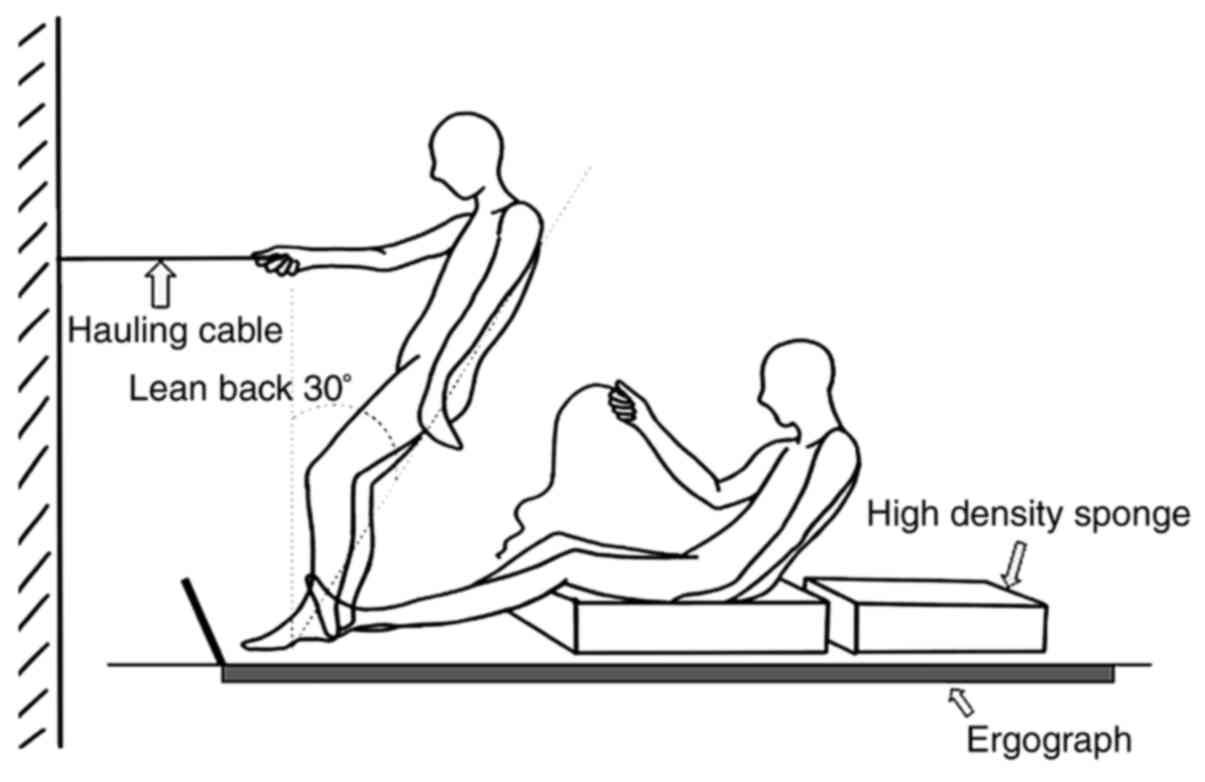
A 3D force table (BP400600; Advanced Mechanical
Technology, Inc.) was used to record the hip force during the
backward fall process. During the test, in order to protect the
volunteer, a 5 cm-thick high-density sponge with a density of 45
kg/m3 was laid above the force table. The size of the
sponge was 60x40x5 cm3, which was required to cover the
surface but not exceed the edge of the platform (6). In addition, the volunteer kept their
body straight, at an angle of 30˚ to the vertical axis, and held a
fixed rope that could be released at will. When the described
initial state was maintained by the volunteer, the rope was
released suddenly under the condition of diverting the volunteer's
attention, so as to achieve the state of simulated unprepared
backward fall (Fig. 1). Data
acquisition frequency of the force platform was set as 50 Hz, and
the precision was 0.01 N. The backward fall was mimicked at least
three times and the impact force on the hip was collected each
time. The average impact force was then calculated.
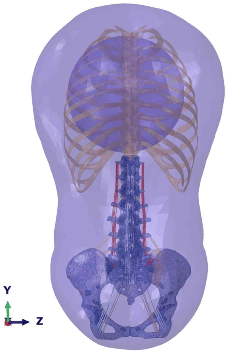
FEA
Abaqus 2016 software (Dassault Systèmes) was used to
simulate the stress state of the human trunk during the process of
backward fall (Fig. 2) (12). Firstly, computed tomography (CT)
scan data of the volunteer were collected from a dual-source
64-slice spiral CT system (Siemens AG) at Jiangxi Provincial
People's Hospital Affiliated to Nanchang University. The scan
parameters were set as 120 kV voltage and 0.625 mm pitch. All
original CT data were stored in digital imaging communications in
medicine format, and then imported into Mimics 19.0 software
(Materialise NV) for reconstruction. The 3D models of overall trunk
surface, vertebrae, thorax and the pelvis were separately divided
and saved in stereolithography (STL) format.
The 3D models in STL format were imported into
Geomagic 2016 software (3D Systems, Inc.) to be modified and
exported as IGES format models. These were then imported into
Hypermesh 14.0 software (Altair Engineering, Inc.) for further
Boolean operation, meshing, addition of truss unit ligament
structures and the endowment of each structure with validated
material property parameters, to establish the final FE model of
the trunk.
The linear and isotropic elasticity property of the
FE model was developed, which included cortical and trabecular
bone, ligament and cartilage structures, intervertebral disc and
surrounding soft tissues. During the mesh procedure, seed sizes
were set to 1-3 mm, and a finer mesh size was used for the regions
of interest of the thoracolumbar junction, including T11, T12 and
the upper lumbar region. An almost 0.5-mm gap was used for the
facet joint of lumbar vertebrae, but the non-contact condition
(within the torque of 7.5 N·m) was defined to avoid nonconvergence
and decrease the expense of the FEA simulation.
The FE model was then imported into Abaqus software
to set the boundary conditions and load accurate force-time
parameters of the sacrococcygeal bony structures obtained using the
Anybody software (Anybody Technology AS) simulation calculation.
The upper thoracic margin was set as a fixed boundary condition,
the loading point was set as the ischial tubercle and the loading
was carried out step-by-step according to the force-time curve. The
material properties and node information of bony and soft-tissue
structures in the FE model are shown in Table I, and material attribute parameters
of each ligament are shown in Table
II. Moreover, all FE models were simulated based on
quasi-static analysis by Abaqus/standard solver on a T7900 tower
workstation (Dell Inc.). Regarding the AVG 75%, if the AVG was set
to 1, it meant that the stress calculation of all nodes in the
display area were used for generating the average; therefore, a
smoother cloud image was created. If AVG was set to 75%, it meant
that when the relative node variable was less than the value and
the result of the node was averaged.
 | Table IMaterial properties of bony and
soft-tissue structures of finite element analysis model. |
Table I
Material properties of bony and
soft-tissue structures of finite element analysis model.
| First author
(year) | Structure | Elasticity modulus
E (MPa) | Poisson's ratio
(υ) | Number of
elements | Number of
nodes | Type of
element | (Refs.) |
|---|
| Denozière and Ku
(2006), El-Rich et al (2009) and Moramarco et al
(2010) | Cortical bone | 12,000 | 0.3 | 756,474 | 217,388 | C3D4 | (31-33) |
| El-Rich et
al (2009) and Moramarco et al (2010) | Cancellous
bone | 100 | 0.2 | 127,110 | 34,110 | C3D4 | (31,32) |
| Denozière and Ku
(2006), El-Rich et al (2009), Moramarco et al (2010)
and Guo and Li (2020) | Fiber ring | 450 | 0.3 | 75,790 | 22,104 | C3D4 | (31-34) |
| Denozière and Ku
(2006) and El-Rich et al (2009) | Nucleus
pulposus | 1 | 0.5 | 35,510 | 10,100 | C3D4 | (31,32) |
| Denozière and Ku
(2006), Moramarco et al (2010) and Guo and Li (2020) | Cartilage | 23.8 | 0.4 | 2,622 | 859 | C3D4 | (31,33,34) |
| Moramarco et
al (2010) and Guo and Li (2020) | Soft tissue | 1.5 | 0.4 | 503,217 | 112,249 | C3D4 | (33,34) |
| Table IIMaterial properties of ligament
tissues. |
Table II
Material properties of ligament
tissues.
| First author
(year) | Ligament
tissue | Elasticity modulus
E (Mpa) | Poisson's ratio
(υ) | Transverse area
(mm2) | Number of
elements | Type of
element | (Refs.) |
|---|
| Guo and Li (2020)
and Kong and Goel (2003) | Anterior
longitudinal ligament | 7.8 | 0.3 | 24 | 42 | Truss (T3D2) | (34,35) |
| Guo and Li (2020)
and Kong and Goel (2003) | Posterior
longitudinal ligament | 10 | 0.3 | 14.4 | 42 | Truss (T3D2) | (34,35) |
| Guo and Li (2020)
and Kong et al (2003) flavum | Ligamentum | 15 | 0.3 | 40 | 36 | Truss (T3D2) | (34,35) |
| Guo and Li (2020)
and Kong et al (2003) | Supraspinous
ligament | 8 | 0.3 | 23 | 7 | Truss (T3D2) | (34,35) |
| Guo and Li
(2020) | Interspinous
ligaments | 10 | 0.3 | 26 | 28 | Truss (T3D2) | (34) |
| Guo and Li
(2020) | Intertransverse
ligament | 10 | 0.3 | 3.6 | 56 | Truss (T3D2) | (34) |
| Guo and Li
(2020) | Iliolumbar
ligament | 75 | 0.3 | 25 | 12 | Truss (T3D2) | (34) |
| Guo and Li
(2020) | Sacrospinous
ligament | 12.6 | 0.2 | 25 | 8 | Truss (T3D2) | (34) |
| Guo and Li
(2020) | Sacrotuberous
ligament | 33 | 0.3 | 539 | 8 | Truss (T3D2) | (34) |
| Guo and Li
(2020) | Inguinal
ligament | 2.6 | 0.3 | 45 | 6 | Truss (T3D2) | (34) |
| Kong and Goel
(2003) | Anterior sacroiliac
ligaments | 208 | 0.2 | 25 | 20 | Truss (T3D2) | (35) |
| Kong and Goel
(2003) | Posterior
sacroiliac ligament | 133 | 0.2 | 25 | 20 | Truss (T3D2) | (35) |
At FEA simulation data collection, 50 representative
nodes in the stress concentration part of the thoracolumbar spine
were selected to calculate the average von Mises stress value as
the overall stress value of the structure.
Results
Backward fall experiment
The results of the backward fall experiment showed
that the sacrococcygeal region of the volunteer landed first, and
that the hip joint was at a state of flexion at this time. During
the process of backward fall, the trunk was flexed at the time of
landing, and the trunk was extended after landing. The total impact
time of landing was 1.14±0.58 sec, and the maximum impact force was
4,056±263 N, which is approximately six-times the body weight of
the volunteer.
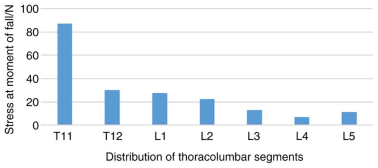
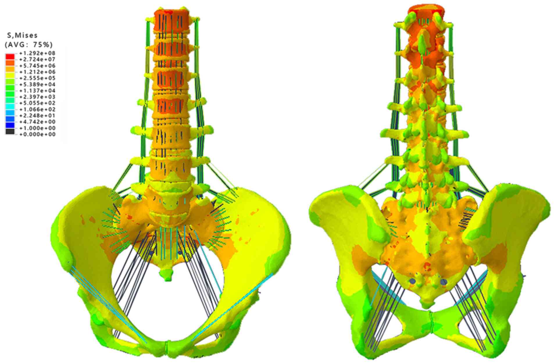
FE simulation of a backward fall
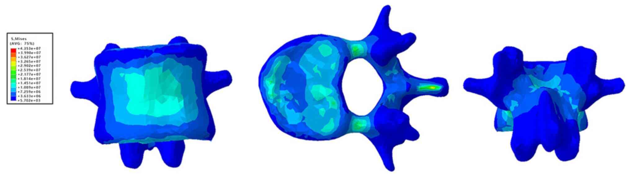
Under static loading, the stress of each vertebral
structure decreased gradually along the spine, following the peak
value of stress, which appeared at 0.4 sec (Fig. 3), and the stress of the T11 front
vertebral body was as high as 42 MPa (Fig. 4). In addition, the stress of the T11
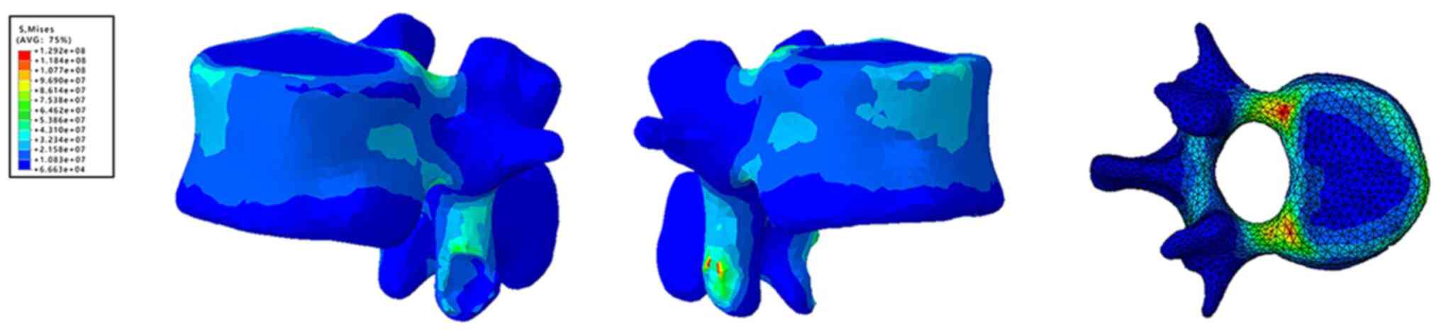
rear edge and pedicle junction was as high as 70.67 MPa (Fig. 5), and the maximum stress value
between the inferior articular process and upper articular process
of T12 was ~128 MPa (Fig. 6).
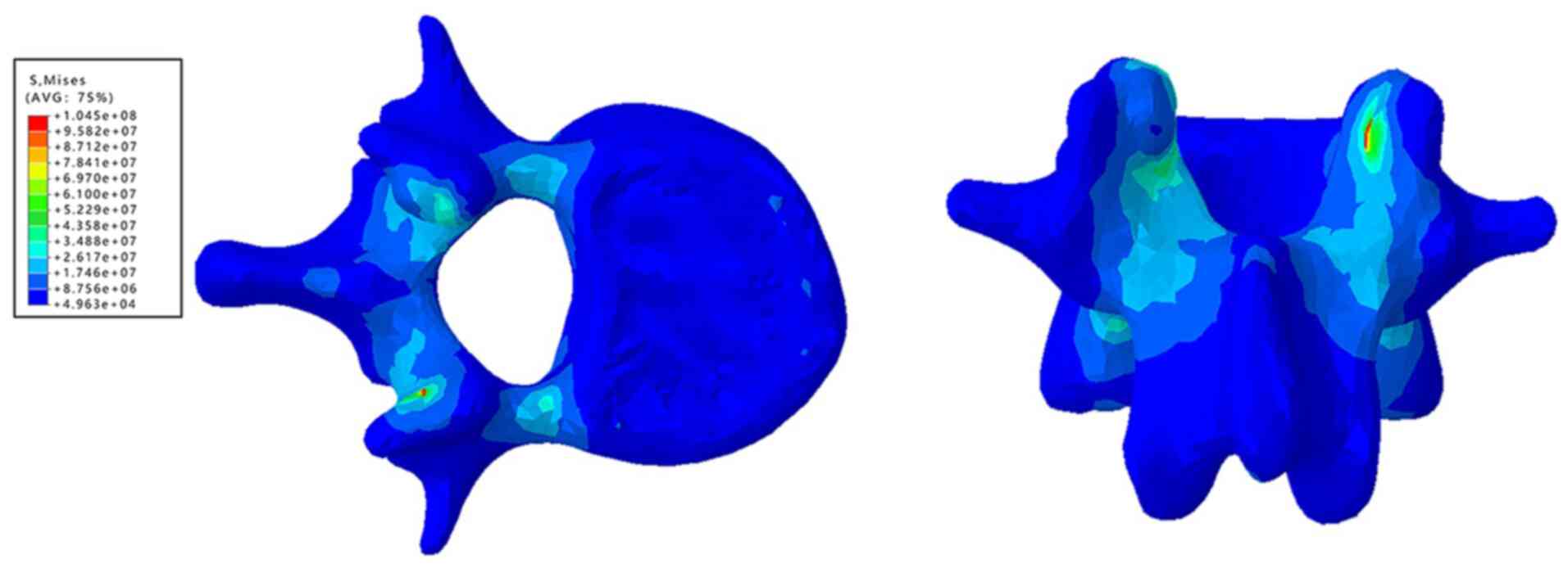
Moreover, the average stress of T12 and the front edge of L1 was 25
MPa, and the maximum stress of the end plate was 21.7 MPa, which
was mainly distributed in the central and posterior part of the end
plate and surrounding cortex (Figs.
6 and 7).
Discussion
Thoracolumbar vertebral compression fractures caused
by OP are a common and difficult problem faced by spinal surgeons,
with the majority of patients being elderly (13). The direct cause of fractures is that
the load acting on the vertebral body causes an internal stress
which exceeds the mechanical strength of the vertebra. Therefore,
regardless of bone condition, the vertebral body load is critical
for the formation of fractures (14,15).
Previous fall experiments have mainly focused on the instantaneous
posture of various parts of the body during the process of a
backward fall (16,17), while the impact force of a backward
fall has not been as well studied. Only the instantaneous posture
and the force value of the force point can be measured using
force-measuring platforms. This means that the mechanical mechanism
of strength transmission to the spine leading to spinal fracture
after a backward fall cannot be accurately determined (18,19).
Khalili et al (20) previously designed an inverted
pendulum model, in which the pine was defined as a single straight
rod. However, this model makes it difficult to directly obtain the
stress condition of the column section during the process of a
backward fall. As an effective way of performing a spinal mechanics
experiment, the FE model can effectively predict the strength of
spinal bone, fracture sites and stress distribution (11). However, most FE studies of fall
experiments have involved simulating stress on the hip (21,22),
and these experimental FE models were not able to simulate the
human postures involved in the process of a backward fall, which
impacted on the results. Similarly, in the process of thoracolumbar
segments during injury, most of the FE research methods were
performed using axial impact (10).
In the setting of loading conditions, due to various reasons such
as limited conditions, the stress situation of subjects was not
able to be added accurately. Hence, in the present study, a healthy
male volunteer participated in simulating a real-life fall before a
simulated fall spine stress FE experiment was carried out to obtain
the accurate fall data. This provided reliable data for further
understanding of the stress mechanism of the spinal structures
during the backward fall process.
In order to obtain accurate fall data, this
experiment simulated the real-life backward fall process. Therein,
it was found that at the moment of impact, the subject was in a
flexion state with the hip joint, the sacrococcygeal region landed
first, then the hip joint extended, and the chest, waist and back
were stressed. The results were consistent with the fall experiment
postures shown by Li et al (23) and Khalili et al (20), and the hip impact force reached
4,056±253 N.
For validation of the FE model, there are few prior
FE studies that simulate the backward fall process to measure the
stress on the spine. In the present study, an FE model was
generated based on the experimental data of a backward fall, in
order to analyze the stress mechanism of each segment of the
thoracolumbar spine during this process. In previous studies, it
was hypothesized that compression fractures accounted for the vast
majority of thoracolumbar fractures caused by falls (20,23).
In the FE results of the present study, the stress concentration
appeared in the anterior and posterior edge of T11, T12 and L1
vertebrae, upper and lower endplates, pedicle and laminae, and the
peak stress appeared in the thoracolumbar segment. This was
consistent with results of Gertzbein et al (24) and Nakashima et al (25) after applying axial load to the
established FE model. The results of the present study complement
those of Qiu et al (26), in
which the established FE model of T12 and L1 segments were measured
under vertical impact load. In the current study, the high stress
areas of the upper and lower endplates were in the nucleus pulposus
center and were parallel to each other, and the stress center of
cancellous bone was largest near the endplate. The stress of
cortical bone was concentrated in the anterior and posterior lower
edge of the T12 vertebral body, and anterior and posterior upper
edge of the L1 vertebral body. These observations were consistent
with the results of lumbar FE demonstrated by Cao et al
(14), which found that >50% of
vertebral body fractures occur between upper and lower
endplates.
It is important to note that there was a clear
stress concentration in the joint process of the lower T11, upper
T12 and upper L1, as well as the transitional part of the pedicle,
and that the stress extended to the posterior edge of the vertebral
body through the pedicle. In addition, some studies have
hypothesized that the interaction of articular processes causes the
increase of stress and the transfer of stress along the pedicle to
the vertebral body, resulting in the local stress concentration at
the posterior edge of the vertebral body (27,28).
Moreover, Wagnac et al (29)
reported that when the load of the facet was high at the level of
L1-L2, the stress concentration of the pedicle root of this segment
also increased, further supporting this hypothesis. In addition,
Fradet et al (30)
hypothesized that fractures of the pedicle could be caused by
multiple loading conditions, such as forward and backward shear
combined with flexion, traction combined with flexion, or traction
combined with flexion and shear. Hence, it was hypothesized in the
present study that the probability of fracture in these stress
areas may be increased. Furthermore, by comparing the stress of
different vertebrae, it has previously been found that the stress
of the L4 vertebrae was significantly lower than that of the
surrounding vertebrae when falling backward (30). Combined with the stress process,
Gertzbein et al (24)
hypothesized that the physiological curvature of vertebrae might
lead to the dispersion of conduction force, resulting in the uneven
distribution of vertebral force. Hence, it could be inferred from
the FE results, that the risk of fracture is increased at the front
vertebral body, and the upper and lower endplates of T11-L2
vertebrae after falling backward, and that fractures may occur due
to the compression of the front of the vertebrae. Due to the power
transmission of the upper and lower articular processes, there is
also the risk of fracture at the junction of the pedicle and
vertebrae of T12 and L1.
The stress distribution of thoracolumbar junction
obtained in the present study showed the injury risk of this
structure during the process of backward fall. Furthermore, the
method of FEA combined in vivo experimental data was shown
to be effective and accurate in this study. Thus, in future
research, the method in the present study could be directly used
for researching protective devices to prevent injury to the
thoracolumbar junction from a backward fall. In addition, the
protective effect and mechanism could be evaluated through an
additional series of studies, which could aid in the development,
optimization and improvement of protective devices for the
thoracolumbar junction in OP.
Indeed, certain limitations in the current study
should also be recognized and addressed. Firstly, the stress
distribution of the thoracolumbar junction obtained in this study
was indirectly obtained by FEA, which could provide a certain
amount of error. Secondly, the sample size in this study was small,
which will need to be increased for further validation. Thirdly,
the current common practice is to select a volunteer of standard
weight and CT scan data for modeling and simulation, which may not
represent the population.
In conclusion, through the combination of FEA and
human body backward fall simulation experiment, the backward fall
process and the force condition of the sacrococcygeal region can be
accurately obtained. The local stress state of the thoracolumbar
junction during the process of a backward fall obtained in FE
simulation is consistent with previous research, and there is a
greater risk of fracture in the front of the T11-L1 vertebral body,
the upper and lower endplates, the junction of the T12 and the L1
pedicle and vertebral body.
Acknowledgements
Not applicable.
Funding
Funding: This work was supported by the National Natural Science
Foundation of China (grant nos. 81460346 and 81702160).
Availability of data and materials
The datasets used and/or analyzed during the current
study are available from the corresponding author on reasonable
request.
Authors' contributions
PDS, XXZ, YWZ, ZW, XYW, YCW, XLY, HRG, XDL, ZZA, JYH
and XPD participated in the data collection, data analysis and data
interpretation. PDS, XXZ, YWZ and XPD wrote and edited the
manuscript. PDS, XXZ, YWZ and XPD contributed to the critical
revision of the manuscript. PDS and XPD confirm the authenticity of
all the raw data. All authors read and approved the final
manuscript.
Ethics approval and consent to
participate
The current study was approved by the Ethics
Committee of Jiangxi Provincial People's Hospital Affiliated to
Nanchang University. Written informed consent was provided by the
involved volunteer.
Patient consent for publication
Written informed consent was obtained from the
volunteer for the publication of his relevant data and images.
Competing interests
The authors declare that they have no competing
interests.
References
|
1
|
Lee H, Jung M, Lee KK and Lee SH: A 3D
human-machine integrated design and analysis framework for squat
exercises with a smith machine. Sensors (Basel).
17(17)2017.PubMed/NCBI View Article : Google Scholar
|
|
2
|
Li B, Sun C, Zhao C, Yao X, Zhang Y, Duan
H, Hao J, Guo X, Fan B, Ning G, et al: Epidemiological profile of
thoracolumbar fracture (TLF) over a period of 10 years in Tianjin,
China. J Spinal Cord Med. 42:178–183. 2019.PubMed/NCBI View Article : Google Scholar
|
|
3
|
Hughes M, Ashour R and Rao J: Treatment of
thoracolumbar burst fractures using orthosis versus no orthosis.
BMJ Evid Based Med. 24:70–71. 2019.PubMed/NCBI View Article : Google Scholar
|
|
4
|
Chang V and Holly LT: Bracing for
thoracolumbar fractures. Neurosurg Focus. 37(E3)2014.PubMed/NCBI View Article : Google Scholar
|
|
5
|
Diaz JJ Jr, Cullinane DC, Altman DT,
Bokhari F, Cheng JS, Como J, Gunter O, Holevar M, Jerome R, Kurek
SJ Jr, et al: EAST Practice Management Guideline Committee:
Practice management guidelines for the screening of thoracolumbar
spine fracture. J Trauma. 63:709–718. 2007.PubMed/NCBI View Article : Google Scholar
|
|
6
|
Sran MM and Robinovitch SN: Preventing
fall-related vertebral fractures: Effect of floor stiffness on peak
impact forces during backward falls. Spine. 33:1856–1862.
2008.PubMed/NCBI View Article : Google Scholar
|
|
7
|
Van Toen C, Sran MM, Robinovitch SN and
Cripton PA: Transmission of force in the lumbosacral spine during
backward falls. Spine. 37:E519–E527. 2012.PubMed/NCBI View Article : Google Scholar
|
|
8
|
Gilbertson LG, Goel VK, Kong WZ and
Clausen JD: Finite element methods in spine biomechanics research.
Crit Rev Biomed Eng. 23:411–473. 1995.PubMed/NCBI View Article : Google Scholar
|
|
9
|
Al Qahtani WMS and El-Anwar MI: Advanced
Computational Methods in Bio-Mechanics. Open Access Maced J Med
Sci. 6:742–746. 2018.PubMed/NCBI View Article : Google Scholar
|
|
10
|
Tyndyka MA, Barron V, McHugh PE and
O'Mahoney D: Generation of a finite element model of the
thoracolumbar spine. Acta Bioeng Biomech. 9:35–46. 2007.PubMed/NCBI
|
|
11
|
Jones AC and Wilcox RK: Finite element
analysis of the spine: Towards a framework of verification,
validation and sensitivity analysis. Med Eng Phys. 30:1287–1304.
2008.PubMed/NCBI View Article : Google Scholar
|
|
12
|
Karimi MT, Rabczuk T and Pourabbas B:
Evaluation of the efficiency of various force configurations on
scoliotic, lordotic and kyphotic curves in the subjects with
scoliosis. Spine Deform. 8:361–367. 2020.PubMed/NCBI View Article : Google Scholar
|
|
13
|
Roy DK, O'Neill TW, Finn JD, Lunt M,
Silman AJ, Felsenberg D, Armbrecht G, Banzer D, Benevolenskaya LI,
Bhalla A, et al: Determinants of incident vertebral fracture in men
and women: results from the European Prospective Osteoporosis Study
(EPOS). Osteoporosis Int. 14:19–26. 2003.PubMed/NCBI View Article : Google Scholar
|
|
14
|
Cao KD, Grimm MJ and Yang KH: Load sharing
within a human lumbar vertebral body using the finite element
method. Spine. 26:E253–E260. 2001.PubMed/NCBI View Article : Google Scholar
|
|
15
|
Imai K, Ohnishi I, Bessho M and Nakamura
K: Nonlinear finite element model predicts vertebral bone strength
and fracture site. Spine. 31:1789–1794. 2006.PubMed/NCBI View Article : Google Scholar
|
|
16
|
Ivancic PC: Hybrid cadaveric/surrogate
model of thoracolumbar spine injury due to simulated fall from
height. Accid Anal Prev. 59:185–191. 2013.PubMed/NCBI View Article : Google Scholar
|
|
17
|
Panjabi MM, Hoffman H, Kato Y and
Cholewicki J: Superiority of incremental trauma approach in
experimental burst fracture studies. Clin Biomech (Bristol, Avon).
15:73–78. 2000.PubMed/NCBI View Article : Google Scholar
|
|
18
|
Lee SC, Ding J, Prosser LA, Wexler AS and
Binder-Macleod SA: A predictive mathematical model of muscle forces
for children with cerebral palsy. Dev Med Child Neurol. 51:949–958.
2009.PubMed/NCBI View Article : Google Scholar
|
|
19
|
Terrier A, Latypova A, Guillemin M, Parvex
V and Guyen O: Dual mobility cups provide biomechanical advantages
in situations at risk for dislocation: A finite element analysis.
Int Orthop. 41:551–556. 2017.PubMed/NCBI View Article : Google Scholar
|
|
20
|
Khalili M, Borisoff JF and Van der Loos
HF: Developing safe fall strategies for lower limb exoskeletons.
IEEE Int Conf Rehabil Robot. 2017:314–319. 2017.PubMed/NCBI View Article : Google Scholar
|
|
21
|
Majumder S, Roychowdhury A and Pal S:
Simulation of hip fracture in sideways fall using a 3D finite
element model of pelvis-femur-soft tissue complex with simplified
representation of whole body. Med Eng Phys. 29:1167–1178.
2007.PubMed/NCBI View Article : Google Scholar
|
|
22
|
Majumder S, Roychowdhury A and Pal S:
Effects of trochanteric soft tissue thickness and hip impact
velocity on hip fracture in sideways fall through 3D finite element
simulations. J Biomech. 41:2834–2842. 2008.PubMed/NCBI View Article : Google Scholar
|
|
23
|
Li J, Chen D, Tang X and Li H: On the
protective capacity of a safety vest for the thoracic injury caused
by falling down. Biomed Eng Online. 18(40)2019.PubMed/NCBI View Article : Google Scholar
|
|
24
|
Gertzbein SD, Khoury D, Bullington A, St.
John TA and Larson AI: Thoracic and lumbar fractures associated
with skiing and snowboarding injuries according to the AO
Comprehensive Classification. Am J Sports Med. 40:1750–1754.
2012.PubMed/NCBI View Article : Google Scholar
|
|
25
|
Nakashima D, Kanchiku T, Nishida N, Ito S,
Ohgi J, Suzuki H, Imajo Y, Funaba M, Chen X and Taguchi T: Finite
element analysis of compression fractures at the thoracolumbar
junction using models constructed from medical images. Exp Ther
Med. 15:3225–3230. 2018.PubMed/NCBI View Article : Google Scholar
|
|
26
|
Qiu TX, Tan KW, Lee VS and Teo EC:
Investigation of thoracolumbar T12-L1 burst fracture mechanism
using finite element method. Med Eng Phys. 28:656–664.
2006.PubMed/NCBI View Article : Google Scholar
|
|
27
|
Langrana NA, Harten RD RD, Lin DC, Reiter
MF and Lee CK: Acute thoracolumbar burst fractures: A new view of
loading mechanisms. Spine. 27:498–508. 2002.PubMed/NCBI View Article : Google Scholar
|
|
28
|
Wilcox RK, Allen DJ, Hall RM, Limb D,
Barton DC and Dickson RA: A dynamic investigation of the burst
fracture process using a combined experimental and finite element
approach. Eur Spine J. 13:481–488. 2004.PubMed/NCBI View Article : Google Scholar
|
|
29
|
Wagnac E, Arnoux PJ, Garo A and Aubin CE:
Finite element analysis of the influence of loading rate on a model
of the full lumbar spine under dynamic loading conditions. Med Biol
Eng Comput. 50:903–915. 2012.PubMed/NCBI View Article : Google Scholar
|
|
30
|
Fradet L, Petit Y, Wagnac E, Aubin CE and
Arnoux PJ: Biomechanics of thoracolumbar junction vertebral
fractures from various kinematic conditions. Med Biol Eng Comput.
52:87–94. 2014.PubMed/NCBI View Article : Google Scholar
|
|
31
|
Denozière G and Ku DN: Biomechanical
comparison between fusion of two vertebrae and implantation of an
artificial intervertebral disc. J Biomech. 39:766–775.
2006.PubMed/NCBI View Article : Google Scholar
|
|
32
|
El-Rich M, Arnoux PJ, Wagnac E, Brunet C
and Aubin CE: Finite element investigation of the loading rate
effect on the spinal load-sharing changes under impact conditions.
J Biomech. 42:1252–1262. 2009.PubMed/NCBI View Article : Google Scholar
|
|
33
|
Moramarco V, Pérez del Palomar A,
Pappalettere C and Doblaré M: An accurate validation of a
computational model of a human lumbosacral segment. J Biomech.
43:334–342. 2010.PubMed/NCBI View Article : Google Scholar
|
|
34
|
Guo LX and Li WJ: Finite element modeling
and static/dynamic validation of thoracolumbar-pelvic segment.
Comput Methods Biomech Biomed Engin. 23:69–80. 2020.PubMed/NCBI View Article : Google Scholar
|
|
35
|
Kong WZ and Goel VK: Ability of the finite
element models to predict response of the human spine to sinusoidal
vertical vibration. Spine. 28:1961–1967. 2003.PubMed/NCBI View Article : Google Scholar
|