Introduction
Experimental dynamics conducted on the lumbar spine
aims to prove that, given that the vertebral bodies are rigid
systems, the pronounced decrease in resistances in the system is a
phenomenon that can be eminently affected by the adaptive changes
that occur at the level of the intervertebral disc at axial
mechanical stresses. Moreover, these considerations are applicable
to the disc where degenerative processes have occurred (1,2).
However, it does not perfect the ways of distributing loads and
demands that occur in the vertebral body and the intervertebral
disc. Such a model, associated with the biodynamic axial load
study, has the advantage of providing relevant information on bone
movements and the changes in soft neighborhood structures. This
approach should not be neglected in specific pathology management
of the area (1). The behavior of
the monitored specimens, especially the results, allows for
objective parallelism between the values obtained in the present
study and the lumbar vertebral segments in vivo
behavior.
Materials and methods
The biomechanical trial included 11 lumbar segments
L1-L5, gathered from adult human cadavers. To obtain various
anatomical preparations with biomechanical characteristics as close
to those, in vivo, specimens were obtained from fresh,
non-formalin-preserved human cadavers, preserved by freezing at
-22˚; after sampling, the anatomical preparations were maintained
at temperatures below freezing. The decision to protect the samples
only by freezing was taken after examining the specialists' studies
from the literature and which confirmed, with arguments, that this
represented the most effective preservation method concerning the
mechanical strength and elasticity of the intervertebral disc for
maintenance in saline solution, which decreases disc resistance
(3). After the isolation, the
adjacent muscularity was removed from the preparations to view the
osteoligamentous spine fully. The dissection considered the
complete keeping of all bone, disc, articulated and ligamentous
elements in their anatomical position to maintain the mechanical
characteristics specific to the region as close as possible to the
physiological conditions. Preparations were frozen 24 h prior to
the performance of the biomechanical measurement (3).
The test device included the following. i) Tensile
strength-compression universal test machine, with the capacity of
200 kN, type LBG 200, with a rigid construction in 2 columns, for
bending, compression, and tensile strength static tests, with an
accuracy that observes the standards of ISO 7500-1, precision class
0.5 and minimal resolution 0.80 microns. The device has
transmission channels of high resolution and synchronized data,
with high precision power cells, with a piston displacement
transducer with double direction. ii) Command system and digital
control with software module through which the machine operation is
performed, setting of work parameters occurs, and reading the
results specific to the bending/compressing/tensile tests is
carried out. The system ensures the real-time follow-up of test
parameters: force, displacement, elongation and allows the printing
of results and test diagrams, achieved using a digital regulator
for force, displacement, deformation with closed-loop control, and
data acquisition. iii) The programmable package on Windows
platform; a programmable package for statistical tests or according
to the operator needs, represented by TC SOFT application software
(Microsoft Corp.).
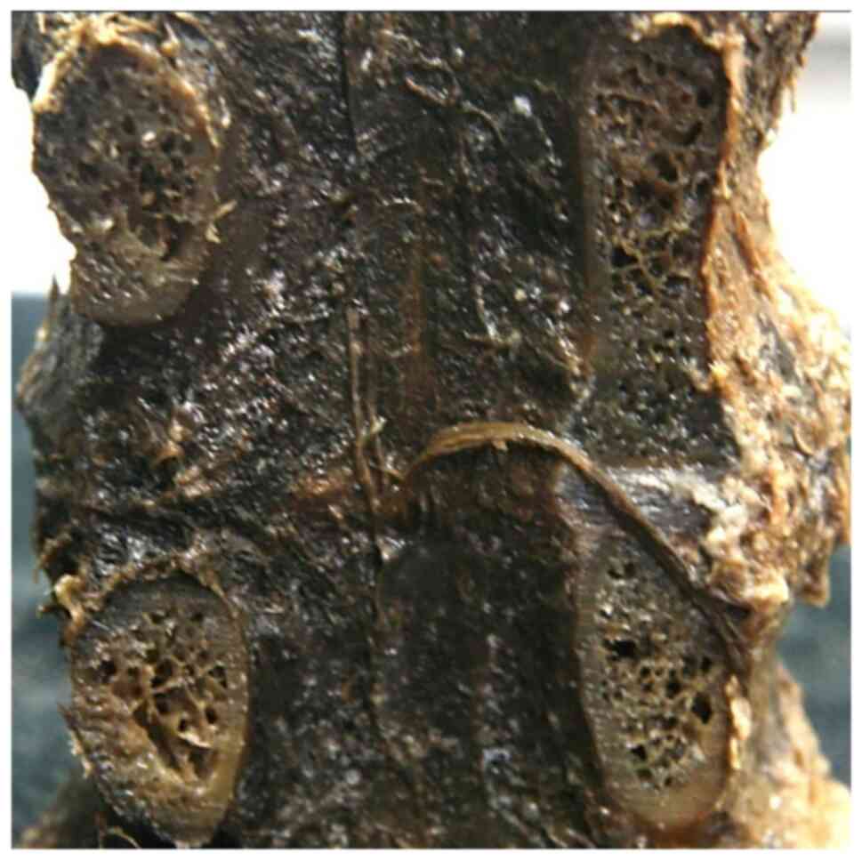
Each vertebral segment under test was comprised of
five vertebrae and four lumbar intervertebral discs prepared
according to the previous description. The specimens were placed in
the testing device, their position being conditioned by the
estimated dimensional values. Thus, to calculate the load and axial
resistance, the samples were placed vertically, central between the
test machine ferries.
The testing was carried out by applying variable
forces and supervision. The displacement interval was represented
by the segment 0-10 mm with maintenance every 2 mm. For each
displacement interval, the force values were recorded, at the
initial moment and the end of displacement, and the ‘return'
resistances encountered after 2, 4, 6 and 8 min, with the
registration of forces at those moments. The force values
(expressed in Nm) reflected the behavior of the assembly
vertebrae-discs-ligaments. To differentially quantify the
resistances from the column of bodies and those induced in the
system by the arches column, separate testing occurred at
compression, control samples, represented by
ligamentous-disc-vertebral blocks (2 lumbar vertebrae and the
corresponding disc) from which all the posterior arch elements were
removed, mainly the zygapophyseal complex (Fig. 1).
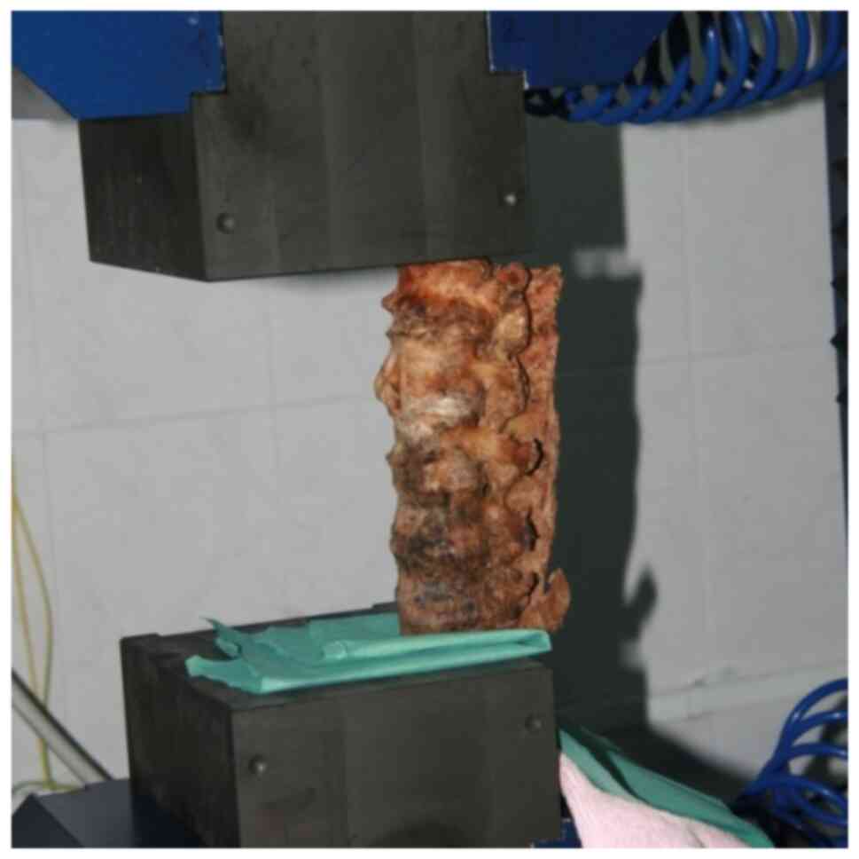
The lateral positioning (eccentric placement)
(Fig. 2) of preparation allowed the
eccentric force application; thus, the flexion/extension movement
could be simulated. The metric displacement monitoring followed the
consistent application of a 5 Nm equal force, quantified
displacement in turn in angular values, which was expressed in
degrees.
Results and Discussion
Axial loading study revealed the existence of three
types of behavior concerning the lumbar segment at compression,
namely:
Type I behavior at the axial loading represented the
most uniform behavior type. As observed from the recorded data, the
lumbar vertebral block showed a fast and pronounced adaptation
after 2 min, an increase of 30-70% of the initial values,
decreasing gradually up to final values.
By the evolution model of system forces, this type
of behavior is deemed optimal for intense and sudden moments and
loads. At the same time, however, the decreased intensity of
response in 4-8 min suggests the efficiency of such a system on
time intervals of relatively low duration.
Type II was characterized by an approximately
constant interval of resistances within 2-4 min. This type of
behavior suggests the existence of an interval in which the
resistance at axial loads remains relatively constant for a more
extended period, a period in which the changes in the
intervertebral disc are minimal. Type II represents the type of
system able to sustain the intense loads (with absolute superior
values to those from type I) for more extended periods without
suffering notable distortions.
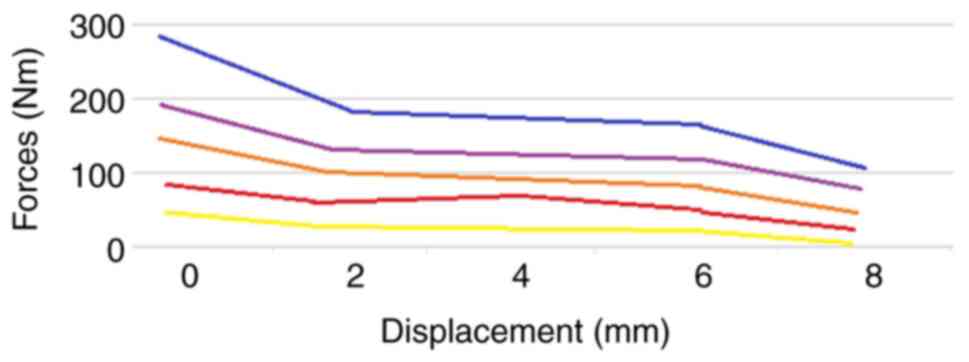
Type III represented the type of ‘intermediary' or
‘compromise' behavior, in which the interval of uniform resistances
is the most extensive. Thus, after a substantial reduction between
0 and 2 min, a relatively uniform interval appears between 2 and 6
min, after which the disc resistance actually ‘collapses' or is
markedly reduced up to the end (Fig.
3).
Type III presents the lowest values of resistance in
the final stages (min 8) for all displacement intervals, except for
the intervals of 0-2 min. Such behavior suggests the possibility of
developing some effective resistances at the axial external loads
on longer intervals. However, when these resistances are exceeded,
the disc-vertebra assembly can no longer face the loads.
Regarding the distribution of the three types within
the tested batch, the overwhelming percentage was represented by
type III, i.e., by the most adjustable type to loads, with 55% of
the cases. It was followed, in order, by type I (27%) and type II
(18%).
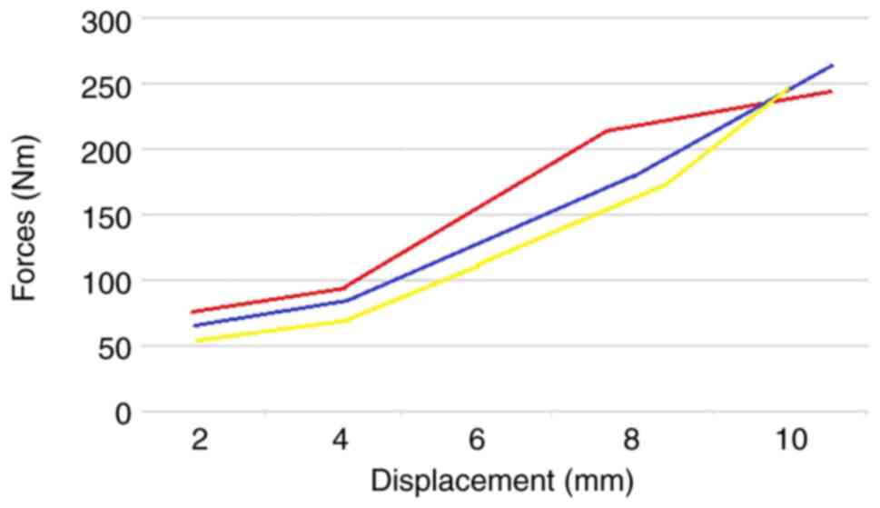
Table I and Fig. 4 summarize the type of behavior of
the three types suggested in terms of the forces [Newton-meters
(Nm)] related to the displacement (mm), a linear model behavior
similar to those provided by literature (4).
 | Table IThree types of axial loading
behavior. |
Table I
Three types of axial loading
behavior.
| Displacement
(mm) | 2 | 4 | 6 | 8 | 10 |
|---|
| Type 1 | 52 | 82 | 122 | 177 | 257 |
| Type 2 | 68 | 91 | 144 | 211 | 246 |
| Type 3 | 61 | 88 | 136 | 184 | 255 |
Thus, type I constantly presents the lowest
resistances, displacement of 10 mm, and type II involves the most
potent displacement forces (i.e., resistances). By contrast, type
III reconfirms the position of the intermediate kind with the
uniform behavior. An exciting aspect of the study was represented
by the comparative analysis of how the three lumbar vertebral types
behaved in the five displacement moments: 2, 4, 6, 8, and
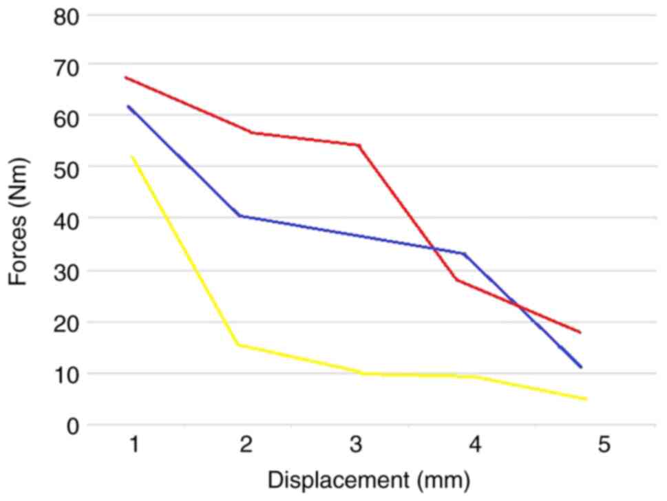
subsequently 10 mm. Thus, at a displacement of 2 mm, type I
presents the lowest values and, at the same time, it ‘concedes’
resistance at the fastest rate, the type II resists to the highest
loads, but on a relatively short interval, and type III is
constantly placed in an intermediate position, both in terms of
values and in terms of dynamics. In addition, in the case of the 2
mm displacement, the highest value differences were recorded
between the three suggested types (Fig.
5).
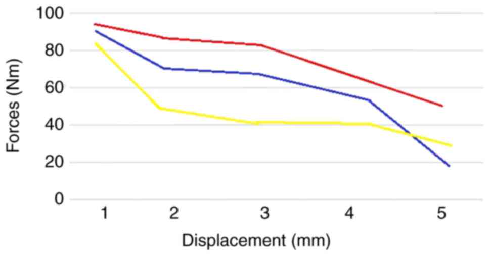
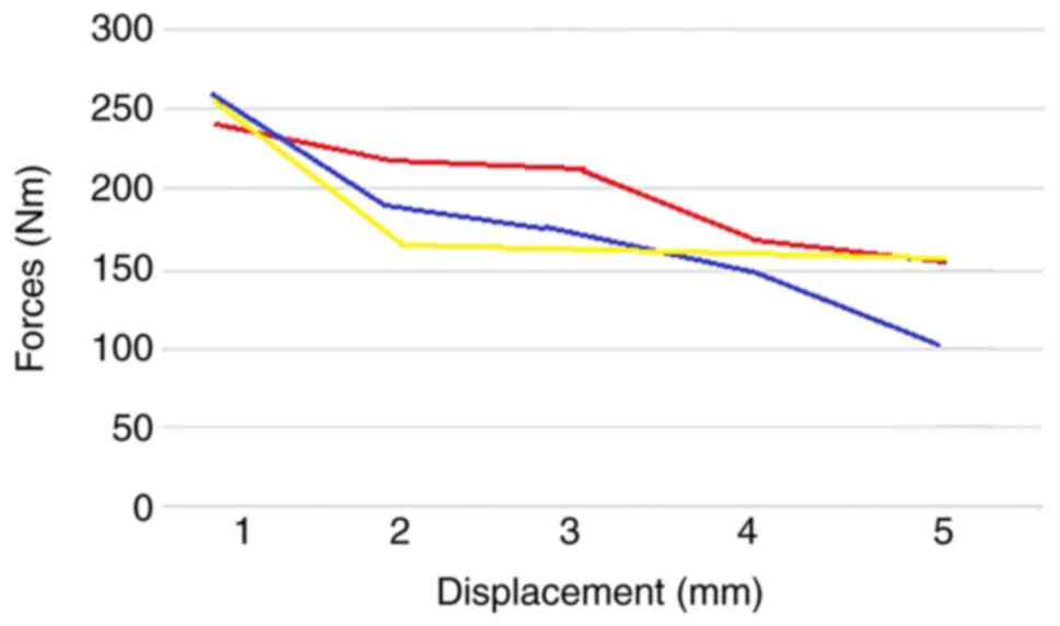
In the case of the 4 mm displacement, type I
maintains the behavior model, descending steeply after min 2; type
II loses the interval between min 2 and 4 but, at the same time, is
reduced less while type III not only constantly maintains the trend
but also manifests a slight peak at min 4. At the same time, the
resistance of this type is decreased significantly after min 6, so
that, at the end of the experiment, it presents the lowest
responsiveness (Fig. 6).
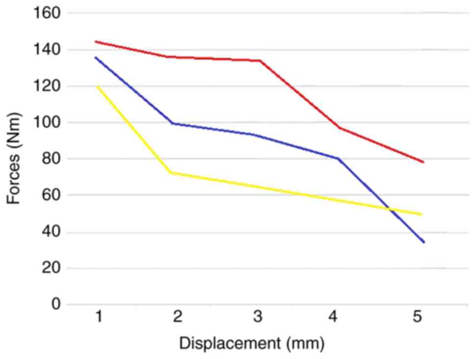
After compression of 6 mm, the behavior of the three
models was maintained, with two exceptions: The value difference
between them was significantly low, and the interval peak
disappeared from min 4 of type III. As in the previous case, at the
end of the load, the resistance of type III was found to be the
lowest (Fig. 7).
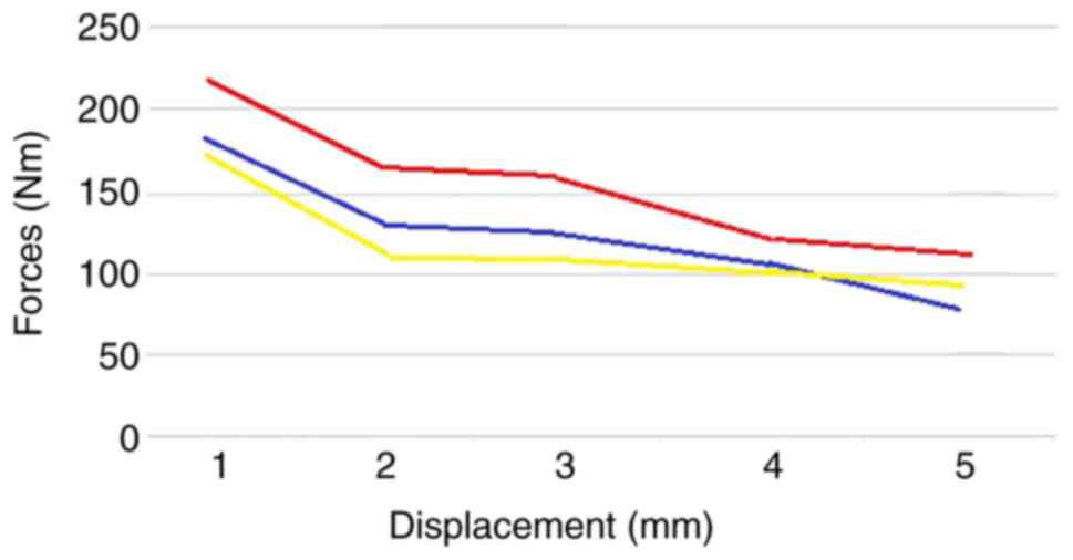
As the displacement interval increased (8 and 10
mm), the value differences of resistances between the three types
was reduced while the evolving trend remained. Moreover, type III
resistance decreased below the one of type I before minute eight
(Fig. 8).
This phenomenon was even more apparent at the 10-mm
displacement when the resistance of type III was the lowest after
only 3 min. Thus, at higher displacements, type III loses
effectiveness. In reality, such a displacement is specifically only
examined in vitro, while in vivo, such amplitudes are
impossible in the case of a simple axial load, more evident in
complex applications, composed and, very often, exceed
physiological limits (Fig. 9).
All the above considerations support that: type I is
the type of adequate response to prolonged and average to
low-intensity loads; type II, responds to maximum loads, but for
relatively reduced intervals; while type III is indeed the type
with the uniform behavior, at least in the load margin and
physiological displacements.
Eccentric loading was focused on the reproduction of
the characteristic movements of flexion in the sagittal and frontal
plane. The eccentric placing of the specimens allowed the bodies'
axial loading to reach the edge of the column, followed by the
corresponding ‘pinching’ of the discs in the relevant plane and
correlation distances with angular values.
The values obtained (Table II) are comparable to those offered
by the existing literature (5),
relative to the loads imposed.
| Table IIEccentric loading values. |
Table II
Eccentric loading values.
| | Sagittal flexion | Frontal flexion |
|---|
| Anatomical
sample | L1-L3 | L3-L5 | L1-L3 | L3-L5 |
|---|
| 1 | 32.9 | 20.7 | 29.7 | 16.4 |
| 2 | 43.2 | 24.1 | 34.5 | 18.4 |
| 3 | 42.1 | 21.4 | 33.4 | 18.6 |
| 4 | 33.2 | 20.1 | 24.6 | 13.6 |
| 5 | 24.9 | 18.4 | 35.4 | 19.7 |
| 6 | 41.2 | 29.1 | 21.5 | 11.2 |
| 7 | 36.5 | 24.1 | 28.7 | 15.4 |
| 8 | 29.7 | 19.7 | 28.4 | 13.7 |
| 9 | 39.7 | 20.8 | 24.3 | 9.1 |
| 10 | 43.2 | 22.7 | 33.5 | 20.4 |
| 11 | 40.1 | 20.8 | 30.1 | 15.7 |
| Median | 36.97273 | 21.99091 | 29.46364 | 15.65455 |
It is noteworthy that the mobility in the sagittal
plane (flexion earlier in our case) is much higher than that in the
frontal plane, clearly indicating limited mobility by the
intervertebral disc and articular complex by the presence of
arches. Statistical analysis demonstrated the lack of any
correlation values between the two types of movements
(R2=0.005507), underlining the absence of any elements
of prediction. An interesting aspect is that correlations appeared
low, statistically insignificant, even within the same movement in
the sagittal plane between the two levels L1-L3 and L3-L5
(R2=0.610427), suggesting the possibility of the
emergence of significant differences in mobility between respective
levels. The only values perfectly correlated were evident in the
case of lateral flexion between the two analysis levels
(R2=0.938386), which suggests an element of uniformity
in this type of mobility. It is an aspect worthy of considering in
the analysis of phase III discopathy etiopathogeny clinically.
The calculation of loads and resistances in the
vertebral-disc system has always been a concern for many authors,
who employed various calculation and testing methods (6). As absolute values, in an orthostatic
position, a weight of 47 kg presses on L4-L5 disc; with any change
in the position due to muscle tension, this weight is converted
into a load intervertebral disc pulpous nucleus between 282 and 726
kg. The longer the levers and the heavier the weight to lift, the
higher the loads on the pulpous nucleus, which may reach up to
1,200 kg. In order to increase the accuracy of the determinations,
center probes were placed at the level of the intervertebral discs
to record intra-disc pressure. Experiments have shown that, on the
third lumbar disc, on a 70 kg individual, each disc carries, in
dorsal decubitus position, a pressure of 21 kg, in lateral
decubitus carries 70 kg, sitting position carries 100 kg, with
trunk reclined forward at 45˚ carries 150 kg, and in sitting
position with the trunk reclined forward at 45˚ holding a weight of
20 kg pressure of 210 kg (6).
Despite the limitations, the current study on
biomechanical resistance and the outcome from investigating the
axial lumbar vertebral level, has highlighted several important
issues.
Linders and Nuckley were among the first to study
the displacement mechanism of the nucleus pulpous substance inside
the IVD and found that, in the flexion of the spine, the front of
adjoining vertebral plateaus approach and their rear part is
separating, and the nuclear substance moves with part of the mass
(most) towards the rear of the IVD, albeit some of it moves towards
the front, while on the right side flexion, most of the nucleus
mass moves to the left and a small part of it moves to the right
(7). The aforementioned studies are
opposed to the hypothesis of the pulpous nucleus as a single mass,
moving like a ‘ball’ inside the IVD.
According to this study, while the vertebral bodies
are rigid systems, the pronounced reduction in the resistance
system is a phenomenon that can be placed exclusively on adaptive
changes that occur in the intervertebral disc.
Resistance-pronounced decrease in the system within minutes is
nothing but a volumetric adaptation to mechanical axial loads of
the intervertebral disc. Decreased mobility in the system increases
stress on the structures, while disc elasticity prevents this
phenomenon. In addition, such considerations are applicable in the
case of the disc at which the degenerative processes occurred
(4,8,9). At
present, it is certain that the resistance of the disc varies
depending on the degree of degeneration, leading, in turn, to a
decreased resistance in cases of moderate degeneration and
increased rigidity, and subsequently to a decrease of the force
absorption power, in case of severe degeneration (6,7,10-19).
Furthermore, the parallelism between the load on the one hand, and
the disc degeneration, on the other hand, can engage the fibrous
annulus in a vicious cycle, which invariably results in
irreversible ruptures (7).
Another aspect worth mentioning is that, while the
preservation of preparations tried, as much as possible, to limit
the biochemical changes that occur in vitro, the experiment
was conducted at ambient temperature and not the average body
temperature, which, in turn, may alter the values obtained.
In the current study, type III, the type considered
most adaptable, represented the majority (55%), i.e., more than
double each of the other two types. It was followed by type I, the
one that, after a swift adjustment to the initial moment,
‘conceded’ resistance in a uniform manner.
In conclusion, the behavior type of the monitored
specimens and especially the results obtained allowed the mapping
of objective parallelism between values found in the current study
and behavior in vivo of the lumbar vertebral segment. A
critical aspect revealing the degree of objectivity of the study is
that this type of behavior manifested throughout the test interval
of 10 mm, an element confirming that preparations behaved
uniformly, a biomechanical behavior induced obviously by the
morphological substrate. Additionally, monitoring the behavior of
the lumbar complex for long intervals of time after application of
forces is an original element that allows the evaluator to
determine as objectively as possible, concerning the evolution of
adaptation, the likely degree of disc degeneration. Examination of
disc loads and especially the behavior of vertebral segments under
various load types and values can contribute on the one hand to
identifying the potential etiologies of pathological manifestations
at this level and, on the other hand, to the development of
effective prevention and/or treatment strategies in the matter.
Thus, correlating the disc behavior and the load makes
biomechanics, besides a prognostic factor, a genuine therapeutic
factor in disc regeneration.
Acknowledgements
Not applicable.
Funding
Funding: No funding was received.
Availability of data and materials
Not applicable.
Authors' contributions
DMI, CI, IB, PB, EG and MGI conceived and designed
the study. DMI, PB, FV, SIM, MGI and BO acquired the data. DMI, PB,
IB and MGI assessed the authenticity of the data. CI, IB, EG, FV
and SIM analyzed the data. DMI, PB, IB, MGI and BO validated the
results. EG, MGI, DMI, PB, FV, CI and IB were responsible for the
preparation of the original draft. SIM, EG, FV and BO, were
responsible for the final manuscript editing. DMI, PB, CI, IB, SIM
and MGI supervised the manuscript publication. All authors read and
approved the final manuscript.
Ethics approval and consent to
participate
Not applicable.
Patient consent for publication
Not applicable.
Competing interests
The authors declare that they have no competing
interests.
References
|
1
|
Iliescu DM, Bordei P, Ionescu EV, Albina
S, Oprea C, Obada B, Lupu AA, Hangan TL and Iliescu MG:
Anatomic-imaging correlations of lumbar disk-vertebral morphometric
indices. Int J Morphol. 35:1553–1559. 2017.
|
|
2
|
Ardeleanu V, Toma A, Pafili K, Papanas N,
Motofei I, Diaconu CC, Rizzo M and Stoian AP: Current
pharmacological treatment of painful diabetic neuropathy: A
narrative review. Medicina (Kaunas). 56(25)2020.PubMed/NCBI View Article : Google Scholar
|
|
3
|
Stefanescu DC, Ciucu AA, Rabinca AA,
Buleandra M, Stoian AP, Jecan CR and Razvan H: An integrative
medical perspective on novel dopamine detection method. Revista de
Chimie. 69:277–281. 2018.
|
|
4
|
Kurowski P and Kubo A: The relationship of
degeneration of the intervertebral disc to mechanical loading
conditions on lumbar vertebrae. Spine (Phila Pa 1976). 11:726–731.
1986.PubMed/NCBI View Article : Google Scholar
|
|
5
|
Barthes X, Walter B, Zeller R and
Dubousset JF: Biomechanical behaviour in vitro of the spine and
lumbosacral junction. Surg Radiol Anat. 21:377–381. 1999.PubMed/NCBI View Article : Google Scholar
|
|
6
|
Nachemson AL, Schultz AB and Berkson MH:
Mechanical properties of human lumbar spine motion segments.
Influence of age, sex, disc level, and degeneration. Spine (Phila
Pa 1976). 4:1–8. 1979.PubMed/NCBI View Article : Google Scholar
|
|
7
|
Linders DR and Nuckley DJ: Deduction of
spinal loading from vertebral body surface strain measurements. Exp
Mechanics. 47:303–310. 2007.
|
|
8
|
Niosi CA and Oxland TR: Degenerative
mechanics of the lumbar spine. Spine J. 4 (6 Suppl):202S–208S.
2004.PubMed/NCBI View Article : Google Scholar
|
|
9
|
Hutton WC, Ganey TM, Elmer WA, Kozlowska
E, Ugbo JL, Doh ES and Whitesides TE Jr: Does long-term compressive
loading on the intervertebral disc cause degeneration? Spine (Phila
Pa 1976). 25:2993–3004. 2000.PubMed/NCBI View Article : Google Scholar
|
|
10
|
Stoian PA, Hainarosi R, Pietrosanu C,
Rusescu A, Andronache LF, Paunica S, Balalau C and Pituru TS:
Modern concepts in non-surgical esthetics; a review. J Mind Med
Sci. 6:190–195. 2019.
|
|
11
|
Best BA, Guilak F, Setton LA, Zhu W,
Saed-Nejad F, Ratcliffe A, Weidenbaum M and Mow VC: Compressive
mechanical properties of the human anulus fibrosus and their
relationship to biochemical composition. Spine (Phila Pa 1976).
19:212–221. 1994.PubMed/NCBI View Article : Google Scholar
|
|
12
|
Gay RE, Ilharreborde B, Zhao K, Zhao C and
An KN: Sagittal plane motion in the human lumbar spine: Comparison
of the in vitro quasistatic neutral zone and dynamic motion
parameters. Clin Biomech (Bristol, Avon). 21:914–919.
2006.PubMed/NCBI View Article : Google Scholar
|
|
13
|
Hansson TH, Keller TS and Spengler DM:
Mechanical behavior of the human lumbar spine. II. Fatigue strength
during dynamic compressive loading. J Orthop Res. 5:479–487.
1987.PubMed/NCBI View Article : Google Scholar
|
|
14
|
Berkson M, Nachemson A and Schultz A:
Mechanical properties of human lumbar spine motion segments. II.
Response in compression and shear, influence of gross morphology. J
Biomech Eng. 101:53–55. 1979.
|
|
15
|
Tofolean DE, Mazilu L, Staniceanu F,
Mocanu L, Suceveanu AI, Baz R, Parepa RI, Suceveanu AP, Bondari S,
Bondari D and Voinea F: Clinical presentation of a patient with
Cutis Laxa with systemic involvement: A case report. Rom J Morphol
Embryol. 56:1205–1210. 2015.PubMed/NCBI
|
|
16
|
Iliescu M, Bordei P, Iliescu DM, Ciobotaru
C, Lucescu V, Covaleov A and Ionescu C: Anatomo-clinical and
imagistic correlations within the lumbar discopathy. Surg Radiol
Anatomy. 31(145)2009.
|
|
17
|
Tuta LA, Iorga I, Azis O and Voinea F:
End-of-life care in patients with end-stage renal disease-ethical
and clinical issues. Bk2: Political Sciences, Law, Finance,
Economics and Tourism Conference Proceedings. pp487-493, 2015.
|
|
18
|
lliescu M, Bordei P, Albina S and Ionescu
C: Morphology of the intervertebral foramen: A direct relation with
low back pain. ARS Medica Tomitana. 18:62–65. 2012.
|
|
19
|
Moraru D, Suceveanu AP, Stoian AP, Nitipir
C, Pituru S, Voinea F, Timofte D and Suceveanu AI: Amyloidosis-the
importance of an early diagnosis. In: Proceeding of 35 Balkan
Medical Week, 25th-27th September 2018, Athens, pp117-121,
2018.
|