Introduction
Diabetes is a metabolic syndrome characterized by
elevated blood glucose levels and is caused by insufficient insulin
production and/or peripheral tissue resistance to the action of
insulin (1).
The ninth edition of the Diabetes Overview,
published by the International Diabetes Federation (IDF), has
estimated that >500 million adults worldwide suffer from
diabetes mellitus, with ~1.2 billion adults with diabetes in China
alone (2). Diabetic retinopathy
(DR) is the most common microvascular complication and a major
cause of blindness in patients with diabetes (3). It has been reported that DR is closely
associated with damage to the retinal microvascular system mediated
by a high-glucose environment. DR can affect the structure of the
retina, leading to its impaired metabolism and dysfunction
(4).
Long non-coding RNAs (lncRNAs) are a class of
non-coding RNA molecules that are >200 nucleotides in length.
lncRNAs can exert their biological functions through several
pathways (5). MicroRNAs
(miRNAs/miRs) are small non-coding single-stranded RNAs, 18-25
nucleotides in length, which act mainly through complementary
pairing with downstream target mRNAs to promote their degradation
or suppression (6,7). miRNAs are involved in several
physiological processes, including cell proliferation, apoptosis,
signal transduction, differentiation, metabolism and hormone
secretion, as well as in the maintenance of the stemness potential
of embryonic stem cells. In addition, miRNAs regulate human growth
and development, and allow the body to adapt to its environment
(8). However, the role of miRNAs in
DR has not been extensively investigated. miR-27b is commonly
dysregulated in human cancers, such as glioma, cervical and breast
cancer, and is downregulated in lung adenocarcinoma, gastric
cancer, acute myeloid leukemia, colorectal, prostate and bladder
cancer (9).
The aim of the present study was to investigate the
effect of the long intergenic non-protein-coding RNA 963
(LINC00963) on the proliferation and migration of high
glucose-induced human retinal capillary endothelial cells
(HRECs).
Materials and methods
Cell culture and establishment of the
DR cell model
HRECs were purchased from the China Infrastructure
of Cell Line Resources, Institute of Basic Medical Sciences,
Chinese Academy of Medical Sciences. Cells were maintained in
endothelial cell medium (ScienCell Research Laboratories, Inc.)
supplemented with 5% FBS (Beijing Solarbio Science & Technology
Co., Ltd.), 100 µg/ml penicillin and 100 µg/ml
streptomycin (Gibco; Thermo Fisher Scientific, Inc.), at 37˚C in a
humidified atmosphere of 5% CO2. The cells were divided
into the following groups: i) Normal glucose (NG) group (5 mM
normal glucose); ii) high glucose (HG) group (30 mM glucose); and
iii) mannitol (MA) group (24.5 mmol/l mannitol + 5.5 mmol/l
glucose).
Cell viability
Cell viability was assessed using a Cell Counting
Kit-8 (CCK-8) assay (Dojindo Molecular Technologies, Inc.),
according to the manufacturer's instructions. Briefly, HRECs were
resuspended in endothelial cell medium (ScienCell Research
Laboratories, Inc.) and seeded into 96-well plates at a density of
5,000 cells/well. Subsequently, a total of 10 µl CCK-8 solution was
added to each well and cells were incubated for 1 h at room
temperature. Finally, the optical density in each well was measured
at a wavelength of 450 nm to determine cell viability.
Prediction of target genes
The Encyclopedia of RNA Interactomes (ENCORI;
starBase v2.0 https://starbase.sysu.edu.cn) is an open-source
platform for evaluating the miRNA-ncRNA, miRNA-mRNA, ncRNA-RNA,
RNA-RNA, RBP-ncRNA and RBP-mRNA interactions from CLIP-seq,
degradome-seq and RNA-RNA interactome data.
Dual-luciferase reporter assay
The sequences of wild-type (WT) LINC00963 (accession
no. NC_000009; 5'-GUGGGGCUGUGUUGACUGUGAG-3') and mutant (MUT)
LINC00963 (5'-GUGGGGCUGUGUUGUGACACUU-3') were amplified by Shanghai
GenePharma Co., Ltd., cloned into a luciferase reporter vector pGL3
(cat. no. E1761; Promega Corporation), named WT LINC00963-Luc and
MUT LINC00963-Luc respectively. Cell were co-transfected with
miR-27b mimic (50 nM, 5'-UUCACAGUGGCUAAGUUCUGC-3'; Shanghai
GenePharma Co., Ltd.) or miR-NC (50 Nm,
5'-UAACACAACUCUAGAGUAGGA-3'; Shanghai GenePharma Co., Ltd.) and WT
or MUT LINC00963-Luc into HRECs using the Lipofectamine®
3000 transfection reagent (Invitrogen; Thermo Fisher Scientific,
Inc.) with 37˚C for 48 h. The Dual-Luciferase® Reporter
Assay System (cat. no. E1910, Promega Corporation) was used to
evaluate the relative luciferase signals. The relative luciferase
activity was measured at a wavelength of 410 nm using a plate
reader (BD Biosciences) and normalized to Renilla luciferase
activity.
Reverse transcription-quantitative PCR
(RT-qPCR)
Total RNA was extracted from HRECs using
TRIzol® reagent (Invitrogen; Thermo Fisher Scientific,
Inc.) and cDNA was synthesized using the PrimeScript RT kit (Takara
Bio, Inc.) at 37˚C for 15 min and at 85˚C for 5 sec. The cDNA
solution was then stored at -80˚C. Subsequently, qPCR was performed
using the iTaq™ Universal SYBR-Green Supermix (Bio-Rad
Laboratories, Inc.) on an ABI 7500 Real-Time qPCR system (Applied
Biosystems; Thermo Fisher Scientific, Inc.) using the following
conditions: Pre-denaturation at 95˚C for 3 min, followed by 40
cycles of denaturation 95˚C for 30 sec, annealing at 58˚C for 30
sec and extension at 72˚C for 30 sec. The relative mRNA expression
levels were calculated using the 2-ΔΔCq method (10) and normalized to the levels of the
internal reference gene GAPDH, and the miRNA levels were normalized
to U6. The primer sequences used were the following: LINC00963
forward, 5'-GGTAAATCGAGGCCCAGAGAT-3' and reverse,
5'-ACGTGGATGACAGCGTGTGA-3'; GAPDH forward,
5'-CACATCGCTCAGACACCATG-3' and reverse, 5'-TGACGGTGCCATTGGAATTT-3';
miR-27b forward, 5'-CCGGCCTTCACAGTGGCTA-3' and reverse,
5'-CGGGTCGGTGGCAGAACTT-3'; and U6 forward,
5'-CTCGCTTCGGCAGCACATATA-3' and reverse,
5'-ACGCTTCACGAATTTGAGTGTC-3'.
Cell transfection
Two short hairpin RNAs (shRNAs) against LINC00963
(shRNA-LINC00963-1 and shRNA-LINC00963-2; 500 ng/µl; Guangzhou
RiboBio Co., Ltd.) were used for the specific knockdown of
LINC00963. The shRNA clone containing non-sense shRNA sequences
served as a negative control (shRNA-NC; 500 ng/µl). In addition,
two miR-27b inhibitors (miR-27b inhibitor-1 and miR-27b
inhibitor-2; 100 pmol) and a corresponding negative control
(miR-NC; 100 pmol) were purchased from Shanghai GenePharma Co.,
Ltd. All shRNAs and inhibitors were separately transfected into
HRECs cells (density, 1x10⁵ cells/well) using the
Lipofectamine® 2000 reagent (Invitrogen; Thermo Fisher
Scientific, Inc.) at 37˚C. Subsequent assays were carried out at 24
h following transfection.
Western blotting
HRECs were harvested and total proteins were
extracted using RIPA lysis buffer (Beyotime Institute of
Biotechnology) supplemented with protease inhibitors (dilution,
1:100; Beyotime Institute of Biotechnology). The lysates were
centrifuged at 850 x g for 15 min at 4˚C. The supernatant was
collected and mixed with a loading buffer (Beyotime Institute of
Biotechnology) containing 100 mM dithiothreitol. Western blot
analysis was subsequently performed. Briefly, total proteins were
quantified using a protein concentration determination kit
(Beyotime Institute of Biotechnology), and proteins (30 µg/lane)
were separated by 15% SDS-PAGE. The separated proteins were
subsequently transferred onto PVDF membranes (EMD Millipore) and
blocked with 5% BSA (Beyotime Institute of Biotechnology) at room
temperature for 2 h. The membranes were then incubated with the
following primary antibodies (all purchased from Abcam) at 4˚C
overnight: Anti-cyclin-dependent kinase 2 (anti-CDK2; cat. no.
ab29; 1:1,000), anti-Ki67 (cat. no. ab15580; 1:1,000) and
anti-GAPDH (cat. no. ab8245; 1:2,000). Following incubation with
the primary antibody, the membranes were washed with TBST (0.1%
Tween-20) and incubated at room temperature for 1.5 h with a
horseradish peroxidase-conjugated goat anti-rabbit IgG secondary
antibody (1:5,000; cat. no. SA00001-9) or a goat anti-mouse IgG
secondary antibody (1:5,000; cat. no. SA00001-8; both from
ProteinTech Group, Inc.). Protein bands were visualized using a
luminol reagent (Santa Cruz Biotechnology, Inc.) and analyzed using
ImageJ software (version 1.48; National Institutes of Health).
Wound healing assay
HRECs were seeded into a 6-well plate (density,
6x10⁴ cells/well) and 24 h following treatment with
different concentrations of glucose, cultured until 80-90% and
subjected to serum-free starvation for 4 h (Gibco; Thermo Fisher
Scientific, Inc.). Subsequently, a wound across the confluent cell
monolayer was created in the cell monolayer using a 200-µl pipette
tip, and the cells were then washed with culture medium to remove
the non-adherent cells. Images were then captured under a light
microscope (Olympus Corporation) at 0 h and after incubation with
serum-free medium for 24 h at 37˚C in a humified atmosphere of 5%
CO2 (magnification, x100). Finally, an image measurement
software (Digimizer, v4.2.6; MedCalc Software bvba) was utilized to
calculate the relative migration distance of cells in each group
according to the following formula: Migration distance=scratch
width at 0 h-scratch width at 24 h.
Transwell invasion assay
A Transwell invasion assay was performed to measure
the cell invasion ability. Briefly, 2x10⁵ HRECs were
resuspended in 100 µl serum-free medium (Thermo Fisher Scientific,
Inc.) and plated into the upper chambers of a 24-well Transwell
plate (8-µm pore size; Corning, Inc.) precoated with Matrigel™ (BD
Biosciences) for 24 h at 37˚C following transfection. RPMI-1640
medium supplemented with 20% FBS was added to the lower chamber to
act as a chemoattractant. Following 24 h of incubation at 37˚C, the
invading HRECs on the bottom surface of the filter were fixed with
100% methanol at 4˚C for 30 min and stained with hematoxylin at
room temperature for 20 min. Cell invasion was evaluated in three
randomly selected fields under a fluorescence microscope
(magnification, x20).
Statistical analysis
All experiments were independently repeated three
times. Statistical analyses were performed using the GraphPad Prism
5 software (GraphPad Software, Inc.). All data are expressed as the
mean ± SEM, unless otherwise specified. The statistical
significance of the differences between two groups were evaluated
using an unpaired two-tailed Student's t-test, while one-way ANOVA
followed by Tukey's post hoc test was performed to analyze
differences among multiple groups. P<0.05 was considered to
indicate a statistically significant difference.
Results
HREC viability and LINC00963
expression in cells treated with different concentrations of
glucose
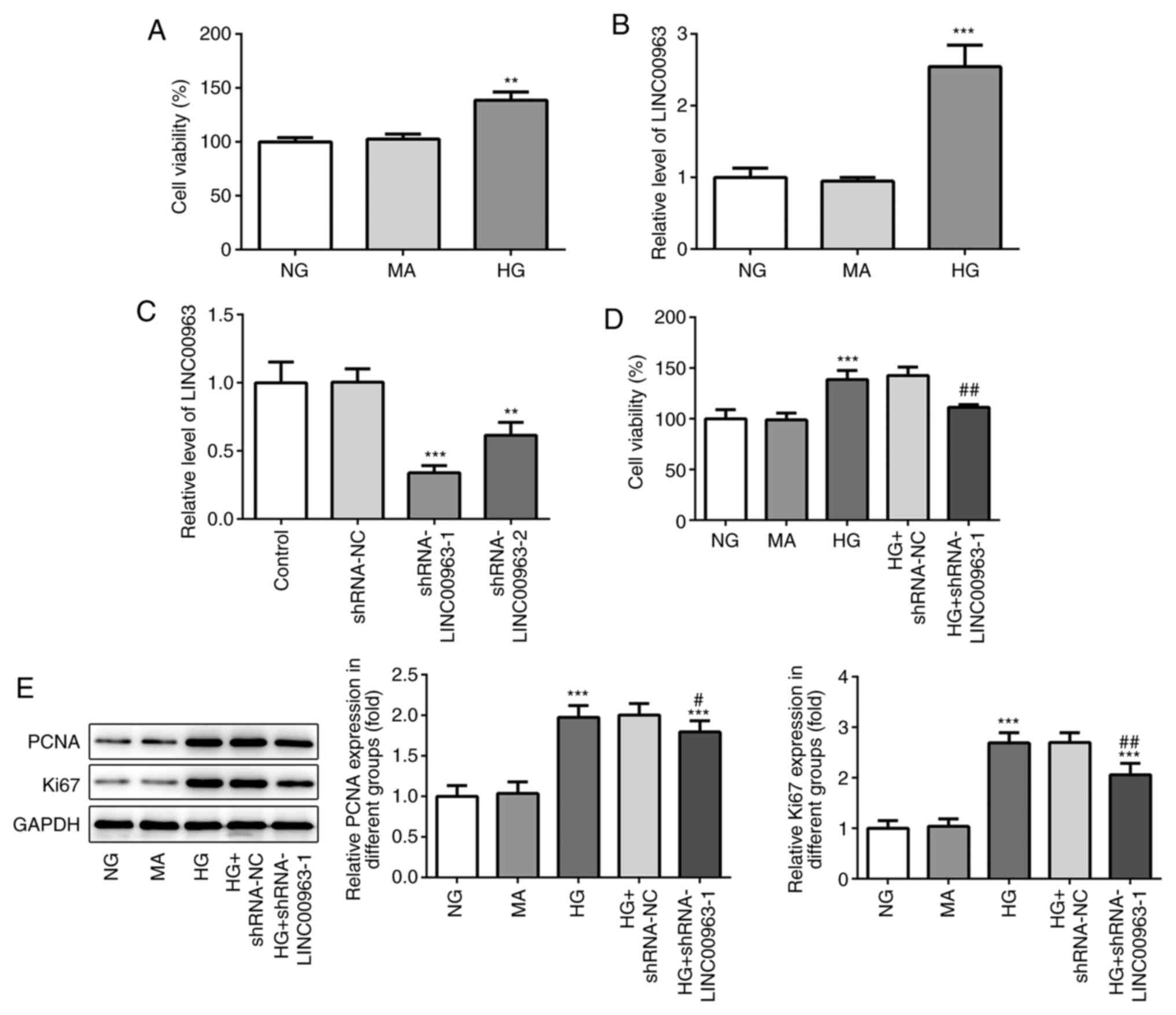
As shown in Fig. 1A,
cell viability was significantly higher in the HG group compared
with that in the NG group. The expression levels of LINC00963 were
evaluated in HRECs treated with high and low concentrations of
glucose and mannitol (Fig. 1B).
Compared with the NG group, the expression levels of LINC00963 were
found to be significantly higher in the HG group.
Effect of LINC00963 knockdown on
HRECs
As shown in Fig. 1C,
transfection of HRECs with shRNA-LINC00963-1 silenced LINC00963
expression more efficiently compared with shRNA-LINC00963-2 (0.33
vs. 0.62, respectively). Therefore, shRNA-LINC00963-1 was utilized
for subsequent experiments. The cell viability in the HG +
shRNA-LINC00963-1 group was notably attenuated compared with the HG
group (Fig. 1D). Furthermore, the
protein expression levels of proliferating cell nuclear antigen
(PCNA) and Ki67 were no significantly different between the NG and
MA groups (Fig. 1E). In addition,
the expression of PCNA and Ki67 was upregulated in the HG group
compared with the NG and MA groups, and it was downregulated in the
HG + shRNA-LINC00963-1 group compared with the HG group (Fig. 1E).
LINC00963 knockdown attenuates the
migration and invasion ability of HRECs
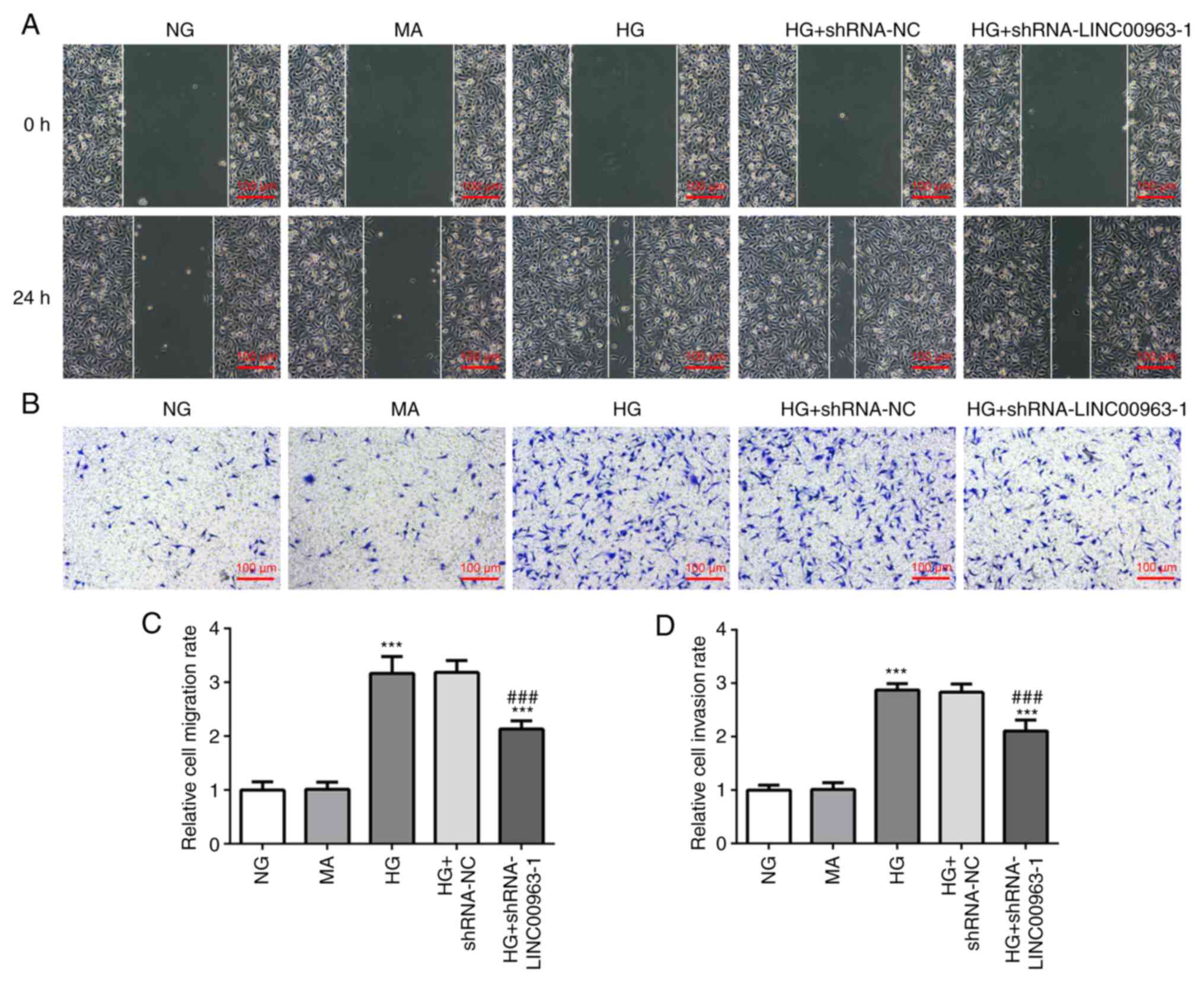
Following incubation for 24 h, the scratches in the
cell monolayer were wider in the NG and MA groups compared with
those in the HG group (Fig. 2A),
indicating that the cell migration ability was enhanced in a
high-glucose environment. Furthermore, compared with the HG group,
the scratches were wider in the HG + shRNA-LINC00963-1 group,
suggesting that LINC00963 knockdown could attenuate the migration
and invasion ability of HRECs. It was also shown that transfection
with shRNA-LINC00963-1 decreased the migration ability of HRECs
(Fig. 2B).
LINC00963 directly targets
miR-27b
To reveal the molecular mechanisms underlying the
role of LINC00963 in HRECs, the ENCORI database (starBase v2.0,
http://starbase.sysu.edu.cn) was
utilized. The analysis predicted that LINC00963 could directly
target miR-27b (Fig. 3A). miR-27b
has been reported to be a tumor suppressor gene. miR-27b can
inhibit epithelial-mesenchymal transition andinhibit the
proliferation and migration of oral squamous cell carcinoma by
targeting integrin subunit α5(11).
In addition, miR-27b inhibited the growth and metastatic behavior
of ovarian cancer cells by targeting C-X-C motif chemokine ligand
1(12). In the present study, the
association between LINC00963 and miR-27b was confirmed by
dual-luciferase reporter assays, demonstrating that co-transfection
of HRECs with WT LINC00963-Luc and miR-27b mimics significantly
reduced the relative luciferase activity of LINC00963-Luc. In
addition, cell viability was enhanced in the HG + shRNA-LINC00963-1
+ miR-27b inhibitor group compared with the HG + shRNA-LINC00963-1
group. Additionally, the expression levels of PCNA and Ki67 were
upregulated in the HG + shRNA-LINC00963-1 + miR-27b inhibitor group
compared with those in the HG + shRNA-LINC00963-1 group. The
aforementioned findings indicated that miR-27b inhibited the
proliferation of HRECs (Fig. 3B-H).
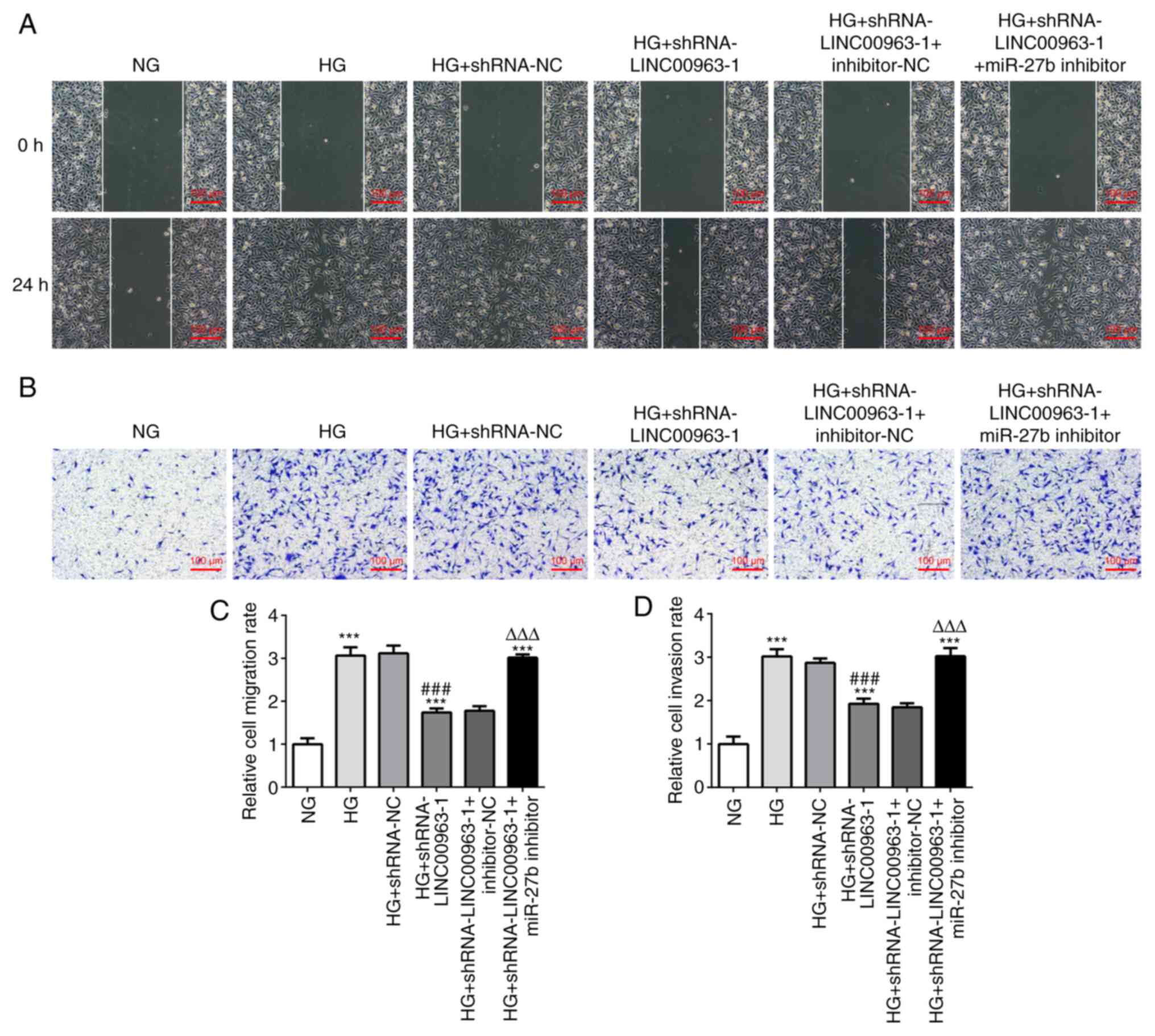
As shown in Fig. 4, miR-27b
attenuated the wound healing and migration rate of HRECs.
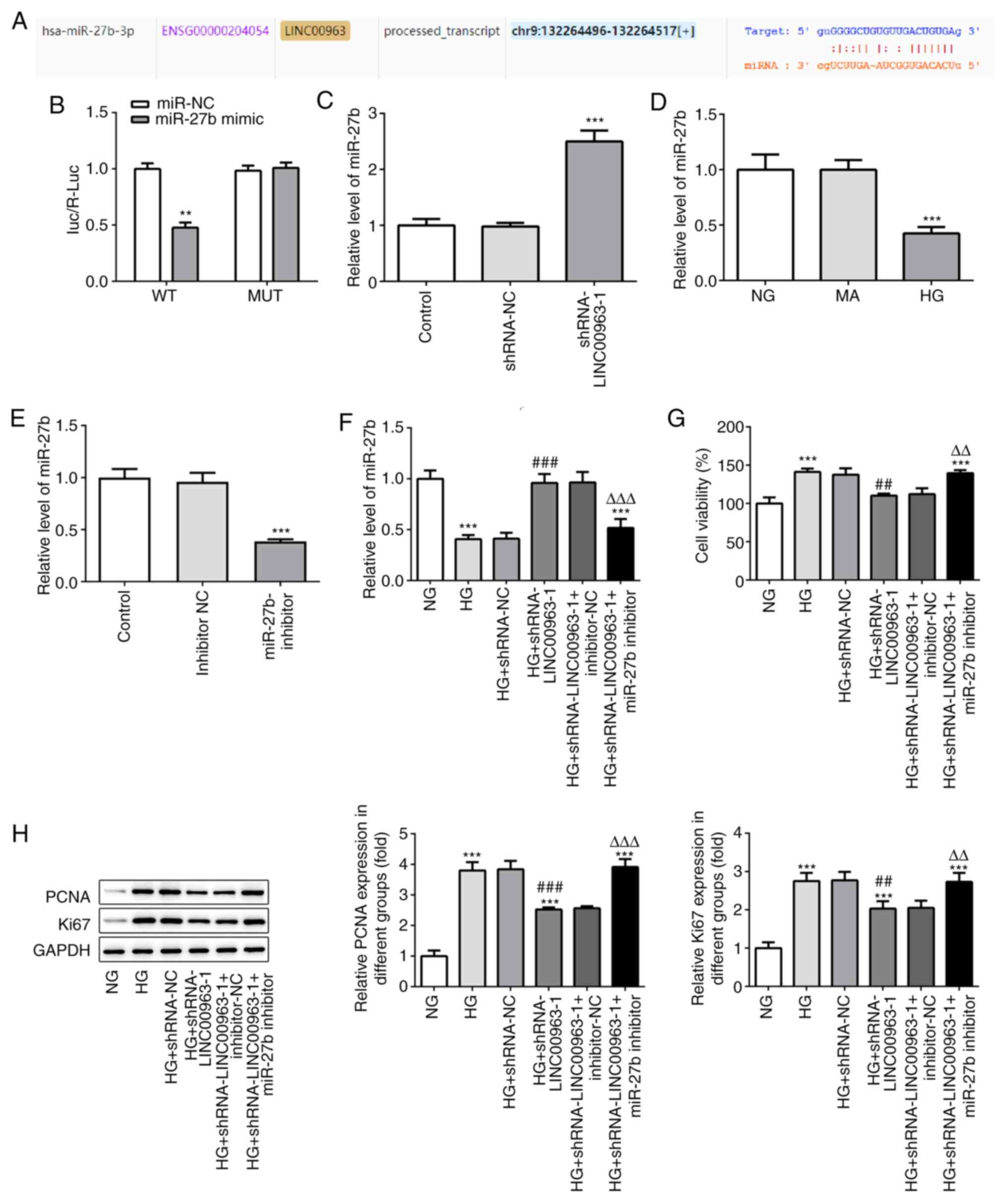 | Figure 3LINC00963 directly targets miR-27b.
(A) Complementary binding site between the 3'-untranslated region
of LINC00963 and miR-27b was predicted using the StarBase database.
(B) Dual-luciferase reporter assay was performed to evaluate the
association between LINC00963 and miR-27b. **P<0.01
vs. miR-NC. (C) RT-qPCR was used to determine the relative
expression levels of miR-27b in HRECs following LINC00963
knockdown. (D) RT-qPCR was used to determine the relative
expression levels of miR-27b in HRECs treated with different
concentrations of glucose. (E) RT-qPCR analysis revealed that the
miR-27b expression levels were downregulated in HRECs following
transfection with miR-27b inhibitor. (F) RT-qPCR was used to
determine the relative expression levels of miR-27b following HG,
HG + shRNA-NC, HG + shRNA-LINC00963-1, HG + shRNA-LINC00963-1 +
inhibitor-NC and HG + shRNA-LINC00963-1 + miR-27b inhibitor. (G)
HREC viability following transfection with miR-27b inhibitor. (H)
Protein expression levels of PCNA and Ki67. All experiments were
repeated three times. ***P<0.001 vs. NG group;
##P<0.01, ###P<0.001 vs. HG group;
ΔΔP<0.01, ΔΔΔP<0.001 vs. HG +
shRNA-LINC00963-1 group. LINC00963, long intergenic
non-protein-coding RNA 963; HRECs, human retinal capillary
endothelial cells; PCNA, proliferating cell nuclear antigen; NG
group, normal glucose group; HG group, high glucose group; MA,
mannitol group; RT-qPCR, reverse transcription-quantitative PCR;
miR, microRNA; shRNA, short hairpin RNA; NC, negative control. |
Discussion
Diabetes, which is caused by the abnormal secretion
and/or inability of the body to use insulin, is one of the most
common non-communicable diseases in clinical practice (13,14).
In 2017, ~425 million individuals, aged 20-79 years, were diagnosed
with diabetes worldwide. This number is estimated to increase to
629 million by 2045(15).
Clinically, diabetes progresses slowly over several years and is
accompanied by several complications. Targeted nursing, including
proper control of patients' blood glucose and real-time monitoring
of physical data, may be effective in improving the cure rate of
patients with diabetes (16,17).
DR is one of the most common complications of diabetes.
Hyperglycemia can damage the retinal vascular system and
endothelial cells and thicken the basement membrane (18,19).
In the present study, the viability of HRECs in a high-glucose
environment was determined by a CCK-8 assay. It was demonstrated
that cell viability was higher in the HG group compared with the NG
group, suggesting that the high-glucose environment may enhance the
viability of HRECs.
LINC00963 is non-coding RNA (20) that is considered to act as a
regulatory factor of cell apoptosis (21). LINC00963 has recently been reported
to promote breast and hepatocellular carcinogenesis (22,23).
Its expression is upregulated in several types of cancer, such as
melanoma and bladder cancer (24,25).
However, the association between LINC00963 and diabetes has not
been reported to date. Thus, the expression of LINC00963 was
measured by RT-qPCR in the present study, and the results
demonstrated that its expression was upregulated under high-glucose
conditions. It has been reported that PCNA, an essential protein
for nuclear DNA synthesis, is widely involved in cell proliferation
and invasion (26,27). In addition, Ki67, a nuclear antigen
protein involved in cell proliferation (28), is closely associated with cell
mitosis, and its expression reflects the normal proliferative
activity of cells (29). Western
blot analysis revealed that, compared with the NG group, the
expression levels of PCNA and Ki67 were elevated in the HG group,
suggesting that cells in the HG group exhibited strong
proliferation ability. However, downregulation of PCNA and Ki67,
mediated by LINC00963 silencing, indicated that LINC00963
downregulation may inhibit cell proliferation. Next, migration and
invasion experiments were performed, and the results demonstrated
that treatment of HRECs with shRNA-LINC00963-1 attenuated cell
migration, indicating that LINC00963 may promote cell
migration.
Subsequently, bioinformatics analysis using the
ENCORI database predicted that miR-27b could directly interact with
the LINC00963 3'-UTR. miR-27 has been proven to play contradictory
roles in different diseases. miR-27b was reported to act as a tumor
suppressor in gastric cancer, exhibiting reduced expression in
human gastric cancer tissues and cells (30). It has been demonstrated that miR-27b
promotes fibroblast-like synovial cell apoptosis (18), as well as endothelial and glioma
cell proliferation (31). In the
present study, the cell proliferation and migration rates, and the
protein expression levels of PCNA and Ki67 were increased in the
miR-27b inhibitor group, suggesting that miR-27b may attenuate cell
proliferation. Furthermore, miR-27b inhibitor reversed the
inhibitory effect of LINC00963 on cell migration in response to
high-glucose stimulation.
In summary, LINC00963 may regulate the proliferation
and apoptosis of HERCs via targeting miR-27b. The findings of the
present study may contribute to the clinical diagnosis of DR and
provide a theoretical basis for research into effective drugs for
the treatment of this condition. However, there were certain
limitations to the present study. First, no in vivo
experiments were performed. Second, the molecular mechanism
underlying the effects of LINC00963 silencing on miR-27b expression
and HREC function was not fully investigated. Therefore, these
issues require further in-depth research and will be addressed in
future studies.
Acknowledgements
Not applicable.
Funding
Funding: No funding was received.
Availability of data and materials
The datasets used and/or analyzed during the current
study are available from the corresponding author on reasonable
request.
Authors' contributions
RZ and CN designed the study and wrote the
manuscript; YG performed the experiments; JW collected and analyzed
the data; LH interpreted the data. RZ and LH can confirm the
authenticity of the raw data. All the authors have read and
approved the final manuscript. All authors confirm the authenticity
of the raw data.
Ethics approval and consent to
participate
Not applicable.
Patient consent for publication
Not applicable.
Competing interests
The authors declare that they have no competing
interests.
References
|
1
|
Dall TM, Yang W, Halder P, Pang B,
Massoudi M, Wintfeld N, Semilla AP, Franz J and Hogan PF: The
economic burden of elevated blood glucose levels in 2012: Diagnosed
and undiagnosed diabetes, gestational diabetes mellitus, and
prediabetes. Diabetes Care. 37:3172–3179. 2014.PubMed/NCBI View Article : Google Scholar
|
|
2
|
Patterson CC, Karuranga S, Salpea P,
Saeedi P, Dahlquist G, Soltesz G and Ogle GD: Worldwide estimates
of incidence, prevalence and mortality of type 1 diabetes in
children and adolescents: Results from the International diabetes
federation diabetes atlas, 9th edition. Diabetes Res Clin Pract.
157(107842)2019.PubMed/NCBI View Article : Google Scholar
|
|
3
|
Yau JW, Rogers SL, Kawasaki R, Lamoureux
EL, Kowalski JW, Bek T, Chen SJ, Dekker JM, Fletcher A, Grauslund
J, et al: Global prevalence and major risk factors of diabetic
retinopathy. Diabetes Care. 35:556–564. 2012.PubMed/NCBI View Article : Google Scholar
|
|
4
|
Savage SR, McCollum GW, Yang R and Penn
JS: RNA-seq identifies a role for the PPARβ/δ inverse agonist
GSK0660 in the regulation of TNFα-induced cytokine signaling in
retinal endothelial cells. Mol Vis. 21:568–576. 2015.PubMed/NCBI
|
|
5
|
Quinn JJ and Chang HY: Unique features of
long non-coding RNA biogenesis and function. Nat Rev Genet.
17:47–62. 2016.PubMed/NCBI View Article : Google Scholar
|
|
6
|
Pan C, Wang X, Shi K, Zheng Y, Li J, Chen
Y, Jin L and Pan Z: MiR-122 reverses the doxorubicin-resistance in
hepatocellular carcinoma cells through regulating the tumor
metabolism. PLoS One. 11(e0152090)2016.PubMed/NCBI View Article : Google Scholar
|
|
7
|
Reichholf B, Herzog VA, Fasching N,
Manzenreither RA, Sowemimo I and Ameres SL: Time-resolved small RNA
sequencing unravels the molecular principles of microRNA
homeostasis. Mol Cell. 75:756–768.e7. 2019.PubMed/NCBI View Article : Google Scholar
|
|
8
|
Ruiz MA, Feng B and Chakrabarti S:
Polycomb repressive complex 2 regulates MiR-200b in retinal
endothelial cells: Potential relevance in diabetic retinopathy.
PLoS One. 10(e0123987)2015.PubMed/NCBI View Article : Google Scholar
|
|
9
|
Ding L, Ni J, Yang F, Huang L, Deng H, Wu
Y, Ding X and Tang J: Promising therapeutic role of miR-27b in
tumor. Tumour Biol. 39(1010428317691657)2017.PubMed/NCBI View Article : Google Scholar
|
|
10
|
Livak KJ and Schmittgen TD: Analysis of
relative gene expression data using real-time quantitative PCR and
the 2(-Delta Delta C(T)) method. Methods. 25:402–408.
2001.PubMed/NCBI View Article : Google Scholar
|
|
11
|
Li T, Wu Q, Liu D and Wang X: MiR-27b
Suppresses tongue squamous cell carcinoma epithelial-mesenchymal
transition by targeting ITGA5. Onco Targets Ther. 13:11855–11867.
2020.PubMed/NCBI View Article : Google Scholar
|
|
12
|
Liu CH, Jing XN, Liu XL, Qin SY, Liu MW
and Hou CH: Tumor-suppressor miRNA-27b-5p regulates the growth and
metastatic behaviors of ovarian carcinoma cells by targeting CXCL1.
J Ovarian Res. 13(92)2020.PubMed/NCBI View Article : Google Scholar
|
|
13
|
Murai N, Saiki R, Hashizume M, Kigawa Y,
Koizumi G, Tadokoro R, Endo K, Iizaka T, Otsuka F, Izumizaki M and
Nagasaka S: Alcohol flushing is independently associated with
lesser degree of carotid atherosclerosis in Japanese type 2
diabetic patients. Diabetol Int. 9:68–74. 2017.PubMed/NCBI View Article : Google Scholar
|
|
14
|
Chong PL, Pisharam J, Abdullah A and Chong
VH: Gestational diabetes insipidus. QJM. 112:123–124.
2019.PubMed/NCBI View Article : Google Scholar
|
|
15
|
Wong TY and Sabanayagam C: The war on
diabetic retinopathy: Where are we now? Asia Pac J Ophthalmol
(Phila). 8:448–456. 2019.PubMed/NCBI View Article : Google Scholar
|
|
16
|
Wilson M, Chen HS and Wood M: Impact of
nurse champion on quality of care and outcomes in type 2 diabetes
patients. Int J Evid Based Healthc. 17:3–13. 2019.PubMed/NCBI View Article : Google Scholar
|
|
17
|
Allen D: The nurse's role in childhood
diabetes. Nurs Child Young People. 28(11)2016.PubMed/NCBI View Article : Google Scholar
|
|
18
|
Mohammad HMF, Sami MM, Makary S, Toraih
EA, Mohamed AO and El-Ghaiesh SH: Neuroprotective effect of
levetiracetam in mouse diabetic retinopathy: Effect on glucose
transporter-1 and GAP43 expression. Life Sci.
232(116588)2019.PubMed/NCBI View Article : Google Scholar
|
|
19
|
Clore JN and Thurby-Hay L:
Glucocorticoid-induced hyperglycemia. Endocr Pract. 15:469–474.
2009.PubMed/NCBI View Article : Google Scholar
|
|
20
|
Xie LB, Chen B, Liao X, Chen YF, Yang R,
He SR, Pei LJ and Jiang R: LINC00963 targeting miR-128-3p promotes
acute kidney injury process by activating JAK2/STAT1 pathway. J
Cell Mol Med. 24:5555–5564. 2020.PubMed/NCBI View Article : Google Scholar
|
|
21
|
Wang L, Han S, Jin G, Zhou X, Li M, Ying
X, Wang L, Wu H and Zhu Q: Linc00963: A novel, long non-coding RNA
involved in the transition of prostate cancer from
androgen-dependence to androgen-independence. Int J Oncol.
44:2041–2049. 2014.PubMed/NCBI View Article : Google Scholar
|
|
22
|
Zhang N, Zeng X, Sun C, Guo H, Wang T, Wei
L, Zhang Y, Zhao J and Ma X: LncRNA LINC00963 promotes
tumorigenesis and radioresistance in breast cancer by sponging
miR-324-3p and inducing ACK1 expression. Mol Ther Nucleic Acids.
18:871–881. 2019.PubMed/NCBI View Article : Google Scholar
|
|
23
|
Wu JH, Tian XY, An QM, Guan XY and Hao CY:
LINC00963 promotes hepatocellular carcinoma progression by
activating PI3K/AKT pathway. Eur Rev Med Pharmacol Sci.
22:1645–1652. 2018.PubMed/NCBI View Article : Google Scholar
|
|
24
|
Jiao H, Jiang S, Wang H, Li Y and Zhang W:
Upregulation of LINC00963 facilitates melanoma progression through
miR-608/NACC1 pathway and predicts poor prognosis. Biochem Biophys
Res Commun. 504:34–39. 2018.PubMed/NCBI View Article : Google Scholar
|
|
25
|
Zhou N, Zhu X and Man L: LINC00963
functions as an oncogene in bladder cancer by regulating the
miR-766-3p/MTA1 axis. Cancer Manag Res. 12:3353–3361.
2020.PubMed/NCBI View Article : Google Scholar
|
|
26
|
Chatzileontiadou DSM, Samiotaki M,
Alexopoulou AN, Cotsiki M, Panayotou G, Stamatiadi M, Balatsos NAA,
Leonidas DD and Kontou M: Proteomic analysis of human angiogenin
interactions reveals cytoplasmic PCNA as a putative binding
partner. J Proteome Res. 16:3606–3622. 2017.PubMed/NCBI View Article : Google Scholar
|
|
27
|
Wang X, Wang D, Yuan N, Liu F, Wang F,
Wang B and Zhou D: The prognostic value of PCNA expression in
patients with osteosarcoma: A meta-analysis of 16 studies. Medicine
(Baltimore). 96(e8254)2017.PubMed/NCBI View Article : Google Scholar
|
|
28
|
Rahmanzadeh R, Huttmann G, Gerdes J and
Scholzen T: Chromophore-assisted light inactivation of pKi-67 leads
to inhibition of ribosomal RNA synthesis. Cell Prolif. 40:422–430.
2007.PubMed/NCBI View Article : Google Scholar
|
|
29
|
Scholzen T and Gerdes J: The Ki-67
protein: From the known and the unknown. J Cell Physiol.
182:311–322. 2000.PubMed/NCBI View Article : Google Scholar
|
|
30
|
Feng Q, Wu X, Li F, Ning B, Lu X, Zhang Y,
Pan Y and Guan W: MiR-27b inhibits gastric cancer metastasis by
targeting NR2F2. Protein Cell. 8:114–122. 2017.PubMed/NCBI View Article : Google Scholar
|
|
31
|
Urbich C, Kaluza D, Fromel T, Knau A,
Bennewitz K, Boon RA, Bonauer A, Doebele C, Boeckel JN,
Hergenreider E, et al: MicroRNA-27a/b controls endothelial cell
repulsion and angiogenesis by targeting semaphorin 6A. Blood.
119:1607–1616. 2012.PubMed/NCBI View Article : Google Scholar
|