Introduction
The ophthalmic artery (OA) is the first intracranial
collateral branch of the internal carotid artery (ICA). It
originates immediately after the emergence of the ICA from the
cavernous sinus, features a short intracranial course, follows the
optic canal, and enters the orbit (1). After entering the orbit it branches
out in a complex pattern and vascularizes the eyeball and
surrounding ophthalmic tissues, contributing indirectly to the
neutralization of the toxic particles resulting from a high degree
of exposure of the eye to external factors (2,3). The
ocular branches provide the blood supply to the optical apparatus
and the orbital branches supply the ocular adnexa (4,5).
According to the Bouthillier classification system,
the OA is the ICA's fifth branch and belongs to its sixth segment.
The ICA is divided into seven segments: cervical, petrous
(emergence of caroticotympanic and vidian arteries), lacerum,
cavernous (that provides the meningohypophyseal and inferolateral
trunks), clinoid, ophthalmic (that gives rise to the OA and the
superior hypophyseal artery), and communicating segment where
before its final division, the ICA leads to the anterior choroidal
and posterior communicating arteries (6).
The first orbital dissection was performed in the
19th century, in particular with the works of Meyer (7) who was the first anatomist who, based
on macroscopic cadaveric dissection, assessed the normal orbital
vascularization and described the anatomy and branching of the
OA.
Due to the significance of OA, numerous authors have
studied its distinct features, such as the caliber, course
variants, distance between its entrance into the optic canal, and
eyeball. An exhaustive study of the literature reveals a solid
number of views held by various researchers, the OA being primarily
studied on cadavers (1,4,8,9) and
classic angiographic studies (10,11),
in line with the tendency to study even smaller caliber vessels
(12-14).
Although digital subtraction angiography (DSA)
remains the gold standard for visualizing smaller vessels, it is an
invasive diagnostic method with significant morbidity represented
mainly by ischemic stroke due to the embolic phenomenon, especially
in older patients with atherosclerotic vessels. Neurological
complications may be reduced if arteriography were restricted to
patients undergoing surgery (e.g., carotid endarterectomy) or if
diagnostic angiography were replaced with a reliable non-invasive
imaging modality (15).
The growing clinical applications of computed
tomography angiographic (CTA) can be attributed to the development
of helical CT scanners with a larger number of detector rows. The
increase in the number of image sections per study allows for the
description of complex vascular axes with small branches and
correct analysis of nearby soft tissues (16,17).
The aim of the present study was to accurately
measure the diameter of the OA and investigate whether bilateral
variations in diameter can be recorded in relation to patient age
and sex. The result showed that variations in OA and ICA caliber
were identified for both sex and age.
Patients and methods
Patients and criteria
Α retrospective study was conducted between
01.03.2017 and 01.03.2019, comprising 80 CTA investigations from
patients aged between 22 and 80 (40 male and 40 female), totaling a
number of 160 arteries. The patients were submitted to a CTA
evaluation of the carotid artery at the attending physician's
indication for various assessments in the context of their
underlying disease. Prior informed consent was obtained from the
patients. The research was conducted in accordance with the
Helsinki Declaration and in compliance with all relevant national
regulations regarding patient studies (approved by the Ethics
Committee of ‘Sf. Apostol Andrei’ County Hospital,
34490/08.08.2019).
The inclusion criteria for the study were: CT
confirmation of the ICA normal anatomy, more precisely, the absence
of atheroma plaques or congenital or tumoral vascular pathologies
affecting the carotid vascular axis, as well as the lack of carotid
aneurysms, changes in vascular structure, or neoplasms affecting
the ICA.
Method
CTAs were performed on a multi-slice CT scanner with
64 detectors (Light Speed VCT; GE Medical Systems). The actual scan
was augmented by automatic intravenous injection of iodinated
contrast material at a dose of 1 ml/kg body weight at an injection
rate of 4 ml/sec. The scan was started individually by tracking the
loading bolus, with a region of interest placed at the aortic arch
level and with automatic triggering at detected values of over 120
Hounsfield units. The obtained images were sent to a dedicated
workstation (Advantage Workstation, GE Medical Systems) for
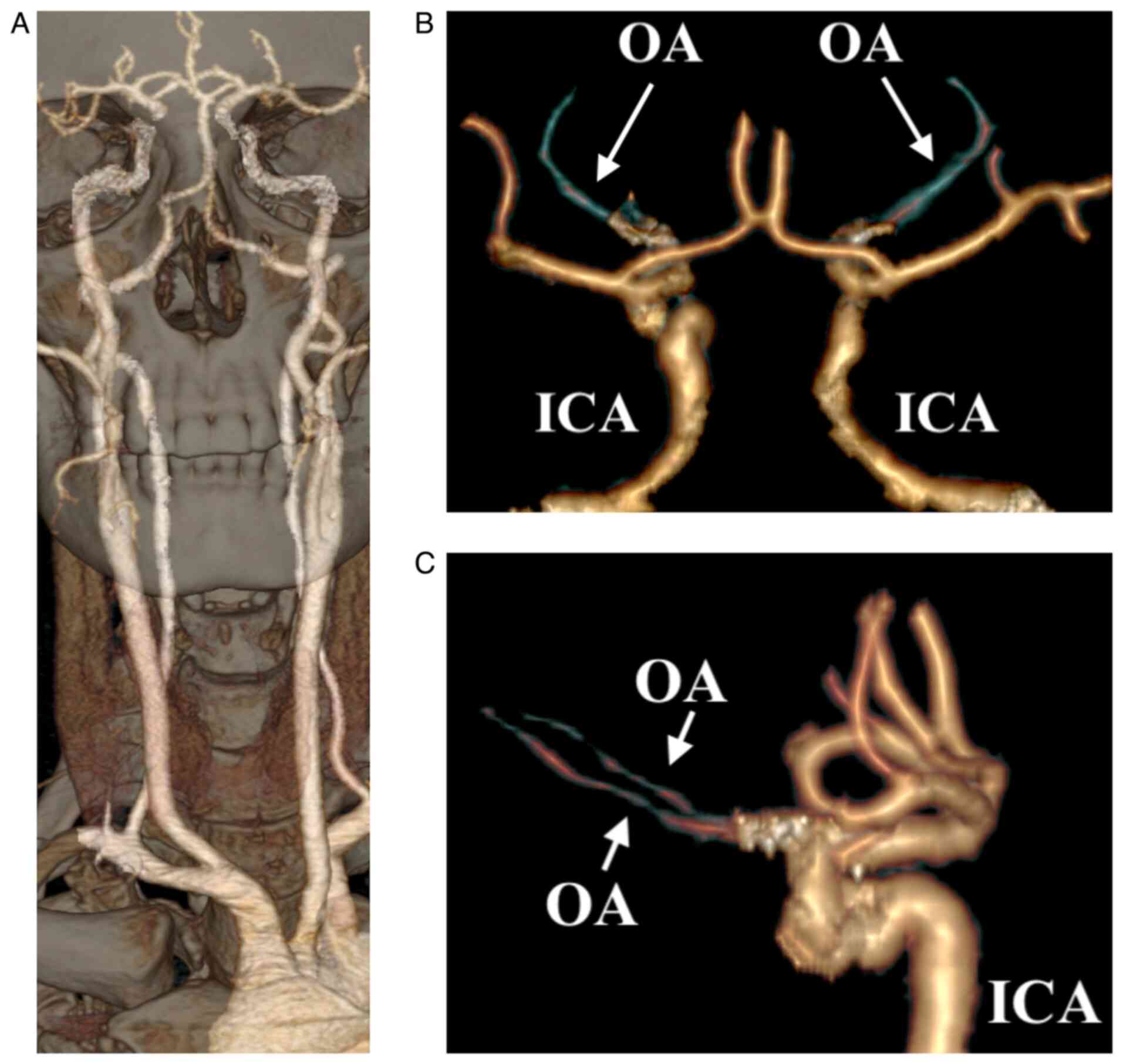
post-processing. From the source images, volume rendering technique
(VRT) reformatted images (Fig. 1)
and multiplanar reconstructions (MPR), respectively, were obtained
in the tri-planar system (axial, sagittal, and coronal), thus
revealing the OA and its origin from the ICA. Maximum intensity
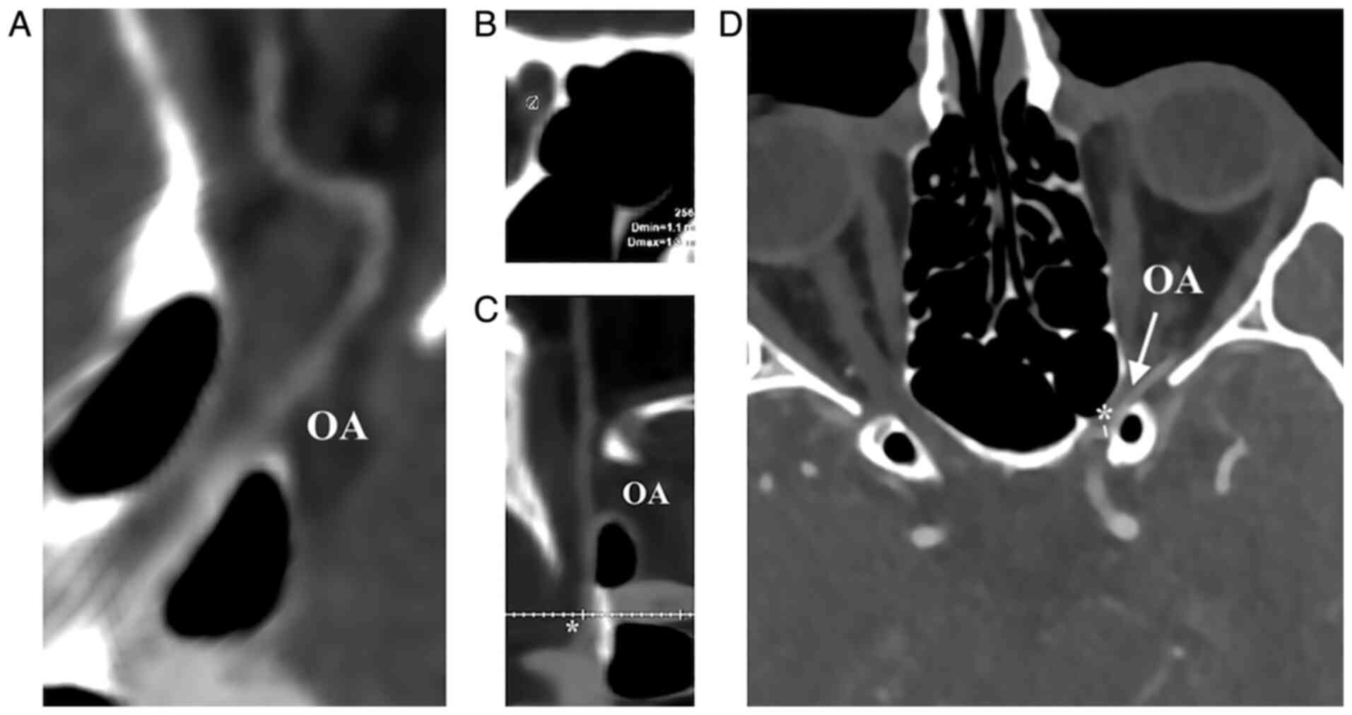
projection (MIP) images were also employed. The OA was measured
semi-automatically at its entrance in the optical canal,
approximately 5 mm from its origin. The ICA was measured with the
same tools caudally and cranially from the OA's origin.
The study aimed to measure the OA diameter
accurately and investigate bilateral variations in diameter related
to age and sex of the patients. The measurement of the OA's
diameter at the point of interest was performed automatically
(Fig. 2).
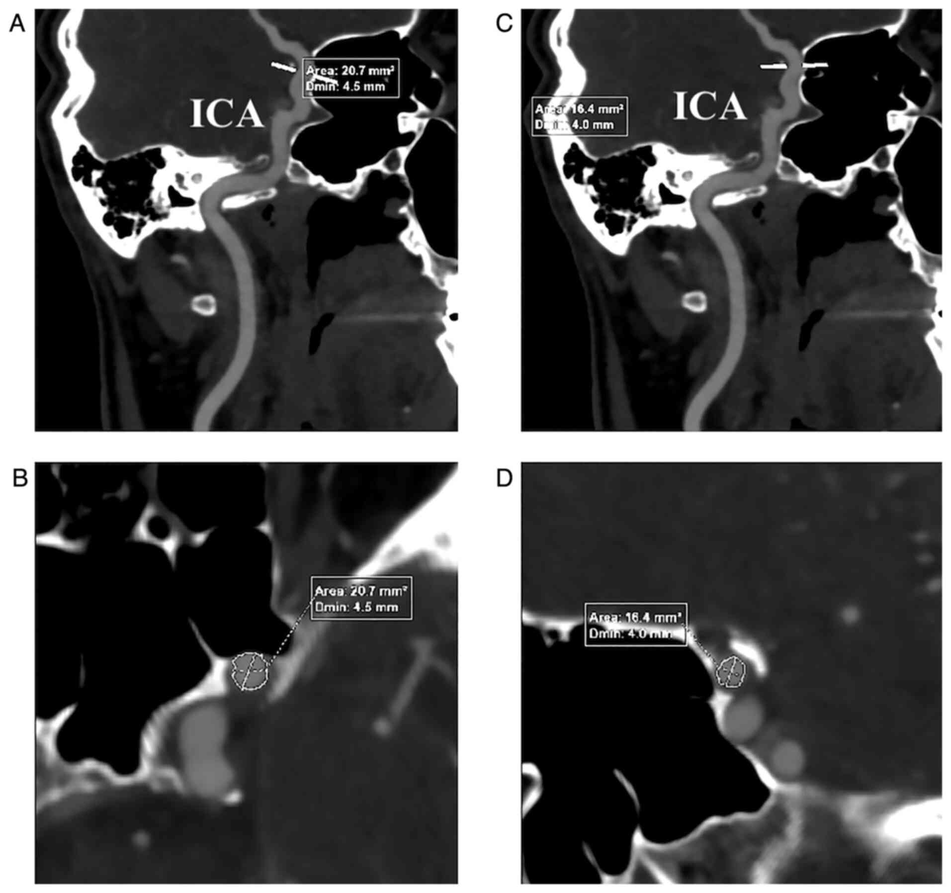
Once the OA was identified and measured, ICA
measurements followed in the pre- and post-emergent portions of the
OA. Oblique MIP images were captured to highlight the ICA regions
of interest, the level at which the measurements were performed
automatically, similar to the previously presented method (Fig. 3).
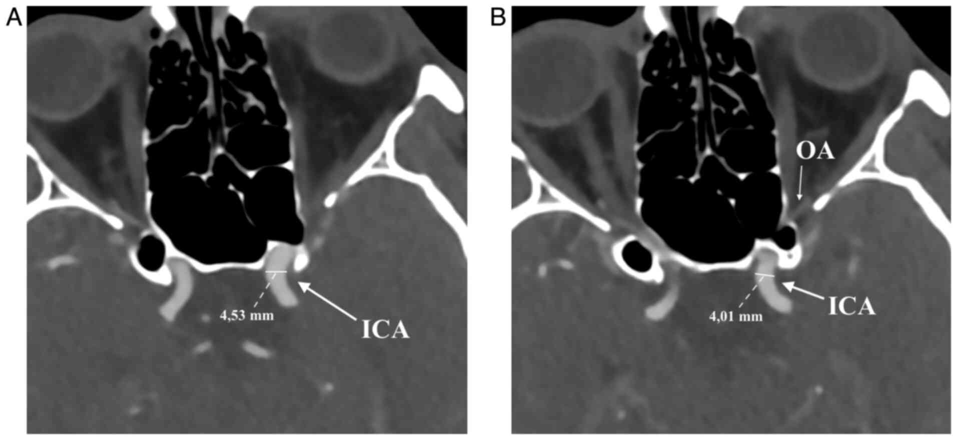
The measurements were also shown axially to have
greater ease in visualizing the regions of interest (Fig. 4). In all cases, the minimum diameter
(Dmin) is presented, according to the automatic measurements.
The influence of age and sex was determined using
the average diameter of the OA (average between the diameter on the
left and right sides). The patients were stratified according to
five age groups: <40 years, 40-49 years, 50-59 years, 60-69
years, and >70 years. The dimensions of the ICA were also
measured before and after the emergence of the OA, and its variable
relation to each part and the sex of the subjects were
recorded.
Statistical analysis
The Student's t-test for two pairs of samples was
used to investigate the differences in right and left diameters, a
Welch t-test was used to determine the influence of sex on the
diameter and to correlate these two factors, the point-biserial
correlation coefficient test was utilized. To investigate the
influence of the age group the ANOVA procedure was used, applying
the Tukey-Kramer post-hoc test. The tests were considered
statistically significant when the P-value was <0.05.
Results
Patients
The study subjects presented normal internal carotid
arteries confirmed by CTA and each OA showed emergence from the
ophthalmic segment of the ipsilateral ICA. CTA images of the ICA
were processed and the OA was identified as originating along the
ICA on MPR reconstructions and MIP images.
From the measurements performed, the results
revealed an average diameter of the OA of 1.38 mm (±0.23), with a
median value of 1.35 mm (Table
I).
 | Table IOA caliber. |
Table I
OA caliber.
| Variable | OA diameter
(mm) | Right OA diameter
(mm) | Left OA diameter
(mm) |
|---|
| Mean ± SD | 1.38±0.23 | 1.38±0.24 | 1.38±0.23 |
| Median (IQR) | 1.35 (0.37) | 1.37 (0.38) | 1.37 (0.34) |
| Min-Max
(range) | 1.02-1.98
(0.96) | 1.01-1.98
(0.97) | 1.03-1.95
(0.92) |
OA caliber and sex
Following analysis of the study data, a slight
sex-related difference was denoted between the diameters of the OA.
In men, the mean average value was 1.43 mm (±0.24), while in women,
the average value was 1.34 mm (±0.20) (Table II). Albeit a difference of
approximately 0.1 mm was recorded in the caliber of the OA between
the two groups; this did not prove to be statistically significant
(P>0.05, Table III).
| Table IIOA caliber by sex. |
Table II
OA caliber by sex.
| OA diameter
(mm) | Females (n=40) | Males (n=40) |
|---|
| Mean ± SD | 1.34±0.20 | 1.43±0.24 |
| Median (IQR) | 1.32 (0.31) | 1.37 (0.36) |
| Min-Max
(range) | 1.02-1.71
(0.69) | 1.08-1.98
(0.90) |
| Table IIIOA mean difference by sex. |
Table III
OA mean difference by sex.
| Mean F (mm) | Mean M (mm) | P-value | Mean difference
(95% CI) |
|---|
| 1.34 | 1.43 | 0.0719 | -0.09
(-0.18-0.01) |
The dimensional variation of the entire group of
patients in terms of OA diameters relative to the laterality was
analyzed using a bidirectional Welch t-test. The results revealed
no significant difference in our study group. The average value of
the diameter on the right side was 1.38 mm (±0.24) with a median of
1.37 mm, and on the left side, we found a value of 1.38 mm (±0.23)
with a median of 1.37 mm. The values varied between 1.01 and 1.98
mm on the right side, respectively, and 1.03-1.95 mm on the left
side at a P-value of 0.96 (Table
I).
The values of the OA diameters were calculated for
each sex and for each side. In the female group, an average
diameter for the OA on the right side of 1.32 mm (±0.22) was
obtained and on the left side of 1.33 mm (±0.2). By contrast, for
males, the results showed a values for the OA diameter on the right
side of 1.43 mm (±0.24) and the left 1.43 mm (±0.26) (Table IV).
| Table IVOA caliber sorted by side and
sex. |
Table IV
OA caliber sorted by side and
sex.
| | rOA (mm) | lOA (mm) |
|---|
| Male | | |
|
Mean ±
SD | 1.43±0.24 | 1.43±0.26 |
|
Median
(IQR) | 1.44 (0.33) | 1.39 (0.41) |
|
Min-Max
(range) | 1.08-1.98
(0.90) | 1.06-1.95
(0.89) |
| Female | | |
|
Mean ±
SD | 1.32±0.22 | 1.33±0.2 |
|
Median
(IQR) | 1.3 (0.34) | 1.32 (0.3) |
|
Min-Max
(range) | 1.01-1.76
(0.75) | 1.03-1.77
(0.74) |
OA caliber and age
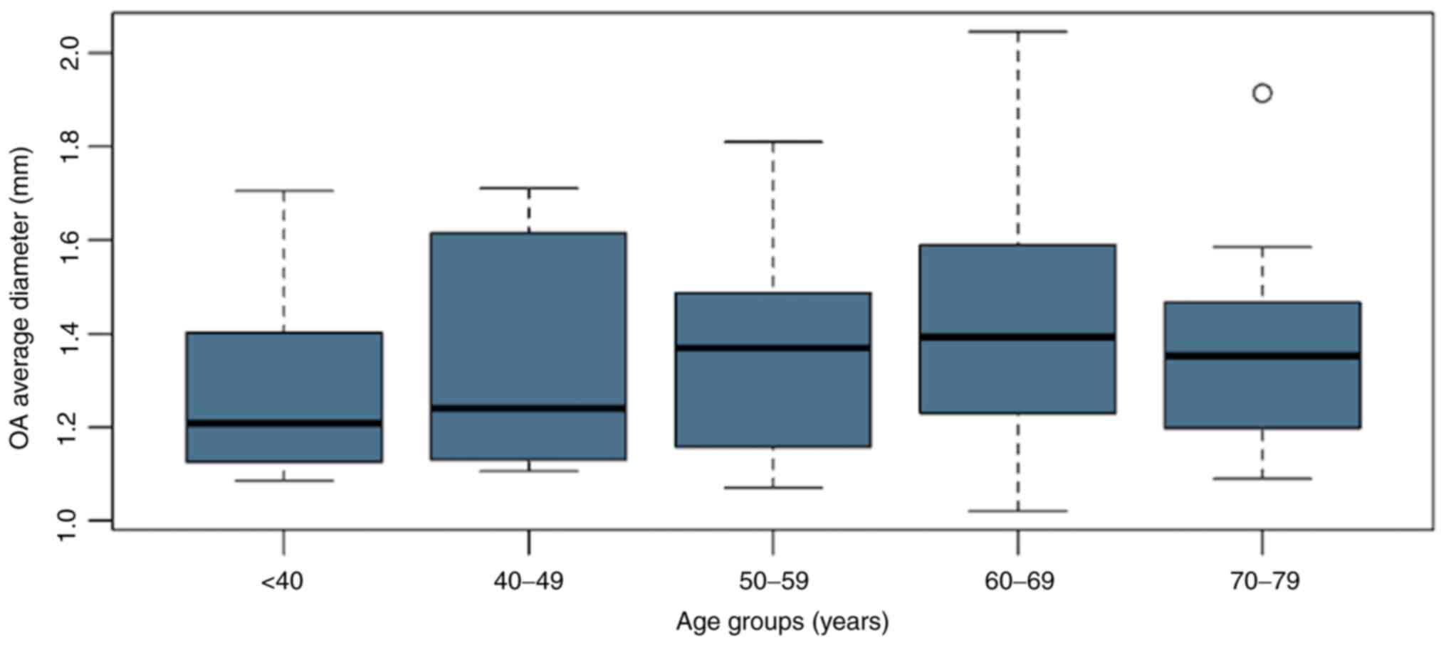
The study group was then subdivided into age groups,
analyzing the average diameter of OA for each subgroup, as follows:
for the subgroup <40 years, a value of 1.28 mm (±0.21) was
obtained, between 40 and 49 years a value of 1.34 mm (±0.26),
between 50 and 59 years a value of 1.37 mm (±0.23), between 60 and
69 years a value of 1.42 mm (±0.23) and in the group 70-79 years a
value of 1.37 mm (±0.23) (Fig.
5).
An ANOVA test was used to verify whether there were
statistically significant differences between age groups. The
results of the ANOVA test showed that the differences were not
statistically significant (F=0.68, degrees of freedom=4, 74;
P=0.6020). The average age was 59.72 years (±0.23) in the studied
group with a median of 63 years and minimum and maximum values
between 22 and 80 years. The global distribution was deviated to
the left, with a negative skewness, and patients under the age of
35 were outliers.
The measurements on the ICA were performed on the
same samples, according to the previously described method.
Diameter variations of the ICA below and above the OA emergence
were analyzed, resulting in an average ICA value below the OA of
4.19 mm (±0.49) with a median of 4.12 mm and, above the OA of 2.84
mm (±0.43) with a median of 2.79 mm, with an average decrease of
the caliber of 1.37 mm (±0.46) with a median of 1.38 mm (±0.675)
(Table V).
| Table VICA caliber before and after OA
origin. |
Table V
ICA caliber before and after OA
origin.
| | ICA below OA
(mm) | ICA above OA
(mm) | Decrease in caliber
(mm) |
|---|
| Mean ± SD | 4.19±0.49 | 2.84±0.43 | 1.37±0.46 |
| Median (IQR) | 4.12 (0.62) | 2.79 (0.66) | 1.38 (0.675) |
| Min-Max
(range) | 2.89-5.41
(2.52) | 2.08-4.05
(1.97) | 0.15-2.52
(2.37) |
Average values
The data analysis on the female patients revealed
the mean average values for the ICA below OA of 4.05 mm (±0.42)
with a median of 3.96 mm, while above the OA the average diameter
was 2.85 mm (±0.4) with a median of 2.81 mm and an average decrease
of the caliber of 1.20 mm. For males, we found mean ICA values
below the OA of an average of 4.35 mm (±0.44) with a median of 4.31
mm and ICA values above the OA of 2.82 mm (±0.46), with a median of
2.79 mm and an average decrease in caliber of 1.53 mm. By comparing
the values, in females, the average ICA measurement below the OA
was lower compared to ICA below the OA in men, but the ICA
measurement above the OA in women had higher minimal values than in
men, thus revealing a less steep narrowing of the ICA (Table VI).
| Table VIICA caliber sorted by side and
sex. |
Table VI
ICA caliber sorted by side and
sex.
| | ICA below OA
(mm) | ICA above OA
(mm) | Decrease in caliber
(mm) |
|---|
| Male average | | | |
|
Mean ±
SD | 4.35±0.44 | 2.82±0.46 | 1.53±(0.43) |
|
Median
(IQR) | 4.31 (0.65) | 2.79 (0.72) | 1.57 (0.53) |
|
Min-Max
(range) | 3.35-5.39
(2.04) | 2.08-4.05
(1.97) | 0.46-2.52
(2.04) |
| Male left side | | | |
|
Mean ±
SD | 4.39±0.5 | 2.80±0.51 | 1.58±0.43 |
|
Median
(IQR) | 4.32 (0.75) | 2.79 (0.78) | 1.59 (0.46) |
|
Min-Max
(range) | 3.56-5.39
(1.83) | 2.08-4.05
(1.97) | 0.67-2.52
(1.85) |
| Male right
side | | | |
|
Mean ±
SD | 4.33±0.38 | 2.84±0.41 | 1.49±0.44 |
|
Median
(IQR) | 4.29 (0.6) | 2.78 (0.63) | 1.57 (0.59) |
|
Min-Max
(range) | 3.35-5.01
(1.66) | 2.25-3.67
(1.42) | 0.46-2.41
(1.95) |
| Female average | | | |
|
Mean ±
SD | 4.05±0.42 | 2.85±0.4 | 1.20±0.43 |
|
Median
(IQR) | 3.96 (0.51) | 2.81 (0.62) | 1.17 (0.57) |
|
Min-Max
(range) | 3.22-5.08
(1.86) | 2.17-3.63
(1.5) | 0.15-2.22
(2.07) |
| Female left
side | | | |
|
Mean ±
SD | 4.11±0.45 | 2.87±0.4 | 1.24±0.41 |
|
Median
(IQR) | 4.04 (0.49) | 2.87 (0.65) | 1.28 (0.55) |
|
Min-Max
(range) | 3.42-5.08
(1.66) | 2.28-3.6
(1.32) | 0.15-2.22
(2.07) |
| Female right
side | | | |
|
Mean ±
SD | 3.98±0.4 | 2.83±0.39 | 1.15±0.45 |
|
Median
(IQR) | 3.91 (0.48) | 2.78 (0.56) | 1.13 (0.66) |
|
Min-Max
(range) | 3.22-4.94
(1.72) | 2.17-3.63
(1.5) | 0.43-1.98
(1.55) |
The group was divided as per sex for a more detailed
study and the ICA diameter below and above the OA emergence was
analyzed according to laterality for each group. In the male group,
on the left side, average dimensional values of ICA below the OA of
4.39 mm (±0.5) with a median value of 4.32 mm were identified and
above the OA of 2.80 mm (±0.51) with a median of 2.79 mm, therefore
noting an average caliber decrease of 1.58 mm. On the right side,
the dimension of ICA below the OA was 4.33 mm (±0.38) with a median
value of 4.29 mm, and the value for ICA above the OA were 2.84 mm
(±0.41) with a median of 2.78 mm, observing a decrease in caliber
of 1.49 mm (Table VI).
Analyzing the values obtained in the male group, on
the left side higher values of the ICA diameter below the OA were
identified, while the ICA values above the OA were higher on the
right side. In the female group, the left side average dimensional
value of the ICA below the OA of 4.11 mm (±0.45) with a median
value of 4.04 mm were observed and the value of the ICA above the
OA of 2.87 mm (±0.4) with a median of 2.87 mm were noted, observing
a decrease in the average caliber of 1.24 mm. Analyzing the average
values for the right side, we found the ICA diameter below the OA
of 3.98 mm (±0.4) with a median value of 3.91 mm and above the OA
of 2.83 mm (±0.39) with a median of 2.78 mm, observing a decrease
in the average caliber of 1.15 mm.
Assessing the values obtained from both groups,
slightly higher values were observed on the left side regarding ICA
diameter below the OA, while ICA values above the OA were higher on
the left side in the female group, whereas for the male group the
dimensions were slightly increased on the right side.
The result of the point biserial correlation
coefficient test (r=0.33) applied to our two variables: sex and
laterality. No significant correlation was found between these two
variables, although slight differences in caliber between sexes and
both sides were evident.
Discussion
The OA arises from the medial ICA of the anterior
clinoid process (18) and in more
than 90% of cases, the OA is the first branch of the intradural C6
segment (19,20). On its anatomical trajectory, it
travels inferior to the optic nerve in the optic canal. In the
orbit, it bypasses the lateral face of the posterior optic nerve of
the ciliary ganglion, then its superior face and has triads between
the superior oblique and medial right muscles, displaying an
anterior trajectory forward under the superior oblique muscle. It
ends in the medial angle of the eye with the dorsal arteries of the
nose and the supratrochlear artery. Anatomic variants are
described, such as emergence from the middle meningeal artery of a
very small percentage, or trajectory that courses inferior to the
optic nerve, in 10% of cases (18).
OA gives rise to multiple fine collateral branches
and these can be grouped into 3 categories: i) arteries of the
first group emerge inferior and external to the optic nerve; ii)
arteries of the second group detach above the optic nerve; iii)
arteries of the third group formed medially by the optic nerve,
along the medial wall of the optic cavity (21).
The OA is a natural source of direct anastomoses
with the external carotid artery (ECA) through its multiple
collateral vessels (22,23). Known anastomoses between branches of
OA and the external carotid artery are found between the lacrimal
artery and the anterior branch of the middle meningeal artery,
progressing through the superior orbital fissure or the
tympanomeningeal fissure (24,25).
Additionally, there are more anastomoses between the lacrimal
artery and the deep temporal, transverse facial, and
orbitozygomatic infraorbital arteries (26,27).
Other ICA-ECA links are known to be found between the dorsal nasal
artery and the facial artery or infraorbital artery and, rarely,
between the supratrochlear branches and the frontal branches of the
superficial temporal artery (22,27).
Thorough knowledge of the microsurgical anatomy of
the OA, the appearance of its branches, and its anastomoses with
the carotid system are critical to proceed with safe and effective
endovascular and other surgical procedures involving this vascular
area (22,25). In the current study, an OA diameter
was identified that varies between 1.02 and 1.98 mm, with an
average of 1.38 mm, while other specialized studies
(angiographic/cadaveric) reported variability of the OA diameter
between 0.7 and 1.4 mm (1), an
average value of its original diameter of 1.6 mm, with a range of
1.5-1.8 mm (25), or a mean average
of 1.50 mm (28,29).
Regarding the analysis of the sex-related results of
the present study, a slight difference between the OA dimensions
was recorded, namely the OA diameter in women was smaller than that
in men. The data, supported by specialized studies where the
numerical values vary, but the ratio is maintained, showed a
difference in size, in favor of males. Literature studies have
reported OA values for males of 1.54 mm (9) or 1.37 mm (28), while for females, the values reach
1.31 mm (9) and 1.35 mm,
respectively (30).
Results similar to the current study regarding
sex-related differences of the average volumes of the OA caliber
were also reported by other studies that also admit that there is
no statistically significant difference between OA diameters
depending on sex, despite minor variations in caliber (30).
We did not find a certain dominant side regarding
the OA caliber, as values between the left side and the right side
varied by less than 0.1 mm, as indicated by other authors (29,31).
The proximal and distal ICA caliber of the OA
emergence was measured, with values of 4.19 and 2.84 mm,
respectively, and caliber variations of 1.37 mm. Our obtained mean
values are approximately 2 mm smaller than those found by
Jiménez-Castellanos et al in their cadaveric study,
reporting values of ICA diameter below OA origin at 7.01±0.92 mm,
and of 5.19±1.08 mm above the OA origin (29).
Regarding the variations of the ICA diameter
above/below the OA emergence, in the present study values of
0.15-2.52 mm were found, with an average of 1.37±0.46 mm.
Sudakevitch emphasized in his study the idea that the carotid
artery suffers a steep narrowing after the emergence of the OA,
ranging between 2 and 3 mm but not more than 4 mm (8). In addition, other specialized studies
found a narrowing of their caliber between 1 and 3 mm (3) or 1.6 mm (32).
Comparing the ICA diameter between males and
females, a higher value in men was found, with a caliber narrowing
of 1.53 mm in men compared to 1.2 mm in women after the emergence
of OA, the results correlating with specialized studies that also
report larger sizes of the ICA in men (33-35).
However, the present study has some limitations.
Besides the relatively low number of studies, only patients with a
normal carotid axis were included in the study as pre-existent
systemic diseases of the included patients were unknown and could
impact the vascular lumen (e.g., diabetes). Another significant
limitation of this study is the origin of the OA; we only included
patients with the OA originating from ICA's ophthalmic segment,
while different origins of the OA have been described, such as the
intracavernous origin (36,37), the double origin from the ICA
(26,38), and middle meningeal artery origin
(1,39), or, in some cases, with emergence
from ICA but not from its C6 segment (40-42)
and even from the anterior cerebral artery (43,44).
The diameter of OA and its relation to the ICA have
rarely been studied in detail on CTAs, and data on this topic are
scarce. Thus, the subject required thorough investigation. We aimed
to analyze morphometric features of the OA, as it is the main
vessel that supplies oxygenated blood to the eye. The study
suggests obtaining further information on the topic aiming to
improve and contribute to the knowledge of the diameters of OA and
ICA above/below the emergence of the OA, data that have an impact
in various medical fields.
The present study revealed an average diameter of
the OA of 1.38 mm, with a median value of 1.35 mm. We analyzed
diameter variations of the ICA below and above the OA emergence,
resulting in an average ICA value below the OA of 4.19 mm,
respectively, above the OA of 2.84 mm, with an average decrease of
the caliber of 1.37 mm. It was observed that the results denoted a
slight sex-related difference between the studied diameters and
only minor differences between both sides but this did not prove to
be statistically significant.
Notable non-pathological aspects and variations of
the OA and the ICA must be familiar for anatomists, radiologists,
and surgeons. The importance of solid knowledge of the anatomy and
morphometry of the OA translates to long-term follow-up of patients
at risk and to planning surgical or interventional procedures. CTA
is a useful tool for monitoring the OA diameter changes in patients
at risk for atherosclerotic carotid disease and could be useful in
patients with other diseases that affect the small vessels. Further
studies may be helpful to evaluate the applicability of CTA for
timely assessment of the OA. Improved non-invasive visualization of
the OA and its origin, course, and branches on CTA may aid the
avoidance of invasive diagnostic methods.
Acknowledgements
Not applicable.
Funding
Funding: No funding was received.
Availability of data and materials
The datasets used and/or analyzed during the current
study are available from the corresponding author on reasonable
request.
Authors' contributions
RAB conceived the presented idea and took lead in
writing the manuscript. SJ developed the theory. RAB, CS, RB
performed the image analysis. RC, and SJ helped supervise the
project. RB was in charge of overall direction and planning. RAB
and RB authenticate the data in this study. All authors read and
approved the final manuscript.
Ethics approval and consent to
participate
All procedures performed in the studies involving
human participants were in accordance with the ethical standards of
the Ethics Committee of ‘Sf. Apostol Andrei’ County Hospital
(34490/08.08.2019) and with the 1964 Helsinki Declaration and its
later amendments or comparable ethical standards.
Patient consent for publication
Informed consent was obtained from all participants
included in the study.
Competing interests
The authors declare that they have no competing
interests.
References
|
1
|
Hayreh SS and Dass R: The ophthalmic
artery: I: Origin and intra-cranial and intra-canalicular course.
Br J Ophthalmol. 46:65–89. 1962.PubMed/NCBI View Article : Google Scholar
|
|
2
|
Jurja S, Coman M and Hîncu M: The
ultraviolet influence upon soft tissues. Rom J Morphol Embriol.
58:45–52. 2017.PubMed/NCBI
|
|
3
|
Jurja S, Hîncu M, Dobrescu MA, Golu AE,
Bălăşoiu AT and Coman M: Ocular cells and light: Harmony or
conflict? Rom J Morphol Embriol. 55:257–261. 2014.PubMed/NCBI
|
|
4
|
Hayreh SS: The ophthalmic artery: III:
Branches. Br J Ophthalmol. 46:212–247. 1962.PubMed/NCBI View Article : Google Scholar
|
|
5
|
Hayreh SS and Dass R: The ophthalmic
artery: II. Intra-orbital course. Br J Ophthalmol. 46:165–185.
1962.PubMed/NCBI View Article : Google Scholar
|
|
6
|
Bouthillier A, van Loveren HR and Keller
JT: Segments of the internal carotid artery: A new classification.
Neurosurgery. 38:425–433. 1996.PubMed/NCBI View Article : Google Scholar
|
|
7
|
Michalinos A, Zogana S, Kotsiomitis E,
Mazarakis A and Troupis T: Anatomy of the ophthalmic artery: A
review concerning its modern surgical and clinical applications.
Anat Res Int. 2015(591961)2015.PubMed/NCBI View Article : Google Scholar
|
|
8
|
Sudakevitch T: The variations in the
system of trunks of the posterior ciliary arteries. Br J
Ophthalmol. 31:738–760. 1947.PubMed/NCBI View Article : Google Scholar
|
|
9
|
Erdogmus S and Govsa F: Anatomic features
of the intracranial and intracanalicular portions of ophthalmic
artery: For the surgical procedures. Neurosurg Rev. 29:213–218.
2006.PubMed/NCBI View Article : Google Scholar
|
|
10
|
Kuru Y: Meningeal branches of the
ophthalmic artery. Acta Radiol Diagn (Stockh). 6:241–251.
1967.PubMed/NCBI View Article : Google Scholar
|
|
11
|
Lang J and Kageyama I: The ophthalmic
artery and its branches, measurements and clinical importance. Surg
Radiol Anat. 12:83–90. 1990.PubMed/NCBI View Article : Google Scholar
|
|
12
|
Vignaud J, Hasso AN, Lasjaunias P and Clay
C: Orbital vascular anatomy and embryology. Radiology. 111:617–626.
1974.PubMed/NCBI View Article : Google Scholar
|
|
13
|
Lasjaunias P, Brismar J, Moret J and
Théron J: Recurrent cavernous branches of the ophthalmic artery.
Acta Radiol Diagn (Stockh). 19:553–560. 1978.PubMed/NCBI View Article : Google Scholar
|
|
14
|
Shimada K, Kaneko Y, Sato I, Ezure H and
Murakami G: Classification of the ophthalmic artery that arises
from the middle meningeal artery in Japanese adults. Okajimas Folia
Anat Jpn. 72:163–176. 1995.PubMed/NCBI View Article : Google Scholar
|
|
15
|
Hankey GJ, Warlow CP and Sellar RJ:
Cerebral angiographic risk in mild cerebrovascular disease. Stroke.
21:209–222. 1990.PubMed/NCBI View Article : Google Scholar
|
|
16
|
Lian K, White JH, Bartlett ES, Bharatha A,
Aviv RI, Fox AJ and Symons SP: NASCET percent stenosis
semi-automated versus manual measurement on CTA. Can J Neurol Sci.
39:343–346. 2012.PubMed/NCBI View Article : Google Scholar
|
|
17
|
Baz RO, Scheau C, Baz RA and Niscoveanu C:
Buhler's arc: An unexpected finding in a case of chronic abdominal
pain. J Gastrointestin Liver Dis. 29(304)2020.PubMed/NCBI View Article : Google Scholar
|
|
18
|
Hayreh S: Orbital vascular anatomy. Eye.
20:1130–1144. 2006.PubMed/NCBI View Article : Google Scholar
|
|
19
|
Picard L, Vignaud J, Lombardi G and Roland
J: Radiological anatomy of the origin of the ophthalmic artery. Mod
Probl Ophthalmol. 14:164–169. 1975.PubMed/NCBI
|
|
20
|
Kyoshima K, Oikawa S and Kobayashi S:
Interdural origin of the ophthalmic artery at the dural ring of the
internal carotid artery. Report of two cases. J Neurosurg.
92:488–489. 2000.PubMed/NCBI View Article : Google Scholar
|
|
21
|
Huynh-Le P, Natori Y and Sasaki T:
Surgical anatomy of the ophthalmic artery: Its origin and proximal
course. Neurosurgery. 57 (Suppl 4):S236–S241. 2005.PubMed/NCBI View Article : Google Scholar
|
|
22
|
Geibprasert S, Pongpech S, Armstrong D and
Krings T: Dangerous extracranial-intracranial anastomoses and
supply to the cranial nerves: Vessels the neurointerventionalist
needs to know. Am J Neuroradiol. 30:1459–1468. 2009.PubMed/NCBI View Article : Google Scholar
|
|
23
|
Bonasia S, Bojanowski M and Robert T:
Embryology and anatomical variations of the ophthalmic artery.
Neuroradiology. 62:139–152. 2020.PubMed/NCBI View Article : Google Scholar
|
|
24
|
Moret J, Lasjaunias P, Théron J and
Merland JJ: The middle meningeal artery. Its contribution to the
vascularisation of the orbit. J Neuroradiol. 4:225–248.
1977.PubMed/NCBI(In English, French).
|
|
25
|
Perrini P, Cardia A, Fraser K and Lanzino
G: A microsurgical study of the anatomy and course of the
ophthalmic artery and its possibly dangerous anastomoses. J
Neurosurg. 106:142–150. 2007.PubMed/NCBI View Article : Google Scholar
|
|
26
|
Bertelli E, Regoli M and Bracco S: An
update on the variations of the orbital blood supply and
hemodynamic. Surg Radiol Anat. 39:485–496. 2017.PubMed/NCBI View Article : Google Scholar
|
|
27
|
Bossi R and Pisani C: Collateral cerebral
circulation through the ophthalmic artery and its efficiency in
internal carotid occlusion. Br J Radiol. 28:462–469.
1955.PubMed/NCBI View Article : Google Scholar
|
|
28
|
Whitnall SE: The anatomy of the human
orbit and accessory organs of vision. Humphrey Milford, Oxford
University Press, London, 1932.
|
|
29
|
Jiménez-Castellanos J, Carmona A,
Castellanos L and Catalina-Herrera CJ: Microsurgical anatomy of the
human ophthalmic artery: A mesoscopic study of its origin, course
and collateral branches. Surg Radiol Anat. 17:139–143.
1995.PubMed/NCBI View Article : Google Scholar : (In English,
French).
|
|
30
|
Zhang T, Fan S, He W, Zhang T and Wang Y:
Ophthalmic artery visualization and morphometry by computed
tomography angiography. Graefes Arch Clin Exp Ophthalmol.
253:627–631. 2015.PubMed/NCBI View Article : Google Scholar
|
|
31
|
Govsa F, Erturk M, Kayalioglu G, Pinar Y,
Ozer MA and Ozgur T: Neuro-arterial relations in the region of the
optic canal. Surg Radiol Anat. 21:329–335. 1999.PubMed/NCBI View Article : Google Scholar
|
|
32
|
Wolff E: The anatomy of the eye and orbit:
Including the central connections, development and comparative
anatomy of the visual apparatus. Nature. 132(767)1933.
|
|
33
|
Harmon L and Boccalandro F: Comparison of
carotid artery dimensions and lesion length measured by B-mode
ultrasonography and quantitative angiography in patients with
severe stenosis undergoing percutaneous revascularization. J Clin
Ultrasound. 42:270–276. 2014.PubMed/NCBI View Article : Google Scholar
|
|
34
|
Krejza J, Arkuszewski M, Kasner SE,
Weigele J, Ustymowicz A, Hurst RW, Cucchiara BL and Messe SR:
Carotid artery diameter in men and women and the relation to body
and neck size. Stroke. 37:1103–1105. 2006.PubMed/NCBI View Article : Google Scholar
|
|
35
|
Gabrielsen TO and Greitz T: Normal size of
the internal carotid, middle cerebral and anterior cerebral
arteries. Acta Radiol Diagn (Stockh). 10:1–10. 1970.PubMed/NCBI View Article : Google Scholar
|
|
36
|
Dilenge D and Ascherl GF Jr: Variations of
the ophthalmic and middle meningeal arteries: Relation to the
embryonic stapedial artery. AJNR Am J Neuroradiol. 1:45–54.
1980.PubMed/NCBI
|
|
37
|
Lasjaunias P, Moret J, Manelfe C, Théron
J, Hasso T and Seeger J: Arterial anomalies at the base of the
skull. Neuroradiology. 13:267–272. 1977.PubMed/NCBI View Article : Google Scholar
|
|
38
|
Curnow J: Two instances of irregular
ophthalmic and middle meningeal arteries. J Anat Physiol. 8 (Pt
1):155–156. 1873.PubMed/NCBI
|
|
39
|
Parlato C, di Nuzzo G, Luongo M, Tortora F
and Briganti F: Anatomical variant of origin of ophthalmic artery:
Case report. Surg Radiol Anat. 33:275–278. 2011.PubMed/NCBI View Article : Google Scholar
|
|
40
|
Toma N: Anatomy of the ophthalmic artery:
Embryological consideration. Neurol Med Chir (Tokyo). 56:585–591.
2016.PubMed/NCBI View Article : Google Scholar
|
|
41
|
Hamada J, Kitamura I, Kurino M, Sueyoshi
N, Uemura S and Ushio Y: Abnormal origin of bilateral ophthalmic
arteries. Case report. J Neurosurg. 74:287–289. 1991.PubMed/NCBI View Article : Google Scholar
|
|
42
|
Baltsavias G, Türk Y and Valavanis A:
Persistent ventral ophthalmic artery associated with supraclinoid
internal carotid artery aneurysm: Case report and review of the
literature. J Neuroradiol. 39:186–189. 2012.PubMed/NCBI View Article : Google Scholar
|
|
43
|
Islak C, Ogüt G, Numan F, Cokyüksel O and
Kuday C: Persistent nonmigrated ventral primitive ophthalmic
artery. Report on one case. J Neuroradiol. 21:46–49.
1994.PubMed/NCBI(In English, French).
|
|
44
|
Li Y, Horiuchi T, Yako T, Ishizaka S and
Hongo K: Anomalous origin of the ophthalmic artery from the
anterior cerebral artery. Neurol Med Chir (Tokyo). 51:579–581.
2011.PubMed/NCBI View Article : Google Scholar
|