Introduction
Severe abdominal crush injury may affect several
organ systems, including the musculoskeletal, urological,
cardiovascular, integumentary and digestive (1,2).
Every system that is damaged by severe abdominal crush injury could
potentially be fatal. A previously published study indicated that
only 10.6% of patients with traumatic cardiac arrest, who were
transported to UK hospitals in Afghanistan and Iraq, survived
(3). It has also been reported
that crush syndrome (CS) and acute kidney injury are two major
causes of death following earthquakes (1). Furthermore, mesenteric laceration may
lead to severe bleeding, resulting in hemorrhagic shock and
multiple organ failure (4). The
fracture caused by an abdominal crush injury may lead to arterial
hemorrhage, which further results in hemorrhagic shock. In the
present case report, the case of a 58-year-old man suffering from a
severe abdominal crush injury is reported. The total diagnosis of
the patient comprised cardiac arrest, intestinal ischemia necrosis,
multiple fractures, hemorrhagic shock, disseminated intravascular
coagulation (DIC) and thrombosis. His treatment experience is
presented in detail.
Case report
A 58-year-old man was hit in the abdomen by a 4-ton
machine tool at 10:00 a.m. on July 27, 2020. The patient
experienced abdominal pain after injury accompanied by
consciousness disturbance. On admission to the Department of
Emergency Medicine, The General Hospital of Western Theater Command
(Chengdu, China), his blood pressure was 86/48 mmHg, heart rate was
114 beats/min, breathing was 24 times/min, demeanor was irritable,
right lower limb was pale, skin temperature was low and pelvic
tenderness was notable. He had a sensitive light reflection,
regular heart rhythm and labored breathing. The patient was
initially treated with rehydration, hemostasis and acid
suppression. A bedside ultrasound revealed intraperitoneal
effusion. Ultrasound-guided diagnostic abdominal paracentesis was
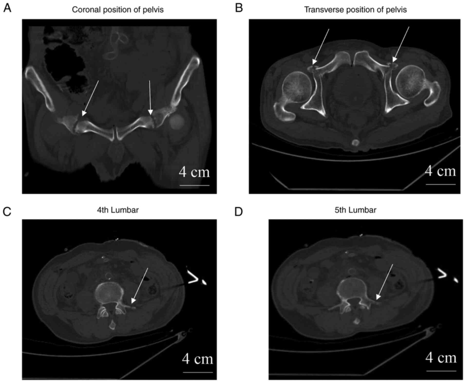
applied and blood without coagulation was observed. Fractures of
the pelvis (Fig. 1A and B) and the 4th and 5th lumbar vertebra
transverse process (Fig. 1C and
D) were observed via chest and
abdomen computed tomography (CT). Blood biochemical parameters were
measured and are presented in Table
I. The levels of D-dimer and creatine kinase were found to be
significantly increased.
 | Table IBlood biochemical parameters
(27/7/2020). |
Table I
Blood biochemical parameters
(27/7/2020).
| Parameters | Values | Normal values | Changing trend |
|---|
| Glucose, mmol/l | 15.6 | 3.3-6.1 | ↑ |
| Lactic acid,
mmol/l | 2.8 | 0.4-2.2 | ↑ |
| White blood cell,
x109/l | 20.42 | 4-10 | ↑ |
| Hemoglobin, g/l | 95 | 120-160 | ↓ |
| Platelet,
x109 | 167 | 100-300 | NC |
| Neutrophil
percentage, % | 89.5 | 50-70 | ↑ |
| Uric acid,
µmol/l | 619 | 100-432 | ↑ |
| Albumin, g/l | 28.7 | 40-55 | ↓ |
| Creatine kinase,
IU/l | 5976.5 | 25-175 | ↑ |
| Lactate
dehydrogenase, IU/l | 341.3 | 95-245 | ↑ |
| Fibrinogen, g/l | 1.87 | 2-4 | NC |
| D-dimer, mg/l | 25.09 | 0.00-0.55 | ↑ |
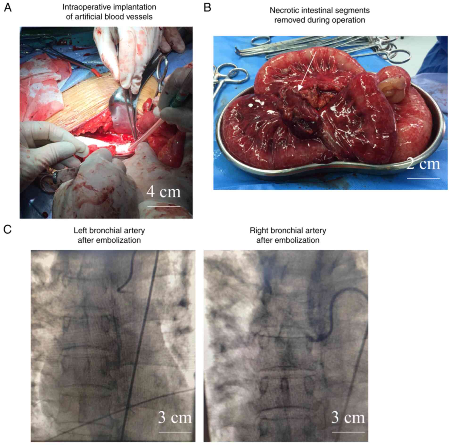
At 13:45 p.m. on July 27, 2020, under general
anesthesia, suture and ligation of multiple abdominal bleeding
vessels, repair of the mesentery of the small intestine, multiple
small bowel resection, enterostomy, implantation of a distal small
intestinal nutrition tube, ascending colon anastomosis of the small
intestine, ileocecal resection, debridement and repair of an
abdominal wall defect, and artificial vascular replacement of the
abdominal aorta and common iliac artery were performed (Fig. 2A). During the operation, a number
of small intestinal mesenteric lacerations with hemorrhage,
multiple segments of small intestine ischemia necrosis (Fig. 2B), small intestine rupture, and
abdominal aorta, and common iliac artery calcification with
thrombosis were observed. The ileocecal region and the adjacent
region of the small intestine were shown to be ischemic and
necrotic; the proximal small intestine was congested and edematous,
and there was no pulsation in the right common iliac artery. The
blood loss during the operation was ~3,000 ml, and 1,220 ml plasma,
10 units of red blood cell suspension and 20 units of
cryoprecipitate were transfused into the patient.
The patient with tracheal intubation was placed in
an emergency intensive care unit after the operation. At 21:10 p.m.
on July 27, 2020 (2 h after surgery), the patient presented with
ventricular fibrillation and was breathing at a rate of 6
breaths/min. Immediate biphasic wave electric defibrillation (200
J) was performed, although the patient did not recover their sinus
rhythm. The electrocardiogram showed that the heart rate of the
patient continued to drop to zero at 21:11 p.m., and chest
compression was performed. The patient was intravenously injected
with epinephrine hydrochloride at regular intervals (1 mg every 5
min). Continuous chest compression and electric defibrillation were
maintained. At 21:25 p.m., the heart rate of the patient recovered.
Epinephrine (1 mg) was injected every 5 min and repeated 2 times.
Subsequently, the patient was treated with a combination of
cefoperazone sulbactam sodium and ornidazole, as an anti-infection
agent, and low molecular weight heparin (100 IU/kg; administered
once every 12 h) as an anticoagulation agent, in addition to
receiving treatment for acid suppression, an albumin supplement,
and a blood transfusion. Treatment with low molecular weight
heparin was discontinued during episodes of hemoptysis and lower
gastrointestinal bleeding. When the bleeding was stable in the
later stages, the patient received antiplatelet drugs (clopidogrel
tablets; 75 mg/day) as anticoagulants on September 2, 2020.
On July 28, 2020 (the 2nd day after the operation),
the urine of the patient was brown and the urine volume decreased
notably. The level of myoglobin increased to 166,094 µg/l, whereas
that of creatine kinase increased to 150,185 IU/l (data not shown).
Acute kidney damage caused by CS was also observed. Continuous
renal replacement therapy (CRRT) treatment was applied to the
patient from July 28, 2020. Anuria was found on July 29, 2020
(i.e., the 3rd day after the operation). CRRT was performed for a
total of 273 h during hospitalization of the patient. The urine
volume began to increase on September 28, 2020 (63 days after the
operation), with >100 ml/24 h produced. Subsequently, the volume
of urine continued to increase, such that the urine volume rose to
>400 ml/24 h from October 12, 2020 onwards (i.e., 77 days after
the operation).
On July 28, 2020 (the 2nd day after the operation),
the patient developed osteofascial compartment syndrome in the
right leg and underwent open decompression. On August 2, 2020 (the
6th day after the operation), the patient received enteral
nutrition solution, although the patient experienced diarrhea. On
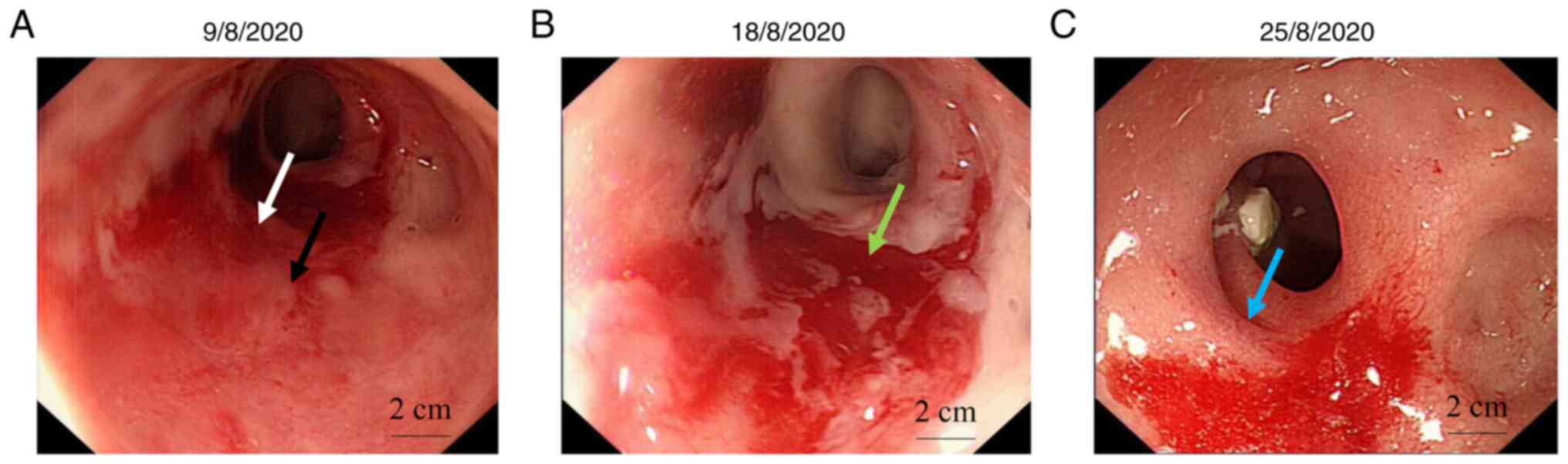
August 9, 2020 (the 13th day after the operation), lower
gastrointestinal bleeding occurred and enteral nutrition was
stopped. A colonoscopy revealed the presence of bright red blood
(100 ml) and necrotic tissue in the rectal cavity (Fig. 3A), which blocked the lumen such
that the endoscope could not pass through. During the laparotomy,
the rectal mucosa was necrotic and exfoliated, measuring ~5 cm in
length. Gauze packing was used for hemostasis. The intraoperative
blood loss was ~200 ml, and the patient was infused with 4 units of
red blood cell suspension.
On August 13, 2020 (the 17th day after the
operation), the patient experienced sudden pulmonary hemorrhage
with hemoptysis (breathing rate, 22 times/min; heart rate, 101
beats/min; blood pressure, 149/79 mmHg and oxygen saturation, 99%).
A total of 12 units of posterior pituitary injection was injected
intravenously. After 2 h, the patient still had hemoptysis with
increased volume, and the oxygen saturation was decreased to 92%.
Blood transfusion was immediately conducted, and a pulmonary
angiography was performed. A thickening of the bronchial and
intercostal arteries was observed (Fig. 2C). with enlarged and disordered
bronchial and intercostal arteries. Polyvinyl alcohol embolization
granules PVA-500 was used to stop the bleeding. After the
operation, the patient still had repeated episodes of hemoptysis
and therefore he was treated with diazepam (10 mg), batroxobin (3
IU) for hemostasis, acid inhibition, somatostatin for suppression
of intestinal secretion, continuous infusion of posterior pituitary
hormone, human prothrombin complex (200 IU), human fibrinogen (0.5
g), and a blood transfusion. Following these therapeutic
interventions, hemoptysis was notably reduced and the vital signs
of the patient were stable.
At 9:50 a.m. on August 18, 2020 (the 22th day after
the operation), the patient discharged a large amount of blood
stool, and was found to be in a state of shock. Emergency blood
transfusion was performed. At 13:51 p.m., a colonoscopy was
performed. Numerous blood clots and blood were found in the
intestinal cavity (Fig. 3B). Blood
scabs were widely attached to the intestinal wall. The rectal
mucosa was observed to be swollen and ulcerated locally. At ~1 cm
away from the anus, pulsatile bleeding was found in the rectum. A
titanium clip was used to stop the bleeding; however, the patient's
anemia was severe, and intermittent blood transfusion was
performed.
On August 25, 2020 (the 29th day after the
operation), a colonoscopy examination was performed. Active
bleeding was found in the rectal cavity, and the rectal mucosa was
congested and swollen (Fig. 3C). A
titanium clip could be seen ~5 cm away from the anus, and another
active bleeding point was observed 5 cm from the anus. An attempt
was made to use another titanium clip for the hemostasis, although
the hemostatic effects were not good. Therefore, a thrombin
injection was administered to the patient for hemostasis under
endoscopy, and the active bleeding was significantly reduced as a
result of this treatment. Thrombus attachments in the deep venous
catheter of the left femoral vein and right internal jugular vein
were observed using color doppler ultrasound. Anti-platelet
aggregation therapy (treatment with clopidogrel, 75 mg) was
initiated on September 2, 2020 (the 37th day after operation).
On September 9, 2020 (the 44th day after the
operation), the patient was diagnosed with nasal septal abscess and
underwent incision and drainage. On September 14, 2020 (the 49th
day after the operation), no rectal bleeding was observed, as
determined from colonoscopy. During hospitalization, the patient
received 60.5 units of red cell suspension, 62.1 units of plasma,
10 units of platelets, 60 units of cryoprecipitate (60 units equate
to 1,800 ml cryoprecipitate coagulation factor) and 1,600 ml human
albumin (20%).
On November 1, 2020 (the 97th day after the
operation), the patient was discharged from hospital. The patient
was in good spirits and was sleeping well. No bloody stools,
abdominal distension, palpitations, chest tightness or other
discomforts were found. The urine volume was 1,300 ml/24 h. The
patient was, however, emaciated. The breath sounds of both lungs
were thick. No obvious dry or wet rales were heard. The abdomen was
in a soft state, without notable tenderness. The small intestine
nutrition tube was fixed in its original place. Muscle atrophy of
both lower limbs, hypoesthesia of the right lower limbs and a
muscle strength of ~3 grades were found. A pelvis CT was performed
on October 24, 2020 (the 89th day after the operation). Fractures
were observed in the left transverse processes of the 4th and 5th
lumbar vertebra, bilateral acetabulum, bilateral pubic superior and
inferior branches. The broken ends were slightly dislocated, and a
small callus was formed. In addition, multiple calcifications in
the abdominal aortic wall and a small amount of effusion in the
pelvic cavity were observed. Color Doppler ultrasound revealed the
formation of atherosclerotic plaques in the bilateral external
iliac arteries and right lower extremity arteries. The results of
the routine blood tests are shown in Table II.
| Table IIBlood biochemical profile
(26/10/2020). |
Table II
Blood biochemical profile
(26/10/2020).
| Parameters | Values | Normal values | Changing trend |
|---|
| Red blood cell,
x1012 | 2.90 | 4-5.5 | ↓ |
| Hemoglobin, g/l | 93 | 120-160 | ↓ |
| Hematocrit, % | 27.9 | 40-50 | ↓ |
| B-type natriuretic
peptide, pg/ml | 131.82 | 0-100 | ↑ |
| Neutrophil
percentage, % | 75.8 | 50-70 | ↑ |
| Myoglobin, µg/l | 117.64 | 3-110 | ↑ |
| Fibrinogen, g/l | 4.45 | 2-4 | ↑ |
| Retinol binding
protein, mg/l | 142.9 | 36-72 | ↑ |
| 5'-ribonucleotide
hydrolase, U/l | 127.2 | 0-10 | ↑ |
| Total bile acid,
µmol/l | 16.8 | 0-12 | ↑ |
| Alkaline phosphatase,
IU/l | 859.8 | 45-125 | ↑ |
| γ-glutamyl
transferase, IU/l | 751.4 | 10-60 | ↑ |
| Glutamic oxaloacetic
transaminase, IU/l | 79.8 | 15-45 | ↑ |
| Alanine
aminotransferase, IU/l | 209.7 | 9-60 | ↑ |
| Potassium,
mmol/l | 3.36 | 3.5-5.3 | ↓ |
| Sodium, mmol/l | 134.2 | 137-147 | ↓ |
| Chlorine, mmol/l | 90.8 | 99-110 | ↓ |
| Phosphorus,
mmol/l | 2.02 | 0.6-1.6 | ↑ |
| Carbon dioxide
binding rate, mmol/l | 31.1 | 21-28 | ↑ |
| Urea, mmol/l | 11.06 | 2.9-7.2 | ↑ |
| Creatinine,
µmol/l | 308 | 44-133 | ↑ |
| High sensitivity
C-reactive protein, mg/l | 7.62 | 0-3 | ↑ |
| Procalcitonin,
ng/ml | 0.4 | 0.00-0.05 | ↑ |
In conclusion, the patient was diagnosed with the
following conditions: i) hemorrhagic shock; ii) cardiac arrest;
iii) ischemia, necrosis and rupture of the ileocecal region and
multiple segments of the small intestine; iv) thrombosis of the
common iliac artery; v) CS; vi) multiple organ dysfunction; vii)
lower gastrointestinal bleeding; viii) pulmonary hemorrhage; ix)
fractures of the pelvis, sacrum, the 4th and 5th lumbar vertebrae,
and the ribs; x) DIC; xi) bilateral pleural effusion and bilateral
pulmonary emphysema and xii) compartment syndrome of the right
leg.
Discussion
Cases of blunt abdominal trauma have been reported
previously. However, most of these cases have reported no more than
three types of diagnoses and the treatments involved were
relatively simple. In one such example, a 4-year-old boy was run
over by a car, sustaining blunt abdominal trauma. Transrectal small
bowel evisceration was performed for this boy, and he was
discharged from hospital on the 9th postoperative day with bed rest
(4). Patients with traumatic
cardiac arrest (TCA) have no pulse, and TCA has been viewed as a
precursor to traumatic death. Very low survival rates (i.e., 10.6%)
following TCA have been reported (3,5). In
the present case report, the patient also suffered cardiac arrest,
which could have been associated with renal insufficiency after CS
and hyperkalemia caused by infusion of excessive blood products
(6.17 mmol/l). Following emergency treatment with continuous chest
compression, electric defibrillation and an injection of
epinephrine hydrochloride, the patient ultimately recovered. It has
been reported that most pelvic injuries may not lead to significant
bleeding, since pelvic injuries are mainly associated with damage
to small arteries (6,7). No damage sustained to large arteries
was found in this case. It is therefore possible to surmise that
the hemorrhagic shock of the patient mainly resulted from
intestinal rupture, traumatic stress, and bleeding of fracture
ends.
The D-dimer levels of the patient were increased on
admission. D-dimer is considered to be a marker for fibrinolysis
and coagulation activation; however, D-dimer has also been used to
predict DIC (8). Furthermore,
D-dimer is considered to be a biomarker for thrombosis, and a
notable increase in its levels is indicative of a poor outcome
(9). The elevated level of D-dimer
in the present case report was therefore a predictor for massive
thrombosis in the abdominal aorta and bilateral common iliac
arteries.
Following the hemostasis and the artificial vessel
replacement surgery, upon the suggestion of the vascular surgeons,
low molecular weight heparin sodium with good absorption, high
selectivity, high bioavailability, less adverse bleeding reactions
and no routine blood sampling monitoring was used. Subsequently,
the dose of the low molecular weight heparin sodium administered
was appropriately reduced over time. In order to cope with the
possible risk of bleeding, the coagulation-associated indicators of
the patient, including the international normalized ratio during
the treatment, were closely monitored.
The patient in the present case report sustained
multiple and serious injuries, with a lengthy operation time. The
patient had an obvious rupture of the small intestine and a
mesenteric injury, and an enterostomy was performed. In addition,
the patient had multiple rib fractures and pleural effusion.
Therefore, the patient was treated with sulperazon empirically for
prophylactic anti-infection purpose, which is indeed worthy of
further discussion. A pulmonary angiography was performed after the
patient experienced sudden pulmonary hemorrhage and hemoptysis.
Because the patient had no history of hypertension, the posterior
pituitary injection was administered to reduce circulatory
pressure. The use of various hemostatic drugs in the later stages
of the operation was due to the persistent bleeding that resulted
from hemoptysis in the lower gastrointestinal region. These
hemostatic measures were proven to be effective in terms of
preventing systemic coagulation dysfunction.
The patient experienced anuria after July 29, 2020
(the 2nd day after the operation). After a total of 273 h of CRRT
treatment, the urine volume was increased from September 28, 2020
(the 63th day after the operation), with the patient producing
>100 ml urine/24 h. The following factors were considered to be
the major reasons leading to acute kidney failure. First, CS
resulted in rhabdomyolysis, which induced the further release of a
large amount of myoglobin into the blood, leading to acute kidney
failure (10). Secondly,
hemorrhagic shock may also have accelerated the kidney damage
(11). In addition, the
application of contrast agent could also have contributed to kidney
damage (12). In this case, timely
diagnosis and active systemic treatment contributed greatly during
the management of the patient's condition, aiding his recovery. The
case has demonstrated that a multidisciplinary treatment strategy
is crucial for the successful treatment of such patients with
multiorgan dysfunction.
Acknowledgements
Not applicable.
Funding
Funding:This study was supported by the National Natural Science
Foundation of China (grant no. 82101467), by the General Hospital
of Western Theater Command (grant no. 2021‑XZYG‑C20) and by the
National Defense Science and Technology and Military Theory
Innovation Program (grant no. JJ2021A03-B023).
Availability of data and materials
All data generated or analyzed during this study are
included in this published article.
Authors' contributions
XY, NT, CHe, KT and CHu took part in the conception
and design of the study. XY and CHe participated in the development
of methodology. GX, JD, LL, KT and CHe contributed to data
acquisition and analysis. NT, KT and CHu wrote and reviewed the
manuscript. XY, LL and CHe contributed to technical and material
support. CHe and CHu supervised the study. XY and KT confirm the
authenticity of all the raw data. All authors have read and
approved the final manuscript.
Ethics approval and consent to
participate
The present study was approved by the Ethics
Committee of The General Hospital of Western Theater Command
(Chengdu, China).
Patient consent for publication
Informed consent was obtained from the patients
included in the study.
Competing interests
The authors declare that they have no competing
interests.
References
|
1
|
He Q, Wang F, Li G, Chen X, Liao C, Zou Y,
Zhang Y, Kang Z, Yang X and Wang L: Crush syndrome and acute kidney
injury in the Wenchuan Earthquake. J Trauma. 70:1213–1217;
discussion 1217-1218. 2011.PubMed/NCBI View Article : Google Scholar
|
|
2
|
Revell MA, Pugh MA and McGhee M:
Gastrointestinal traumatic injuries: Gastrointestinal perforation.
Crit Care Nurs Clin North Am. 30:157–166. 2018.PubMed/NCBI View Article : Google Scholar
|
|
3
|
Barnard EBG, Hunt PAF, Lewis PEH and Smith
JE: The outcome of patients in traumatic cardiac arrest presenting
to deployed military medical treatment facilities: Data from the UK
Joint Theatre Trauma Registry. J R Army Med Corps. 164:150–154.
2018.PubMed/NCBI View Article : Google Scholar
|
|
4
|
Gelas T, Combet S, Perinel J, Javouhey E
and Mure PY: Transrectal small bowel evisceration after abdominal
crush injury. J Pediatr Surg. 47:e53–e56. 2012.PubMed/NCBI View Article : Google Scholar
|
|
5
|
Rosemurgy AS, Norris PA, Olson SM, Hurst
JM and Albrink MH: Prehospital traumatic cardiac arrest: The cost
of futility. J Trauma. 35:468–473; discussion 473-464.
1993.PubMed/NCBI
|
|
6
|
Totterman A, Madsen JE and Roise O:
Multifocal arterial haemorrhage in a partially stable pelvic
fracture after a crush injury: A case report. Arch Orthop Trauma
Surg. 126:113–117. 2006.PubMed/NCBI View Article : Google Scholar
|
|
7
|
Miller PR, Moore PS, Mansell E, Meredith
JW and Chang MC: External fixation or arteriogram in bleeding
pelvic fracture: Initial therapy guided by markers of arterial
hemorrhage. J Trauma. 54:437–443. 2003.PubMed/NCBI View Article : Google Scholar
|
|
8
|
Bates SM: D-dimer assays in diagnosis and
management of thrombotic and bleeding disorders. Semin Thromb
Hemost. 38:673–682. 2012.PubMed/NCBI View Article : Google Scholar
|
|
9
|
Ichkawa Y, Wada H, Ezaki M, Tanaka M,
Hiromori S, Shiraki K, Moritani I, Yamamoto A, Tashiro H, Shimpo H
and Shimaoka M: Elevated D-Dimer levels predict a poor outcome in
critically Ill patients. Clin Appl Thromb Hemost.
26(1076029620973084)2020.PubMed/NCBI View Article : Google Scholar
|
|
10
|
Sever MS and Vanholder R: Crush syndrome:
A case report and review of the literature. J Emerg Med.
48:730–731. 2015.PubMed/NCBI View Article : Google Scholar
|
|
11
|
Harrois A, Libert N and Duranteau J: Acute
kidney injury in trauma patients. Curr Opin Crit Care. 23:447–456.
2017.PubMed/NCBI View Article : Google Scholar
|
|
12
|
Gordon E and Kamath S: Fulminant liver
failure associated with abdominal crush injury in an eleven-year
old: A case report. Case Reports Hepatol.
2013(524371)2013.PubMed/NCBI View Article : Google Scholar
|