Introduction
Early pregnancy loss or missed abortion observed in
the first trimester of pregnancy is a situation that can be noticed
late since the miscarriage event does not occur, despite the embryo
has lost its vitality in the uterus. Missed abortion is the most
common complication of all pregnancies (1,2).
Impairment of the implantation process of an active trophoblast
with placental cells at 5-6 weeks of pregnancy can lead to numerous
serious pregnancy complications such as abortion in the first
trimester, rupture of prenatal membranes, preterm labor, fetal
growth restriction and preeclampsia in the third trimester
(1-4).
While chemokines are generally involved in
homeostasis of the immune system, inducible chemokines may also
play a role mainly in inflammatory processes. Chemokines are
divided into four subfamilies based on the number and spacing of
the first two cysteine residues in a conserved cysteine structure
(4). These four subfamilies are
named C, CC, CXC and CX3C with C being a cysteine and X an amino
acid residue. Among the proteins in the CX3C subclass, CX3CL1 (also
called fractalkine) is synthesized as a transmembrane molecule that
can be released into the circulation as a soluble isoform by
metalloprotease-dependent shedding (5,6).
Among the chemokines, CX3CL1, CCL17, CXCR4, chemokine ligand 12
(CXCL12) and intercellular adhesion molecule (ICAM) 5 are proteins
that can be released from tissues and mediate the migration of
immune system cells to tissues (5-7).
Further research is needed to show that not all other markers from
the cytokine family are equally affected. The thymus and
activation-regulated chemokine CCL17 is one of the inducible
chemokines and is produced by endometrial gland cells during
pregnancy. CXCL12 and its receptors CXCR4 are widely produced at
the maternal-fetal interface and play a regulatory role in
materno-fetal dialogue and immune tolerance during early pregnancy.
It has been reported that CX3CL1 is increased in the first
trimester of pregnancy and in term pregnancy complicated with
preeclampsia (6-11).
In the present study, it was aimed to examine the
levels of CX3CL1, CCL17, CXCR4, CXCL12 and ICAM5 in decidual tissue
and to show their relationship with missed abortion and their
utility as a marker for pregnancy outcomes.
Materials and methods
Patients
The present prospectively designed study was
approved (approval no. 06) by the Local Ethics Committee of Yeni
Yuzyil University (Istanbul, Turkey). Written informed consent was
obtained from all patients. The study was performed with patients
admitted to the Department of Obstetrics and Gynecology of İstanbul
Gaziosmanpasa Hospital (Yeni Yuzyil University). A total of 15
patients aged 18-40 years, who presented with a negative fetal
heartbeat between 6 and 10 gestational weeks to the obstetrics and
gynecology outpatient clinics of the hospital and who had no
additional systemic disease were included. The control group
consisted of 13 patients between 6 and 10 gestational weeks, who
presented with a positive fetal heartbeat and requested a voluntary
abortion. Women <18 years of age, with multiple pregnancies,
hypertension, diabetes, active maternal infection, chronic renal
failure, retroplacental hematoma or systemic lupus erythematosus
were excluded from the study.
Fresh materials recovered after therapeutic
curettage were stored at -20˚C until the analysis. The materials
were sent to Adnan Menderes University Faculty of Medicine
Biochemistry Research Laboratory for the analysis of markers.
CX3CL1, CCL17, CXCR4, CXCL12 and ICAM5 protein levels were
determined by ELISA and gene expression levels by reverse
transcription-quantitative (RT-q)PCR in fresh materials.
Gene expression of CX3CL1, CCL17,
CXCR4, CXCL12, ICAM5 by RT-qPCR
A total of 5 tissue sections (each 10-µm thick) were
collected from each material and put into 1.5-ml microfuge tubes.
RNA isolation from tissues was performed in duplicate through the
use of a commercially available FFPE RNA isolation kit (cat. no.
K156002; Invitrogen; Thermo Fisher Scientific, Inc.) following the
manufacturer's instructions. A total of 1 µg RNA was reverse
transcribed using High Capacity cDNA Reverse Transcription kit
(Applied Biosystems; Thermo Fisher Scientific, Inc.) according to
the manufacturer's protocol. The amplification was carried out
using ready-to-order primers. The following primer sequences were
used for amplification: CX3CL1 forward, 5'-CTTCCCAGGAAGCACAGAGG-3'
and reverse, 5'-CCTCCATCCTGAGCCTTTGG-3'; CCL17 forward,
5'-ACTTCAAGGGAGCCATTCCC-3' and reverse, 5'-CATCCCTGGAGCAGTCCTCA-3';
CXCR4 forward, 5'-TGACGGACAAGTACAGGCTGC-3' and reverse
5'-CCAGAAGGGAAGCGTGATGA-3'; CXCL12 forward,
5'-TGCCAGAGCCAACGTCAAG-3' and reverse, 5'-CAGCCGGGCTACAATCTGAA-3';
glyceraldehyde-3-phosphate dehydrogenase (GAPDH) forward,
5'-AGGGCTGCTTTTAACTCTGGT-3', and reverse,
5'-CCCCACTTGATTTTGGAGGGA-3, and ICAM5 forward,
5'-AGATCGCGTAGAGCTGATGC-3' and reverse,
5'-ACCCTACAGCTCAGGGTGAA-3'.
A total of 100 ng of cDNA were amplified using
SYBRGreen PCR Master Mix (Applied Biosystems; Thermo Fisher
Scientific, Inc.) on the ABI StepOne Plus detection system. The
thermocycling conditions for qPCR were as follows: 95˚C for 10 min,
then 40 cycles of: 95˚C for 15 sec and 60˚C for 1 min. The results
were analyzed using StepOne Software v2.3 (Applied Biosystems;
Thermo Fisher Scientific, Inc.), using the 2-ΔΔCq method
(12) and normalized to GAPDH mRNA
results. Data are expressed as fold induction relative to the
controls.
Protein levels of CX3CL1, CCL17,
CXCR4, CXCL12 and ICAM5 by ELISA
A total of 4 tissue sections (each 10-15 µm thick)
were collected from each material and put into a 1.5 ml centrifuge
tube. The samples were incubated in a 250 µl buffer (pH 7.5, 0.05 M
Tris, 1 mM EDTA, and 0.5% Tween-20). Tissue samples were
homogenized thoroughly and kept samples on ice for 30 min. Samples
were Centrifuged at 10,000 x g for 20 min at 4˚C and the
supernatant was used for measurement. Protein concentrations were
measured with Bradford method (13). The levels of CX3CL1, CCL17, CXCR4,
CXCL12 and ICAM5 were determined with the sandwich ELISA method in
accordance with the manufacturer's protocols (cat. no. EH0255;
Wuhan Fine Biotech Co., Ltd.) with inter-assay cv: <12% and
intra-assay cv: <10%, respectively. IFN-γ and IRF5 values were
presented as ng/µg protein. All ELISA measurements were performed
using a microplate reader (BioTek Epoch). Results were presented as
ml/mg of protein.
Statistical analysis
The sample size of the study was calculated using
G*Power software (version 3.1.9.7; http://www.gpower.hhu.de/). While calculating the
sample size, the effect size was taken as 0.2, the type1 error as
0.05, and the power as 0.8. Statistical analyses were performed
using SPSS 25.0 software (IBM Corp.). Descriptive data are
expressed as numbers and percentages. Normality of continuous
variables was verified with the Kolmogorov-Smirnov Test.
Differences between groups in terms of normally-distributed
continuous variables were evaluated with an independent samples
t-test. The differences between the groups in terms of median
values of the non-normally distributed variables were analyzed with
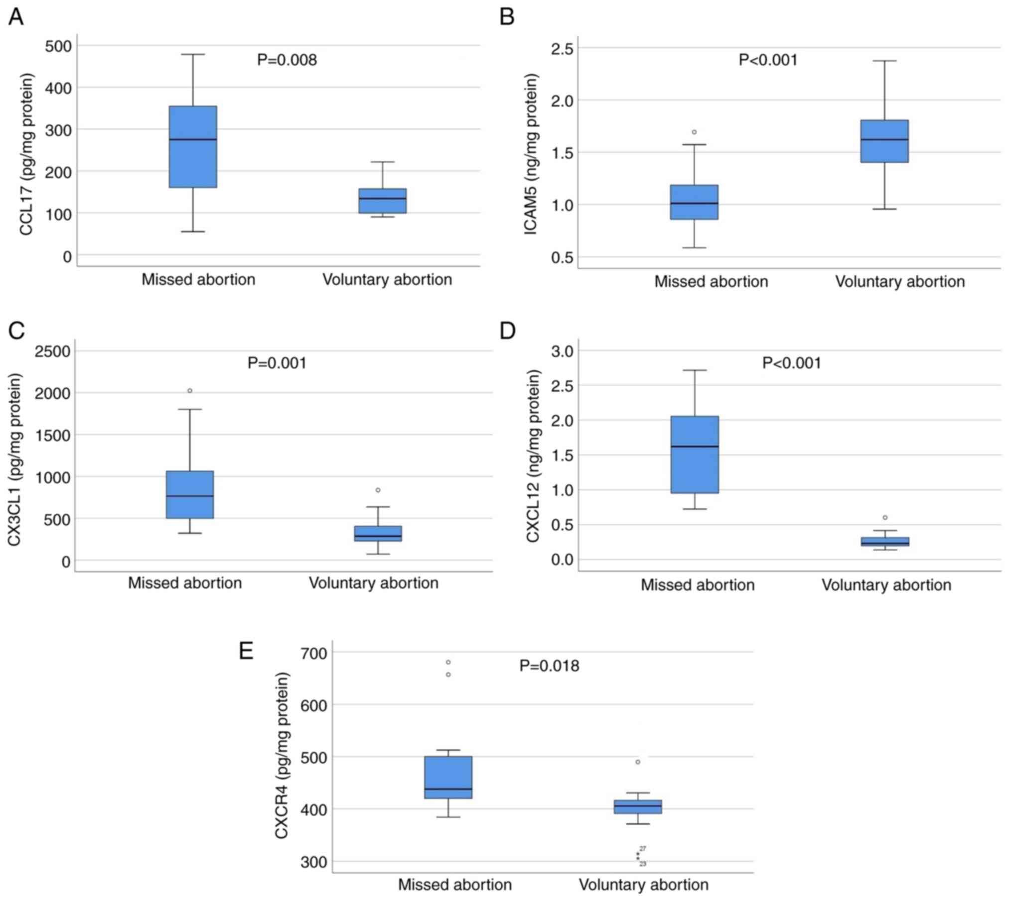
the Mann-Whitney U test. Box-and-whisker plots were used to show to
compare the medians and quartiles of CCL17, ICAM5, CX3CL1, CXCL12
and CXCR4 between the groups (Fig.
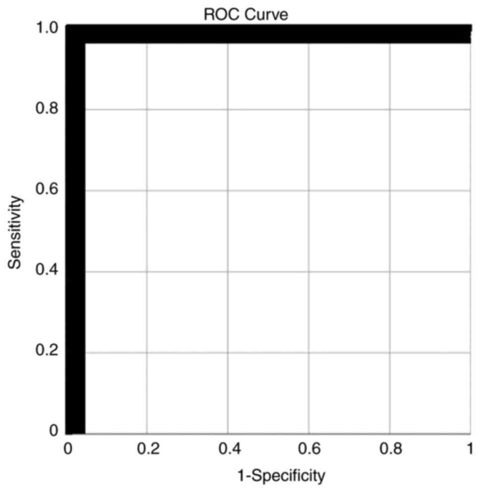
1). Receiver operating characteristic (ROC) analysis was
conducted to determine a threshold value for CXCL12 for
discrimination of missed and voluntary abortion. The results were
evaluated within the 95% confidence interval and P<0.05 was
considered to indicate a statistically significant difference.
Results
The mean age of the pregnant women was 29.7±4.7
years (Table I). There was no
difference for age between groups. Patient characteristics were
shown in Table I. The median CCL17
(274.9 vs. 133.8 pg/mg protein; P=0.008), CX3CL1 (765.2 vs. 284.8
pg/mg protein; P=0.001), CXCL12 (1.6 vs. 0.2 ng/mg protein;
P<0.001) and CXCR4 (473.8 vs. 405.6 pg/mg protein; P=0.018)
protein levels were statistically significantly higher in the
pregnant women with missed abortion compared with those with
voluntary abortion (Fig. 1). The
median ICAM5 protein level was significantly lower in the missed
abortion group than in the voluntary abortion group (1.0 vs. 1.6
ng/mg protein; P<0.001) (Table
II) (Fig. 1).
 | Table IDemographic characteristics of the
studied population. |
Table I
Demographic characteristics of the
studied population.
| | Missed abortion
(n=15) | Voluntary abortion
(n=13) |
|---|
| Characteristics | Median (IQR) | Mean ± SD | Median (IQR) | Mean ± SD | P-value |
|---|
| Age, years | 30(7) | 29.5±4.1 | 31(8) | 30.2±6 | 0.749a |
| Gravida | 2(1) | 1.8±0.7 | 1(2) | 1.8±1 | 0.65b |
| Parity | 1(1) | 0.6±0.6 | 1(2) | 0.8±0.8 | 0.683b |
| Weight (kg) | 60(10) | 60.8±10.9 | 63(17) | 64.8±9.8 | 0.325a |
| Height (cm) | 160(2) | 159.4±5.2 | 161(6) | 162.5±4 | 0.095b |
| Gestational week | 8(1) | 8.1±0.7 | 6 (0) | 6±0.6 |
<0.001b |
| BMI
(kg/m2) | 22.7 (3.2) | 23.9±3.9 | 23.4 (5.5) | 24.6±4.4 | 0.504b |
| Table IIComparison of mean gene and protein
expression levels of chemokines in decidual tissues of patients
between groups. |
Table II
Comparison of mean gene and protein
expression levels of chemokines in decidual tissues of patients
between groups.
| | Missed abortion
(n=15) | Voluntary abortion
(n=13) |
|---|
|
Characteristics | Median (IQR) | Mean ± SD | Median (IQR) | Mean ± SD | P-value |
|---|
| CCL17 (pg/mg
protein) | 274.9(239) | 264.9±130.3 | 133.8(75) | 140.3±45.7 | 0.008a |
| ICAM5 (ng/mg
protein) | 1.0 (0) | 1.03±0.3 | 1.6(1) | 1.6±0.4 |
<0.001b |
| CX3CL1 (pg/mg
protein) | 765.2(726) | 874.1±521.7 | 284.8(199) | 349.1±202.2 | 0.001a |
| CXCL12 (ng/mg
protein) | 1.6 [1] | 1.57±0.68 | 0.2 (0) | 0.3±0.1 |
<0.001a |
| CXCR4 (pg/mg
protein) | 473.8(93) | 469.7±89.5 | 405.6(42) | 408.6±68.8 | 0.018a |
| CCL17/GAPDH | 2.9 (0) | 2.9±0.3 | 1.2 (0) | 1.2±0.2 |
<0.001a |
| ICAM5/GAPDH | 0.8 (0) | 0.9±0.2 | 1.3 (0) | 1.3±0.23 |
<0.001b |
| CX3CL1/GAPDH | 2.9(1) | 3.0±0.8 | 1.1 (0) | 1.2±0.2 |
<0.001a |
| CXCL12/GAPDH | 2.7(2) | 2.8±1 | 1.1 (0) | 1.1±0.2 |
<0.001a |
| CXCR4/GAPDH | 1.2 (0) | 1.3±0.4 | 1.0 (0) | 0.9±0.3 | 0.013b |
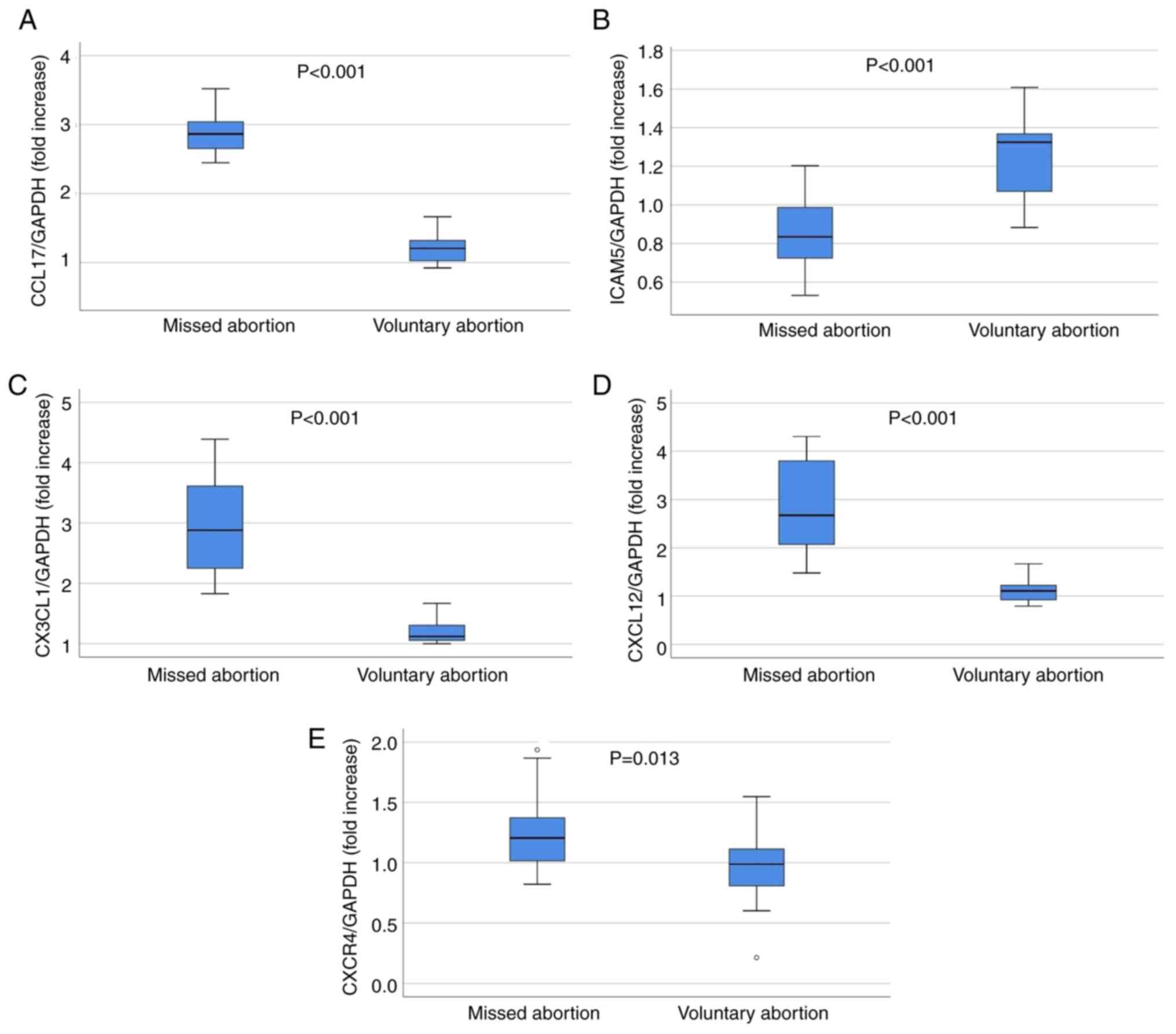
The median amount of increase in gene expression
levels of CCL17/GAPDH (2.9 vs. 1.2 fold increase; P<0.001),
CX3CL1/GAPDH (2.9 vs. 1.1 fold increase; P<0.001), CXCL12/GAPDH
(2.7 vs. 1.1 fold increase; P<0.001) and the mean CXCR4/GAPDH
(1.3 vs. 0.9 fold increase; P=0.013) was found to be significantly
higher in tissues of pregnant women with missed abortion than that
of the pregnant women with voluntary abortion. The mean amount of
increase in ICAM5/GAPDH tissue gene expression level was
significantly lower in the missed abortion group than in the
voluntary abortion group (0.9 vs. 1.3 fold increase; P<0.001)
(Table II) (Fig. 2). ROC analysis revealed that a
threshold value of 0.66 ng/mg protein for CXCL12 had a sensitivity
and specificity of 100% for determination of missed abortion (AUC:
1; lower bound: 1.0; upper bound: 1.0; P<0.001) (Fig. 3).
Discussion
In the present study, it was found that median
CCL17, CX3CL1, CXCL12 and CXCR4 protein levels were significantly
higher in pregnant women with missed abortion than in those with
voluntary abortion. The mean ICAM5 protein level was identified to
be significantly lower in the missed abortion group compared with
the voluntary abortion group. When chemokines were examined in
terms of gene expression levels, the median amounts of increase in
gene expression levels of CCL17, CX3CL, CXCL12 and CXCR4 were
significantly higher in the missed abortion group compared with the
voluntary abortion group. The mean amount of increase in gene
expression levels of ICAM5 protein was significantly lower in the
missed abortion group compared with the voluntary abortion group.
The maternal component of the placenta was used as the material.
The maternal component of the placenta is known as the decidua
basalis. Oxygen and nutrients from maternal blood diffuse through
the villus walls in the intervillous spaces and enter the fetal
capillaries. Also decidua secretes hormones, growth factors and
cytokines. It has receptors for estrogen, progesterone, growth
hormone, and others. If missed abortion is not diagnosed, it may be
accompanied by maternal complications such as septic abortion or
excessive bleeding (4). In
addition, the obtained data will shed light on recurrent
spontaneous pregnancy losses. Certain studies have shown that these
5 chemokines (CX3CL1, CCL17, CXCR4, CXCL12 and ICAM5) that were
studied play a role in the implantation process of the embryo
(9,8,14-16).
CX3CL1 has recently attracted attention in the field
of reproductive research by regulating adhesion and migration
processes in fetal-maternal interaction at different stages of
human pregnancy (8). In addition,
an increasing body of evidence has suggested that a number of
pregnancy pathologies are associated with increased placental
CX3CL1 expression, including chorioamnionitis (17), diabetic pregnancy (18), and severe early-onset preeclampsia
(19,20). In a previous study by Hannan et
al (21) investigating the
CX3CL1 level in the serum of 34 first trimester abortions and 44
normal births, CXCL3 was not detected in term patients, but it was
found in the aborted group. Li et al (22) detected significantly upregulated
expression of CX3CL1 and CX3CL1 receptor in the uterus of
IFN-γ-induced aborted mice. In the present study, the median CX3CL1
protein and gene expression levels were found to be significantly
higher in pregnant women with missed abortion compared with
pregnant women with voluntary abortion. The abortion week in the
materials received was at most 10 weeks, and it was observed in
only 1 patient. Brain tissue is not considered to be dominant for
such a small fetus. Similarly, the brain tissue is damaged in
voluntary abortion cases. The present data are consistent with
previous studies showing the role of CX3CL1 in the feto-maternal
interaction in the process of placental invasion and indicated that
CX3CL1 may be associated with missed abortion (20,21).
Chemokines are unique proteins with the ability to
control immune cell chemoattraction and participate in cell
proliferation, migration and apoptosis. CXCL12 can modulate the
proliferation, invasion and survival of human trophoblasts that are
crucial for establishing a successful pregnancy (23-25).
CXCL12 and its receptor CXCR4 are involved in uterine natural
killer cell recruitment, placentation, vascular remodeling,
embryogenesis, and cardiovascular and central nervous system
organogenesis (23-29).
Suppression of CXCL12/CXCR4 signaling during the small window of
conceptus implantation reduces placental vascularization, induces
autophagy, and moistens the inflammatory placental environment
(30-33).
In addition, recent evidence has suggested that suppression of
CXCL12-induced actions at the fetal-maternal interface reduces
trophoblast invasion into the maternal endometrium and delays
uterine remodeling (30). Chao
et al (34) found that the
expression of CXCL12 chemokine and its receptor CXCR4 increased in
the abortion material induced in mice with IFN-gamma. In another
study, Dimova et al revealed an increased expression of
CXCL12 chemokine and its receptor CXCR4 in the decidua of 30
patients with spontaneous abortion compared with 15 patients with
elective abortion (35). In the
present study, the median protein level and gene expression of
CXCL12 and CXCR4 were found to be significantly higher in pregnant
women with missed abortion than in pregnant women with voluntary
abortion. In addition, the present findings revealed a highly
reliable threshold value for CXCL12 (with a sensitivity and
specificity of 100%) in discrimination of missed and voluntary
abortions. These findings showed the association of these molecules
with missed abortion.
It has been reported that CCL17 level does not
change in cases of spontaneous abortion (11). In the present study, the mean
protein level and gene expression of CCL17 were identified to be
significantly higher in pregnant women with missed abortion than in
pregnant women with voluntary abortion. Furthermore, the mean ICAM5
protein level was found to be significantly lower in pregnant women
with missed abortion than in the those with voluntary abortion. As
ICAM5 is solely formed in the brain tissue, its level may be
reduced in abortion cases due to lack of brain development
(36). However, it was planned to
discriminate the abortion whether it was missed or voluntary, thus
the voluntary abortion cases were used as the control group.
According to the present findings, it was observed that brain
tissue is damaged in missed abortion significantly more than the
voluntary abortion cases.
Since the present study was planned as a cohort
study, the pregnant women were not followed up for a long time
after abortion and the long-term changes in the levels of these
molecules were not examined, which can be considered as the major
limitation of the present study. Again, there is a need for further
studies on whether the amount of gene expression in the tissue
changes on the day of missed abortion diagnosis. Another limitation
is the relatively small number of pregnant women included in the
study, although it was sufficient for statistical analysis. In
addition, other markers from the family of cytokines also need to
be investigated to reveal that not all are equally affected. In
future studies, broader genetic analyzes can be obtained by
examining the relationship between karyotype analyses and
chemokines in missed abortion.
In conclusion, the findings of the present study
suggested that CCL17, CX3CL1, CXCL12, CXCR4 and ICAM5 may be
associated with missed abortion and may play an important role in
placental invasion and the continuation of pregnancy. However,
further comprehensive studies are needed to draw more definitive
conclusions.
Acknowledgements
Not applicable.
Funding
Funding: No funding was received.
Availability of data and materials
The datasets used and/or analyzed during the current
study are available from the corresponding author on reasonable
request.
Authors' contributions
DH was involved in project development and
manuscript writing. SG was responsible for data collection,
management and analysis. OC and DH confirm the authenticity of all
the raw data. OC and VT contributed to data analysis, manuscript
writing and editing. All authors have read and approved the
manuscript.
Ethics approval and consent to
participate
The present prospectively designed study was
approved (approval no. 06) by the local ethics committee of Yeni
Yuzyil University (Istanbul, Turkey). Written informed consent was
obtained from all patients.
Patient consent for publication
Not applicable.
Competing interests
The authors declare that they have no competing
interests.
References
|
1
|
Hanschmidt F, Linde K, Hilbert A,
Riedel-Heller SG and Kersting A: Abortion stigma: A systematic
review. Perspect Sex Reprod Health. 48:169–177. 2016.PubMed/NCBI View
Article : Google Scholar
|
|
2
|
Kapp N and Lohr PA: Modern methods to
induce abortion: Safety, efficacy and choice. Best Pract Res Clin
Obstet Gynaecol. 63:37–44. 2020.PubMed/NCBI View Article : Google Scholar
|
|
3
|
Conti J and Cahill EP: Self-managed
abortion. Curr Opin Obstet Gynecol. 31:435–440. 2019.PubMed/NCBI View Article : Google Scholar
|
|
4
|
Griebel CP, Halvorsen J, Golemon TB and
Day AA: Management of spontaneous abortion. Am Fam Physician.
72:1243–1250. 2005.PubMed/NCBI
|
|
5
|
Hughes CE and Nibbs RJB: A guide to
chemokines and their receptors. FEBS J. 285:2944–2971.
2018.PubMed/NCBI View Article : Google Scholar
|
|
6
|
Palomino DC and Marti LC: Chemokines and
immunity. Einstein (Sao Paulo). 13:469–473. 2015.PubMed/NCBI View Article : Google Scholar : (İn English,
Portuguese).
|
|
7
|
Legler DF and Thelen M: Chemokines:
Chemistry, biochemistry and biological function. Chimia (Aarau).
70:856–859. 2016.PubMed/NCBI View Article : Google Scholar
|
|
8
|
Kervancioglu Demirci E, Salamonsen LA and
Gauster M: The role of CX3CL1 in fetal-maternal interaction during
human gestation. Cell Adh Migr. 10:189–196. 2016.PubMed/NCBI View Article : Google Scholar
|
|
9
|
Ao D, Li DJ and Li MQ: CXCL12 in normal
and pathological pregnancies: A review. Am J Reprod Immunol.
84(e13280)2020.PubMed/NCBI View Article : Google Scholar
|
|
10
|
Gahmberg CG, Ning L and Paetau S: ICAM-5:
A neuronal dendritic adhesion molecule involved in immune and
neuronal functions. Adv Neurobiol. 8:117–132. 2014.PubMed/NCBI View Article : Google Scholar
|
|
11
|
Rasmark ER, Bruno V, Nedstrand E, Boij R,
Strid CP, Piccione E, Berg G, Svensson-Arvelund J, Jenmalm MC,
Rubér M and Ernerudh J: Low-molecular-weight-heparin increases Th1-
and Th17-associated chemokine levels during pregnancy in women with
unexplained recurrent pregnancy loss: A randomised controlled
trial. Sci Rep. 9(12314)2019.PubMed/NCBI View Article : Google Scholar
|
|
12
|
Livak KJ and Schmittgen TD: Analysis of
relative gene expression data using real-time quantitative PCR and
the 2(-Delta Delta C(T)) method. Methods. 25:402–408.
2001.PubMed/NCBI View Article : Google Scholar
|
|
13
|
Bradford MM: A rapid and sensitive method
for the quantitation of microgram quantities of protein utilizing
the principle of protein-dye binding. Anal Biochem. 72:248–254.
1976.PubMed/NCBI View Article : Google Scholar
|
|
14
|
Zlotnik A and Yoshie O: Chemokines: A new
classification system and their role in immunity. Immunity.
12:121–127. 2000.PubMed/NCBI View Article : Google Scholar
|
|
15
|
Garton KJ, Gough PJ, Blobel CP, Murphy G,
Greaves DR, Dempsey PJ and Raines EW: Tumor necrosis
factor-alpha-converting enzyme (ADAM17) mediates the cleavage and
shedding of fractalkine (CX3CL1). J Biol Chem. 276:37993–38001.
2001.PubMed/NCBI View Article : Google Scholar
|
|
16
|
Hundhausen C, Misztela D, Berkhout TA,
Broadway N, Saftig P, Reiss K, Hartmann D, Fahrenholz F, Postina R,
Matthews V, et al: The disintegrin-like metalloproteinase ADAM10 is
involved in constitutive cleavage of CX3CL1 (fractalkine) and
regulates CX3CL1-mediated cell-cell adhesion. Blood. 102:1186–1195.
2003.PubMed/NCBI View Article : Google Scholar
|
|
17
|
Szukiewicz D, Kochanowski J, Mittal TK,
Pyzlak M, Szewczyk G and Cendrowski K: Chorioamnionitis (ChA)
modifies CX3CL1 (fractalkine) production by human amniotic
epithelial cells (HAEC) under normoxic and hypoxic conditions. J
Inflamm (Lond). 11(12)2014.PubMed/NCBI View Article : Google Scholar
|
|
18
|
Szukiewicz D, Kochanowski J, Pyzlak M,
Szewczyk G, Stangret A and Mittal TK: Fractalkine (CX3CL1) and its
receptor CX3CR1 may contribute to increased angiogenesis in
diabetic placenta. Mediators Inflamm. 2013(437576)2013.PubMed/NCBI View Article : Google Scholar
|
|
19
|
Siwetz M, Dieber-Rotheneder M,
Cervar-Zivkovic M, Kummer D, Kremshofer J, Weiss G, Herse F,
Huppertz B and Gauster M: Placental fractalkine is up-regulated in
severe early-onset preeclampsia. Am J Pathol. 185:1334–1343.
2015.PubMed/NCBI View Article : Google Scholar
|
|
20
|
Usta A, Turan G, Sancakli Usta C, Avci E
and Adali E: Placental fractalkine immunoreactivity in preeclampsia
and its correlation with histopathological changes in the placenta
and adverse pregnancy outcomes. J Matern Fetal Neonatal Med.
33:806–815. 2020.PubMed/NCBI View Article : Google Scholar
|
|
21
|
Hannan NJ, Bambang K, Kaitu'u-Lino TJ,
Konje JC and Tong S: A bioplex analysis of cytokines and chemokines
in first trimester maternal plasma to screen for predictors of
miscarriage. PLoS One. 9(e93320)2014.PubMed/NCBI View Article : Google Scholar
|
|
22
|
Li ZY, Chao HH, Liu HY, Song ZH, Li LL,
Zhang YJ, Yang Y and Peng JP: IFN-γ induces aberrant CD49b+ NK cell
recruitment through regulating CX3CL1: A novel mechanism by which
IFN-γ provokes pregnancy failure. Cell Death Dis.
5(e1512)2014.PubMed/NCBI View Article : Google Scholar
|
|
23
|
Kumar A, Kumar S, Dinda AK and Luthra K:
Differential expression of CXCR4 receptor in early and term human
placenta. Placenta. 25:347–351. 2004.PubMed/NCBI View Article : Google Scholar
|
|
24
|
Schanz A, Red-Horse K, Hess AP,
Baston-Büst DM, Heiss C and Krüssel JS: Oxygen regulates human
cytotrophoblast migration by controlling chemokine and receptor
expression. Placenta. 35:1089–1094. 2014.PubMed/NCBI View Article : Google Scholar
|
|
25
|
Jaleel MA, Tsai AC, Sarkar S, Freedman PV
and Rubin LP: Stromal cell-derived factor-1 (SDF-1) signalling
regulates human placental trophoblast cell survival. Mol Hum
Reprod. 10:901–909. 2004.PubMed/NCBI View Article : Google Scholar
|
|
26
|
Wu X, Jin LP, Yuan MM, Zhu Y, Wang MY and
Li DJ: Human first-trimester trophoblast cells recruit
CD56brightCD16-NK cells into decidua by way of expressing and
secreting of CXCL12/stromal cell-derived factor 1. J Immunol.
175:61–68. 2005.PubMed/NCBI View Article : Google Scholar
|
|
27
|
Barrientos G, Tirado-González I, Freitag
N, Kobelt P, Moschansky P, Klapp BF, Thijssen VL and Blois SM:
CXCR4(+) dendritic cells promote angiogenesis during embryo
implantation in mice. Angiogenesis. 16:417–427. 2013.PubMed/NCBI View Article : Google Scholar
|
|
28
|
Virgintino D, Errede M, Rizzi M, Girolamo
F, Strippoli M, Wälchli T, Robertson D, Frei K and Roncali L: The
CXCL12/CXCR4/CXCR7 ligand-receptor system regulates
neuro-glio-vascular interactions and vessel growth during human
brain development. J Inherit Metab Dis. 36:455–466. 2013.PubMed/NCBI View Article : Google Scholar
|
|
29
|
Zhu Y and Murakami F: Chemokine CXCL12 and
its receptors in the developing central nervous system: Emerging
themes and future perspectives. Dev Neurobiol. 72:1349–1362.
2012.PubMed/NCBI View Article : Google Scholar
|
|
30
|
McIntosh SZ, Maestas MM, Dobson JR, Quinn
KE, Runyan CL and Ashley RL: CXCR4 signaling at the fetal-maternal
interface may drive inflammation and syncytia formation during
ovine pregnancy†. Biol Reprod. 104:468–478. 2021.PubMed/NCBI View Article : Google Scholar
|
|
31
|
McIntosh SZ, Maxam CJ, Maestas MM, Quinn
KE and Ashley RL: Intrauterine inhibition of chemokine receptor 4
signaling modulates local and systemic inflammation in ovine
pregnancy. Am J Reprod Immunol. 82(e13181)2019.PubMed/NCBI View Article : Google Scholar
|
|
32
|
Quinn KE, Prosser SZ, Kane KK and Ashley
RL: Inhibition of chemokine (C-X-C motif) receptor four (CXCR4) at
the fetal-maternal interface during early gestation in sheep:
Alterations in expression of chemokines, angiogenic factors and
their receptors. J Anim Sci. 95:1144–11153. 2017.PubMed/NCBI View Article : Google Scholar
|
|
33
|
Runyan CL, McIntosh SZ, Maestas MM, Quinn
KE, Boren BP and Ashley RL: CXCR4 signaling at the ovine
fetal-maternal interface regulates vascularization, CD34+ cell
presence, and autophagy in the endometrium†. Biol Reprod.
101:102–111. 2019.PubMed/NCBI View Article : Google Scholar
|
|
34
|
Chao HH, Li L, Gao X, Wang C and Yue W:
CXCL12 expression in aborted mouse uteri induced by IFN-γ:
Potential anti-inflammatory effect involves in endometrial
restoration after abortion in mice. Gene. 700:38–46.
2019.PubMed/NCBI View Article : Google Scholar
|
|
35
|
Dimova I, Rizov M, Giragosyan S,
Koprinarova M, Tzoneva D, Belemezova K, Hristova-Savova M,
Milachich T, Djonov V and Shterev A: Molecular pathogenesis of
spontaneous abortions-Whole genome copy number analysis and
expression of angiogenic factors. Taiwan J Obstet Gynecol.
59:99–104. 2020.PubMed/NCBI View Article : Google Scholar
|
|
36
|
Gahmberg CG, Tian L, Ning L and
Nyman-Huttunen H: ICAM-5-a novel two-facetted adhesion molecule in
the mammalian brain. Immunol Lett. 117:131–135. 2008.PubMed/NCBI View Article : Google Scholar
|