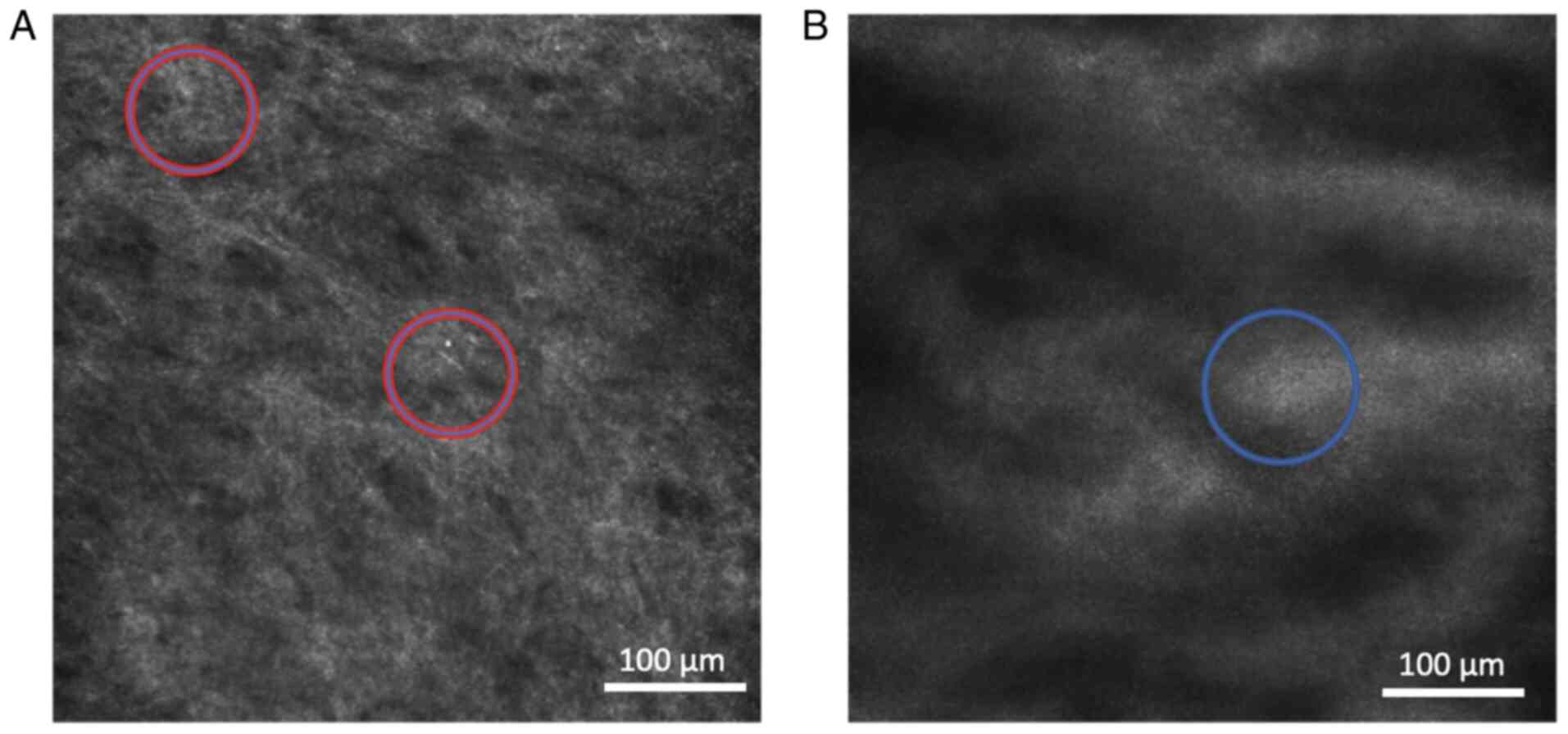
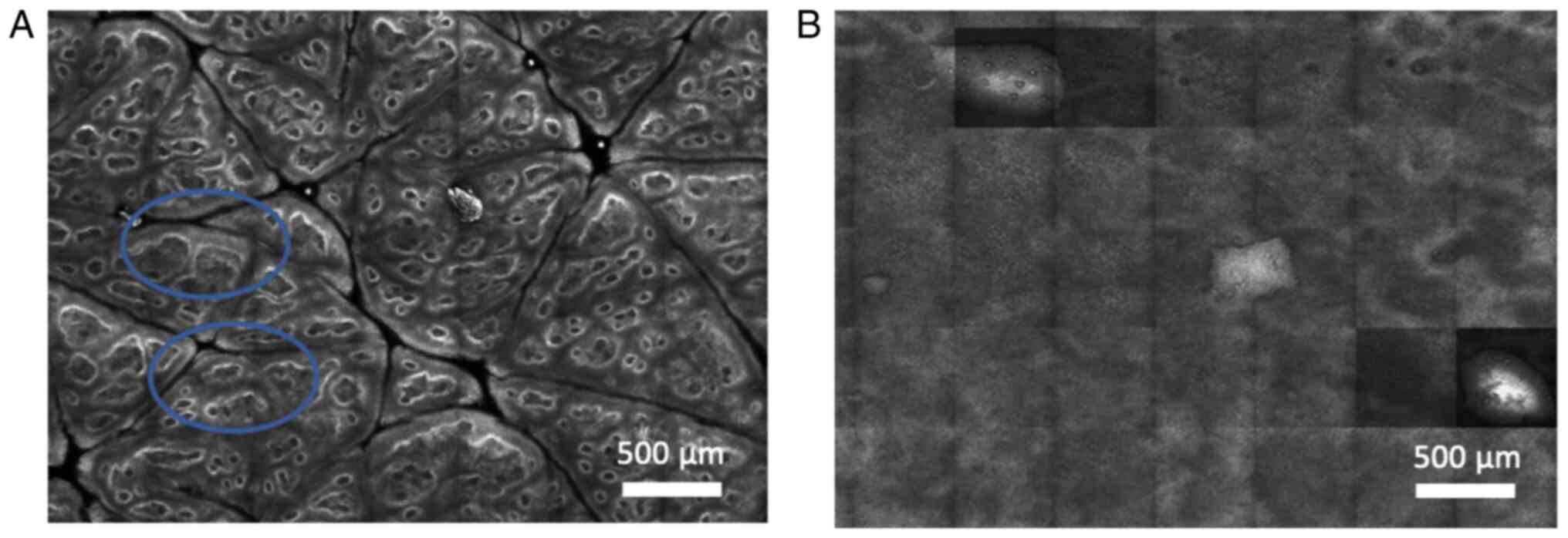
|
1
|
Wallace HJ: Lichen sclerosus et
atrophicus. Trans St Johns Hosp Dermatol Soc. 57:9–30.
1971.PubMed/NCBI
|
|
2
|
Tasker GL and Wojnarowska F: Lichen
sclerosus. Clin Exp Dermatol. 28:128–133. 2003.PubMed/NCBI View Article : Google Scholar
|
|
3
|
Lipscombe TK, Wayte J, Wojnarowska F,
Marren P and Luzzi G: A study of clinical and aetiological factors
and possible associations of lichen sclerosus in males. Australas J
Dermatol. 38:132–136. 1997.PubMed/NCBI View Article : Google Scholar
|
|
4
|
Kantere D, Löwhagen GB, Alvengren G,
Månesköld A, Gillstedt M and Tunbäck P: The clinical spectrum of
lichen sclerosus in male patients-a retrospective study. Acta Derm
Venereol. 94:542–546. 2014.PubMed/NCBI View Article : Google Scholar
|
|
5
|
Bleeker MC, Visser PJ, Overbeek LI, van
Beurden M and Berkhof J: Lichen sclerosus: Incidence and risk of
vulvar squamous cell carcinoma. Cancer Epidemiol Biomarkers Prev.
25:1224–1230. 2016.PubMed/NCBI View Article : Google Scholar
|
|
6
|
Nasca MR, Innocenzi D and Micali G: Penile
cancer among patients with genital lichen sclerosus. J Am Acad
Dermatol. 41:911–914. 1999.PubMed/NCBI View Article : Google Scholar
|
|
7
|
Lewis FM, Tatnall FM, Velangi SS, Bunker
CB, Kumar A, Brackenbury F, Mohd Mustapa MF and Exton LS: British
association of dermatologists guidelines for the management of
lichen sclerosus, 2018. Br J Dermatol. 178:839–853. 2018.PubMed/NCBI View Article : Google Scholar
|
|
8
|
Lacarrubba F, Borghi A, Verzi AE, Corazza
M, Stinco G and Micali G: Dermoscopy of genital diseases: A review.
J Eur Acad Dermatol Venereol. 34:2198–2207. 2020.PubMed/NCBI View Article : Google Scholar
|
|
9
|
Larre Borges A, Tiodorovic-Zivkovic D,
Lallas A, Moscarella E, Gurgitano S, Capurro M, Apalla Z, Bruno J,
Popovic D, Nicoletti S, et al: Clinical, dermoscopic and
histopathologic features of genital and extragenital lichen
sclerosus. J Eur Acad Dermatol Venereol. 27:1433–1439.
2013.PubMed/NCBI View Article : Google Scholar
|
|
10
|
Pan ZY, Lin JR, Cheng TT, Wu JQ and Wu WY:
In vivo reflectance confocal microscopy of Basal cell carcinoma:
Feasibility of preoperative mapping of cancer margins. Dermatol
Surg. 38:1945–1950. 2012.PubMed/NCBI View Article : Google Scholar
|
|
11
|
Jain M, Robinson BD, Shevchuk MM, Aggarwal
A, Salamoon B, Dubin JM, Scherr DS and Mukherjee S: Multiphoton
microscopy: A potential intraoperative tool for the detection of
carcinoma in situ in human bladder. Arch Pathol Lab Med.
139:796–804. 2015.PubMed/NCBI View Article : Google Scholar
|
|
12
|
Calzavara-Pinton P, Longo C, Venturini M,
Sala R and Pellacani G: Reflectance confocal microscopy for in vivo
skin imaging. Photochem Photobiol. 84:1421–1430. 2008.PubMed/NCBI View Article : Google Scholar
|
|
13
|
Gerger A, Koller S, Kern T, Massone C,
Steiger K, Richtig E, Kerl H and Smolle J: Diagnostic applicability
of in vivo confocal laser scanning microscopy in melanocytic skin
tumors. J Invest Dermatol. 124:493–498. 2005.PubMed/NCBI View Article : Google Scholar
|
|
14
|
Balu M, Kelly KM, Zachary CB, Harris RM,
Krasieva TB, König K, Durkin AJ and Tromberg BJ: Distinguishing
between benign and malignant melanocytic nevi by in vivo
multiphoton microscopy. Cancer Res. 74:2688–2697. 2014.PubMed/NCBI View Article : Google Scholar
|
|
15
|
Ardigo M, Cota C, Berardesca E and
González S: Concordance between in vivo reflectance confocal
microscopy and histology in the evaluation of plaque psoriasis. J
Eur Acad Dermatol Venereol. 23:660–667. 2009.PubMed/NCBI View Article : Google Scholar
|
|
16
|
Ardigò M, Maliszewski I, Cota C, Scope A,
Sacerdoti G, Gonzalez S and Berardesca E: Preliminary evaluation of
in vivo reflectance confocal microscopy features of Discoid lupus
erythematosus. Br J Dermatol. 156:1196–1203. 2007.PubMed/NCBI View Article : Google Scholar
|
|
17
|
Contaldo M, Di Stasio D, Petruzzi M,
Serpico R and Lucchese A: In vivo reflectance confocal microscopy
of oral lichen planus. Int J Dermatol. 58:940–945. 2019.PubMed/NCBI View Article : Google Scholar
|
|
18
|
Mills SE (ed): Histology for pathologists.
4th edition. Lippincott Williams & Wilkins, Philadelphia, PA,
2012.
|
|
19
|
Watt FM and Green H: Involucrin synthesis
is correlated with cell size in human epidermal cultures. J Cell
Biol. 90:738–742. 1981.PubMed/NCBI View Article : Google Scholar
|
|
20
|
Lacarrubba F, Verzì AE, Ardigò M and
Micali G: Handheld reflectance confocal microscopy, dermatoscopy
and histopathological correlation of common inflammatory balanitis.
Skin Res Technol. 24:499–503. 2018.PubMed/NCBI View Article : Google Scholar
|
|
21
|
Mazzilli S, Giunta A, Galluzzo M, Garofalo
V, Campione E, DI Prete M, Orlandi A, Ardigò M and Bianchi L:
Therapeutic monitoring of male genital lichen sclerosus: Usefulness
of reflectance confocal microscopy. Ital J Dermatol Venerol.
156:718–719. 2021.PubMed/NCBI View Article : Google Scholar
|
|
22
|
Zipfel WR, Williams RM and Webb WW:
Nonlinear magic: Multiphoton microscopy in the biosciences. Nat
Biotechnol. 21:1369–1377. 2003.PubMed/NCBI View
Article : Google Scholar
|
|
23
|
Hoogedoorn L, Peppelman M, van de Kerkhof
PC, van Erp PE and Gerritsen MJ: The value of in vivo reflectance
confocal microscopy in the diagnosis and monitoring of inflammatory
and infectious skin diseases: A systematic review. Br J Dermatol.
172:1222–1248. 2015.PubMed/NCBI View Article : Google Scholar
|
|
24
|
Edwards SJ, Mavranezouli I, Osei-Assibey
G, Marceniuk G, Wakefield V and Karner C: VivaScope®
1500 and 3000 systems for detecting and monitoring skin lesions: A
systematic review and economic evaluation. Health Technol Assess.
20:1–260. 2016.PubMed/NCBI View
Article : Google Scholar
|
|
25
|
Jacquemus J, Debarbieux S, Depaepe L,
Amini M, Balme B and Thomas L: Reflectance confocal microscopy of
extra-genital lichen sclerosus atrophicus. Skin Res Technol.
22:255–258. 2016.PubMed/NCBI View Article : Google Scholar
|
|
26
|
Wang CQF, Akalu YT, Suarez-Farinas M,
Gonzalez J, Mitsui H, Lowes MA, Orlow SJ, Manga P and Krueger JG:
IL-17 and TNF synergistically modulate cytokine expression while
suppressing melanogenesis: Potential relevance to psoriasis. J
Invest Dermatol. 133:2741–2752. 2013.PubMed/NCBI View Article : Google Scholar
|
|
27
|
Rishpon A, Kim N, Scope A, Porges L,
Oliviero MC, Braun RP, Marghoob AA, Fox CA and Rabinovitz HS:
Reflectance confocal microscopy criteria for squamous cell
carcinomas and actinic keratoses. Arch Dermatol. 145:766–772.
2009.PubMed/NCBI View Article : Google Scholar
|
|
28
|
Ahlgrimm-Siess V, Cao T, Oliviero M,
Hofmann-Wellenhof R, Rabinovitz HS and Scope A: The vasculature of
nonmelanocytic skin tumors on reflectance confocal microscopy:
Vascular features of squamous cell carcinoma in situ. Arch
Dermatol. 147(264)2011.PubMed/NCBI View Article : Google Scholar
|
|
29
|
Arzberger E, Komericki P, Ahlgrimm-Siess
V, Massone C, Chubisov D and Hofmann-Wellenhof R: Differentiation
between balanitis and carcinoma in situ using reflectance confocal
microscopy. JAMA Dermatol. 149:440–445. 2013.PubMed/NCBI View Article : Google Scholar
|
|
30
|
Romano A, Di Stasio D, Petruzzi M, Fiori
F, Lajolo C, Santarelli A, Lucchese A, Serpico R and Contaldo M:
Noninvasive imaging methods to improve the diagnosis of oral
carcinoma and its precursors: State of the art and proposal of a
three-step diagnostic process. Cancers (Basel).
13(2864)2021.PubMed/NCBI View Article : Google Scholar
|
|
31
|
Balu M, Zachary CB, Harris RM, Krasieva
TB, König K, Tromberg BJ and Kelly KM: In vivo multiphoton
microscopy of basal cell carcinoma. JAMA Dermatol. 151:1068–1074.
2015.PubMed/NCBI View Article : Google Scholar
|
|
32
|
Ring HC, Mogensen M, Hussain AA, Steadman
N, Banzhaf C, Themstrup L and Jemec GB: Imaging of collagen
deposition disorders using optical coherence tomography. J Eur Acad
Dermatol Venereol. 29:890–898. 2015.PubMed/NCBI View Article : Google Scholar
|
|
33
|
Kose K, Bozkurt A, Alessi-Fox C, Brooks
DH, Dy JG, Rajadhyaksha M and Gill M: Utilizing machine learning
for image quality assessment for reflectance confocal microscopy. J
Invest Dermatol. 140:1214–1222. 2020.PubMed/NCBI View Article : Google Scholar
|


