Introduction
Endometriosis is a common female gynecological
disease that is estrogen-dependent and is characterized by the
presence of functional endometrial tissue outside the uterine
cavity (1,2). Dysmenorrhea, dyspareunia and
infertility caused by endometriosis seriously affect the physical
and mental health and quality of life of women worldwide (3). Although there are many existing
theories, the understanding of the pathogenesis of endometriosis is
relatively poor (4,5). In addition, the recurrence rate
following drug therapy and surgical treatment is high. The ethical
basis for studying the developmental process of the disease in
human trials is not sufficient, and invasive observational studies
cannot be carried out. Hence, animal models play an important role
in the study of the occurrence, development, pathophysiology and
treatment of this disease.
At present, many animal models, such as rabbits,
mice, rats and primates, have been established domestically and
overseas. Primates have pelvic anatomical structure and
reproductive physiological characteristics similar to those of
humans, and their regular menstrual cycle can spontaneously form
endometriosis, which are ideal animal models with which to study
the pathogenesis, pathophysiology and treatment of endometriosis
(6-8).
However, due to the low molding rate, long cycle, limited quantity
and high price of such models, they are difficult to popularize in
experiments. In addition, the rodent model is the most commonly
used model to study endometriosis. Since rodents cannot
spontaneously form ectopic lesions, most of these models are
induced by surgical transplantation or intraperitoneal injection of
endometrium, uterine fragments, decidua, or menstrual blood
(9,10). Among them, the nude mouse model
with congenital thymus deficiency is the most widely used due to
the lower immune rejection response to transplanted human tissues
(11-13).
However, previous studies consistently use human endometrial tissue
implanted in the subcutaneous or abdominal cavity for modeling and
rarely use endometrial cells (14-16).
Thus, in the present study, we aimed to identify a
superior animal model for endometriosis by using mixed cultures of
stromal and epithelial endometrial cells. Our aim was to determine
the ability of a mixed population of human endometrial stromal
cells (T HESCs; ATCC CRL-4003) and epithelial cells (EECs) to
induce subcutaneous endometriosis-like lesions in nude mice.
Furthermore, we used hematoxylin and eosin (H&E) staining and
histo-immunofluorescence to identify and compare the
histomorphology of these induced lesions in nude mice with
spontaneous endometriosis lesions in women.
Materials and methods
Experimental animals
A total of 20 female nude mice, approximately six
weeks of age and weighing 14-16 g, were purchased from Hunan SJA
Laboratory Animal Co. Ltd (China). Animals were housed in specific
pathogen-free conditions at a monitored ambient temperature of
22-24˚C and a humidity of 40-70%. Animals were maintained under a
light/dark cycle of 12/12 h and fed sterile maintenance fodder.
Animal care and procedures were performed following the approval of
the Laboratory Animal Ethics Committee of Nanchang Royo Biotech
Co., Ltd. (Nanchang, China) (approval no. RYE2019051001).
Injection of human immortalized
endometrial cells into nude mice
In the present study, we used the human immortalized
endometrial stromal cells (T HESCs; ATCC CRL-4003) and immortalized
human endometriosis epithelial cells (EECs; GuangZhou Jennio
Biotech Co., Ltd.) to induce subcutaneous endometriosis in nude
mice (17). The T HESCs was
maintained in DMEM/F12 medium (Beijing Solarbio Science &
Technology Co., Ltd.) and EECs were maintained in MEM (Gibco;
Thermo Fisher Scientific, Inc.) supplemented with 10% FBS (Gibco;
Thermo Fisher Scientific, Inc.) and 1% penicillin-streptomycin
(Beijing Solarbio Science & Technology Co., Ltd.). Both cell
lines were cultured at 37˚C in a humidified incubator containing 5%
CO2.
One week before injection, a sterile 60-day-release
pellet of E2 (estradiol; Innovative Research of America) was
subcutaneously implanted on the back of the nude mouse. On the day
of transplantation, the endometriosis epithelial cells (EECs) and
stromal cells (T HESCs) were trypsinized and counted, and then
distributed into three groups: Group 1, 2x10⁶ T HESCs
only; Group 2, 2x10⁶ EECs only; Group 3,
2x10⁶ T HESCs+2x10⁶ EECs. The cells in each
group were resuspended in medium and mixed with Matrigel (BD
Biosciences) at a 1:1 ratio and made into a final volume of 100 µl.
The cells were then maintained on ice and quickly transplanted into
the nude mice. All the mixed cells were subcutaneously injected on
the left flank of each nude mouse. Each mouse in each group
received only one type of implantation in a single injection.
Histological analysis of endometriosis
lesions in nude mice
Nude mice were placed in a separate ventilation
system for observation and reared for 30 days, and then they were
euthanasia by cervical dislocation, according to the AVMA
Guidelines for Euthanasia (18).
The ectopic tissues formed subcutaneously (only from group 3, n=8)
were carefully stripped and then immersed in 10% formalin fluid.
The tissues were sent to the pathology department for embedding,
sectioning, and hematoxylin and eosin (H&E) staining. Briefly,
the entire tissue was sectioned serially at a thickness of 5 µm,
and then tissue sections were deparaffinized, rehydrated, stained
with hematoxylin for 10 min and eosin for 2 min. Images were
captured with the use of a Digital Slide Scanner Pannoramic Scan
(3DHISTECH, Inc.).
Immunofluorescence
Paraffin-embedded sections were heated for antigen
retrieval in citrate buffer (0.01 M, pH 6.0), and they were
incubated with 5% (v/v) goat serum (Zhongshan Jinqiao Biotec) for
30 min to block non-specific binding sites. The primary antibodies
including mouse anti-cytokeratin 7 (1:500 dilution, 66483-1,
ProteinTech Group, Inc.), mouse anti-E-cadherin (1:500 dilution,
ab40772, Abcam), rabbit anti-human CD10 antibodies (1:250 dilution,
18008-1-AP, ProteinTech Group, Inc.) and rabbit anti-human vimentin
(1:500 dilution, ab45939, Abcam) were used to identify stromal and
epithelial cells and incubated overnight at 4˚C. The slides were
incubated with 10 µg/ml FITC-conjugated goat anti-rabbit secondary
antibody (1:1,000 dilution, P0186, Beyotime Institute of Biotech)
or 10 µg/ml Cy3-conjugated goat anti-mouse secondary antibody
(1:1,000 dilution, P0186, Beyotime Institute of Biotech) for 1 h at
37˚C. Nuclei were counterstained with DAPI. Negative control
included sections stained with a nonimmune serum in the absence of
the primary antibody. Fluorescent images were captured with an
inversed fluorescent microscope (IX-71, Olympus Corp.) at room
temperature. All images were evaluated with the same setting for
brightness and contrast at original magnifications of x100 and
x200.
DNA extraction and PCR
DNA was extracted from the paraffin-embedded tissue
sections using the DNA NucleoSpin Tissue Kit (Omega) as described
previously (19). DNA
concentration and quality were quantified by absorbance readings
taken at 260 and 280 nm using a Nanodrop One Spectrophotometer
(Thermo Fisher Scientific, Inc.).
In this study, we respectively designed two
species-specific primers of the mouse and human to determine the
origin of cells detected in the endometriotic-like lesions in mice
(Table I). For amplification
condition, we used regular and touchdown PCR techniques to amplify
the purified DNA. For touchdown PCR, the cycle condition was as
follows: an initial denaturation step of 95˚C for 10 min, 20 cycles
of 95˚C for 30 sec, touchdown 65-55˚C for 30 sec (decrease 0.5˚C by
per cycle), 72˚C for 30 sec, followed by 15 cycles of denaturation
at 95˚C for 30 sec, annealing at 55˚C for 30 sec, elongation at
72˚C for 30 sec, and a final extension step of 72˚C for 10 min. For
regular PCR, the cycle condition was as follows: an initial
denaturation step of 95˚C for 10 mins, 30 cycles of 95˚C for 30
sec, annealing 60˚C for 30 sec, elongation 72˚C for 30 sec, and a
final extension step of 72˚C for 10 min. All the reagents used for
regular and touchdown PCR were from TaKaRa LA Taq (RR02MA, TaKaRa).
The PCR products were examined by electrophoresis on 2% agarose
gels w/v, stained with ethidium bromide and visualized under a UV
illumination system (Chemi DOC XRS, Bio-Rad Laboratories,
Inc.).
 | Table IPrimer sequences for PCR. |
Table I
Primer sequences for PCR.
| Primer | Sequence | PCR product
(bp) | Note |
|---|
| Mouse
GAPDH-F1: |
5'-CAGGTTGTCTCCTGCGACTT-3' | 571 | Touchdown PCR |
| Mouse
GAPDH-R1: |
5'-CAGCTGGATGTCAGAGCCAA-3' | | |
| Mouse
GAPDH-F2: |
5'-AAGGGCATCTTGGGCTACAC-3' | 549 | Touchdown PCR |
| Mouse
GAPDH-R2: |
5'-CCTGCTTCACCTCCCCATAC-3' | | |
| Human
GAPDH-F1: |
5'-GGCTCTTAAAAAGTGCAGGGTC-3' | 327 | Touchdown PCR |
| Human
GAPDH-R1: |
5'-ATGGTACATGACAAGGTGCGG-3 | | |
| Human
GAPDH-F2: |
5'-TAACTGTCTGCTTCTCTGCTGTAGGC-3' | 772 | Regular PCR |
| Human
GAPDH-R2: |
5'-GCTTCACCACCTTCTTGATGTCATCA-3 | | |
Statistical analysis
The percentage of induced endometriosis-like lesions
was calculated. Student's t-test has been applied to the results.
P<0.05 was considered as indicative of a statistically
significant result.
Results
Formation of endometriosis-like
lesions requires both glandular epithelial and stromal cells
To evaluate the capacity of cell line T HESCs, EEC
or mixed epithelial and stromal cells to form endometriosis-like
lesions in vivo, 2 million cells were injected
subcutaneously to E2-supplemented mice. The results shown in
Table II suggest that both T
HESCs and epithelial cells needed to be injected subcutaneously in
the nude mice for the successful construction of subcutaneous
endometriosis-like lesions. If only a single cell line, either the
mesenchymal cells or epithelial cells, inoculated subcutaneously
into nude mice, none of the model were successfully constructed.
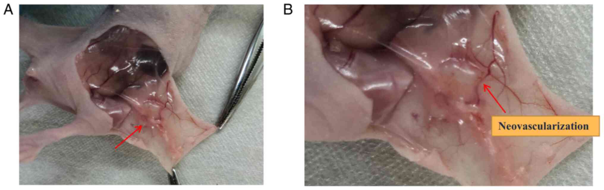
The survival rate of the nude mice and induction of subdermal
endometriosis in group 3 was 100% (Table II), and the subcutaneous anatomy
of nude mice is shown in Fig. 1.
As showed in Fig. 1B, the
endometriosis-like lesion was accompanied by the growth of blood
vessels that supplied the lesion.
| Table IIInjection of immortalized human
endometriosis epithelial and stromal cells into nude mice. |
Table II
Injection of immortalized human
endometriosis epithelial and stromal cells into nude mice.
| Group | Injected cells | Total number of
nude mice | Number of
endometriosis lesions |
|---|
| 1 | T HESCs | 6 | 0 |
| 2 | EECs | 6 | 0 |
| 3 | T HESCs + EECs | 8 | 8
(100%)a,b |
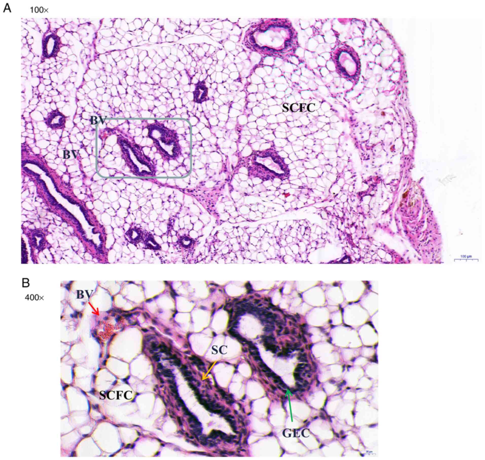
Histomorphology of endometriosis-like
lesions
In group 3, the endometriotic-like lesions observed
in the nude mice consisted of endometriosis-like glands (grey-lined
area) lined with columnar epithelial cell and surrounded by stromal
cells in the fibrous fatty connective tissue (Fig. 2A). Blood vessels were observed
around the glands (Fig. 2B; red
arrow). Histomorphologic analyses demonstrated that most of the
endometriosis glands were developed and fully organized glands
consisting of typical glandular structure (acini) lined with
glandular epithelial cells (green arrow) (Fig. 2B) surrounded by stromal cells
(yellow arrow).
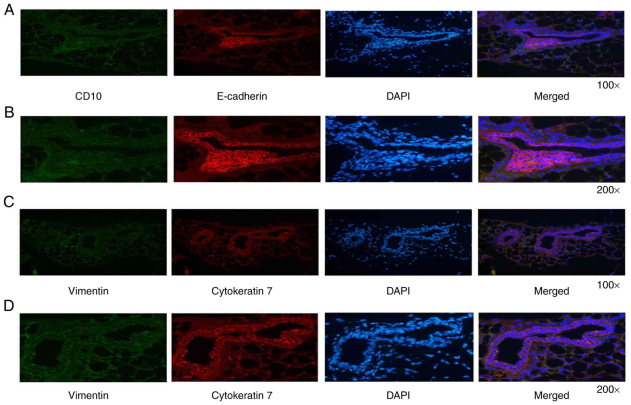
Immunofluorescence analysis of
endometriosis-like lesions
The human origin of cells detected in the
endometriotic-like structure in nude mice was demonstrated by
specific staining with anti-E-cadherin, anti-cytokeratin 7,
anti-vimentin and anti-CD10 antibodies (Fig. 3). Either cytokeratin or E-cadherin
represents a specific epithelial marker protein (20,21),
whereas vimentin and CD10 are both considered as specific stromal
cell marker proteins (22,23). As expected, glandular epithelial
cells were intensely stained for E-cadherin and cytokeratin 7
(Fig. 3A and B), and surrounding stromal cells were
mildly stained for CD10 and vimentin (Fig. 3C and D). These results confirm that the
subcutaneous nodules were endometriotic-like lesions with the
presence of endometriosis epithelial and stromal cells.
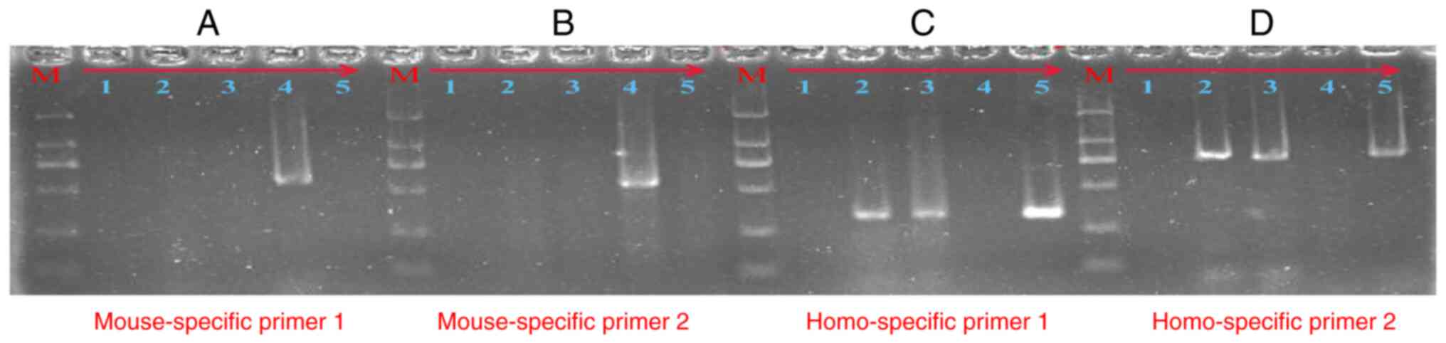
Identification of the cell origin of
endometriosis-like lesions
To confirm that the cells present in the
endometriosis-like lesions were of human origin, we respectively
designed species-specific primers of the mouse and human. The
source of the cells present in the endometriosis-like lesions was
determined by PCR technology. As showed in Fig. 4, the 571-bp (A) and 549-bp (B) PCR
products were amplified by the mouse-specific GAPDH primers, and
the 327-bp (C) and 772-bp (D) PCR products were amplified by the
human-specific GAPDH primers. Lanes 1-5 respectively represent:
no-template control, DNA from endometriosis-like lesion tissue (no.
2), DNA from endometriosis-like lesion tissue (no. 3), positive
control of mouse DNA, and positive control of human peripheral
blood DNA. As detailed in Fig. 4C
and D, the PCR product in lane 2
and lane 3 yielded a clear band at the expected size for human
GAPDH in the agarose gel electrophoresis following the regular and
touchdown PCR protocol. However, there were no bands in lane 2 and
lane 3 in Fig. 4A and B. These results confirm that the cells
present in the endometriosis-like lesions were of human origin.
Discussion
Endometriosis is formed by the growth and spread of
endometrial tissues (glands and stroma) outside the uterine body,
most commonly in the ovaries, with an incidence as high as 6-10%,
which seriously affects the quality of life of these patients
(3,24). At present, many animal models such
as rabbits, rats and primates have been established at home and
abroad (25-27).
Animal models can be divided into two categories, according to the
etiology of endometriosis. One is spontaneous animal models, which
only occur in primates, but its application has been limited due to
the high cost, high feeding requirements and low mold production
rate (28,29). Another is induced animal models, in
which mice are the most commonly used animal model for the study of
endometriosis. Rodents have short and regular estrus cycle, early
sexual maturity, strong fecundity, spontaneous ovulation, but no
endometrial shedding, thus this can only be used to establish an
induced endometriosis model (30-32).
In the induced endometriosis animal model,
endometrial tissues or cells are transplanted to parts outside the
uterine cavity of animals through various surgical procedures or
endometrial fragmentation injection methods to form endometriosis
lesions and induce the occurrence of disease (33). Autologous transplantation can be
used in the research of immunology and drug therapy (34,35).
However, there are differences between animal and human endometrium
in term of biochemical characteristics and other aspects, thus the
histological characteristics and biological response of autologous
transplantation animals are not completely applicable to humans
(26,36). Xenotransplantation involves
transplanting human endometrial tissue or endometrial cells into
immunocompromised mice. This method can preserve the biological
characteristics of human endometrium, and it can be used to study
the invasive ability of human endometrium, drug efficacy, side
effects and so on (37,38).
The results of the present study demonstrated for
the first time that single human immortalized endometriosis
epithelial cells or stromal cells cannot form endometriosis-like
lesions when transplanted subcutaneous into recipient nude mice.
Subcutaneous ectopic endometriosis was established when the two
types of cells were mixed and transplanted subcutaneously into nude
mice. It can be seen that the interaction between glandular
epithelial cells and stromal cells is essential in the formation of
endometriosis lesions (39). Our
results showed a 100% success rate in inducing endometriosis using
mixed human immortalized endometriosis cells, which is better than
other induced endometriosis models (40-42).
These lesions are characterized by the presence of endometriosis
glands lined with cylindrical and flattened epithelial cells and
surrounded by dense stromal cells in the subcutaneous adipose
tissue. Moreover, the cells present in the endometriosis-like
lesions were of human origin identified by PCR technology. All of
these results indicate the stability and reliability of this
experimental model.
This experiment has its advantages and disadvantages
compared with the animal models of ectopic abdominal cavity
constructed by predecessors (43).
Although intraperitoneal implantation creates an abdominal
environment similar to human disease conditions for the growth of
lesions, multiple exploratory laparotomy interfers with the
formation of lesions to a certain extent, leading to the decline of
animal life vitality, and it is difficult to evaluate the final
results with scattered and variable-sized lesions. The subcutaneous
implant used in this study can facilitate the continuous and
intuitive observation of the growth and change of the lesions,
which is convenient for operation and measurement. The success of
the model can be determined by touching the subcutaneous injection
site of the nude mouse. For the in vivo experiment of drug
treatment for endometriosis, it is convenient to observe changes in
the lesions on a daily basis. Human endometrial cells isolated and
cultured in vitro are more similar to the characteristics of
human pathology than animal models of homotransplantation, which
can not only be used to observe the growth and angiogenesis of
endometrial cells, but also study the cytochemistry and molecular
biology. The subcutaneous xenotransplantation model constructed in
this study can be used as a potential experimental model to
understand the molecular mechanism of human endometriosis. For
example, this model can be used to understand the heredity genes or
abnormal expression of key functional proteins, including the MAPK
signaling pathway and WNT signaling pathway, which improve our
understanding of endometriosis (3). In this model, we can overexpression,
knockdown or knockout the key genes in endometrial stromal cells,
and then observe the influence on the formation of endometriosis
lesions. However, the disadvantage of this animal model of
endometriosis is that it cannot be used in the immunological study
of endometriosis.
In summary, a mixture of human immortalized
endometriosis stromal cells and epithelial cells was able to
establish subcutaneous endometriosis lesions in nude mice. The
model built in this study can be used as a valuable tool today to
understand the molecular and cellular behavior of the pathogenesis
of endometriosis. Furthermore, this model has good application
value for the study of gene modification or abnormal protein
expression in the pathogenesis of endometriosis and could be used
to develop potential targeted therapy to treat endometriosis in
women.
Acknowledgements
Not applicable.
Funding
Funding: This work was supported by the National Natural Science
Foundation of China (no. 82060274) and the Natural Science
Foundation of Jiangxi Province (nos. 20202BABL216009 and
20181BAB215009).
Availability of data and materials
The datasets used and/or analyzed during the current
study are available from the corresponding author on reasonable
request.
Authors' contributions
YL and YZ conceived and designed the study. LPL and
ZML wrote the manuscript. ZZW, DMH, LPL, GC and BNC collected and
analyzed the data. YL, ZML, YFC and YZ analyzed and interpreted the
results. YL, YZ, YFC and ZML revised the manuscript in light of the
findings. YL and LPL confirm the authenticity of all the raw data.
All authors have been involved in revising the manuscript. All
authors read and approved the final manuscript.
Ethics approval and consent to
participate
All animal experiments and animal care were
performed under the approval of the Laboratory Animal Ethics
Committee of Nanchang Royo Biotech Co. Ltd (Nanchang, China;
approval no. RYE2019051001; May 9, 2019).
Patient consent for publication
Not applicable.
Competing interests
The authors declare that they have no competing
interests.
References
|
1
|
Koninckx PR, Ussia A, Adamyan L, Wattiez
A, Gomel V and Martin DC: Pathogenesis of endometriosis: The
genetic/epigenetic theory. Fertil Steril. 111:327–340.
2019.PubMed/NCBI View Article : Google Scholar
|
|
2
|
Rolla E: Endometriosis: Advances and
controversies in classification, pathogenesis, diagnosis, and
treatment. F1000Res 8: F1000 Faculty Rev-529, 2019.
|
|
3
|
Zondervan KT, Becker CM and Missmer SA:
Endometriosis. N Engl J Med. 382:1244–1256. 2020.PubMed/NCBI View Article : Google Scholar
|
|
4
|
Czyzyk A, Podfigurna A, Szeliga A and
Meczekalski B: Update on endometriosis pathogenesis. Minerva
Ginecol. 69:447–461. 2017.PubMed/NCBI View Article : Google Scholar
|
|
5
|
Vercellini P, Vigano P, Somigliana E and
Fedele L: Endometriosis: Pathogenesis and treatment. Nat Rev
Endocrinol. 10:261–275. 2014.PubMed/NCBI View Article : Google Scholar
|
|
6
|
D'Hooghe TM, Bambra CS, Cornillie FJ,
Isahakia M and Koninckx PR: Prevalence and laparoscopic appearance
of spontaneous endometriosis in the baboon (Papio anubis,
Papio cynocephalus). Biol Reprod. 45:411–416.
1991.PubMed/NCBI View Article : Google Scholar
|
|
7
|
Hastings JM and Fazleabas AT: A baboon
model for endometriosis: Implications for fertility. Reprod Biol
Endocrinol. 4 (Suppl 1)(S7)2006.PubMed/NCBI View Article : Google Scholar
|
|
8
|
Taylor HS, Alderman Iii M, D'Hooghe TM,
Fazleabas AT and Duleba AJ: Effect of simvastatin on baboon
endometriosis. Biol Reprod. 97:32–38. 2017.PubMed/NCBI View Article : Google Scholar
|
|
9
|
Burns KA, Pearson AM, Slack JL, Por ED,
Scribner AN, Eti NA and Burney RO: Endometriosis in the Mouse:
Challenges and progress toward a ‘Best Fit’ murine model. Front
Physiol. 12(806574)2022.PubMed/NCBI View Article : Google Scholar
|
|
10
|
Fan H: In-vitro models of human
endometriosis. Exp Ther Med. 19:1617–1625. 2020.PubMed/NCBI View Article : Google Scholar
|
|
11
|
Bruner-Tran KL, Webster-Clair D and Osteen
KG: Experimental endometriosis: The nude mouse as a xenographic
host. Ann NY Acad Sci. 955:328–342, 396-406. 2002.PubMed/NCBI View Article : Google Scholar
|
|
12
|
Perello M, Gonzalez-Foruria I, Castillo P,
Martínez-Florensa M, Lozano F, Balasch J and Carmona F: Oral
administration of pentoxifylline reduces endometriosis-like lesions
in a nude mouse model. Reprod Sci. 24:911–918. 2017.PubMed/NCBI View Article : Google Scholar
|
|
13
|
Wang N, Hong S, Tan J, Ke P, Liang L, Fei
H, Liu B, Liu L, Liu Y and Yu B: A red fluorescent nude mouse model
of human endometriosis: Advantages of a non-invasive imaging
method. Eur J Obstet Gynecol Reprod Biol. 176:25–30.
2014.PubMed/NCBI View Article : Google Scholar
|
|
14
|
Ni HJ, Zhang Z, Dai YD and Zhang SY:
Establishment of endometriosis subcutaneous model in
immunodeficient nude mice. Zhonghua Yi Xue Za Zhi. 96:2675–2677.
2016.PubMed/NCBI View Article : Google Scholar : (In Chinese).
|
|
15
|
Wang J and Ma X: Effects of estrogen and
progestin on expression of MMP-2 and TIMP-2 in a nude mouse model
of endometriosis. Clin Exp Obstet Gynecol. 39:229–233.
2012.PubMed/NCBI
|
|
16
|
Wu D, Lu P, Mi X and Miao J: Exosomal
miR-214 from endometrial stromal cells inhibits endometriosis
fibrosis. Mol Hum Reprod. 24:357–365. 2018.PubMed/NCBI View Article : Google Scholar
|
|
17
|
Zeitvogel A, Baumann R and
Starzinski-Powitz A: Identification of an invasive,
N-cadherin-expressing epithelial cell type in endometriosis using a
new cell culture model. Am J Pathol. 159:1839–1852. 2001.PubMed/NCBI View Article : Google Scholar
|
|
18
|
American Veterinary Medical Association
2020. AVMA Guidelines for the Euthanasia of Animals. 9th edition.
American Veterinary Medical Association, Schaumburg, IL, 2020.
|
|
19
|
Luo Y, Zou Y, Wu J, Zhang ZY, Liu FY, Li
LP and Huang OP: The mitochondrial DNA 4977-bp deletion and copy
number alteration in Han Chinese samples with uterine fibroids. Ann
Hum Genet. 83:220–230. 2019.PubMed/NCBI View Article : Google Scholar
|
|
20
|
Guerrieri C, Franlund B and Boeryd B:
Expression of cytokeratin 7 in simultaneous mucinous tumors of the
ovary and appendix. Mod Pathol. 8:573–576. 1995.PubMed/NCBI
|
|
21
|
Mishra A, Galvankar M, Vaidya S, Chaudhari
U and Modi D: Mouse model for endometriosis is characterized by
proliferation and inflammation but not epithelial-to-mesenchymal
transition and fibrosis. J Biosci. 45(105)2020.PubMed/NCBI
|
|
22
|
Atkins HM, Lombardini ED, Caudell DL, Appt
SE, Dubois A and Cline JM: Decidualization of endometriosis in
macaques. Vet Pathol. 53:1252–1258. 2016.PubMed/NCBI View Article : Google Scholar
|
|
23
|
Greaves E, Cousins FL, Murray A,
Esnal-Zufiaurre A, Fassbender A, Horne AW and Saunders PT: A novel
mouse model of endometriosis mimics human phenotype and reveals
insights into the inflammatory contribution of shed endometrium. Am
J Pathol. 184:1930–1939. 2014.PubMed/NCBI View Article : Google Scholar
|
|
24
|
Giudice LC: Clinical practice.
Endometriosis. N Engl J Med. 362:2389–2398. 2010.PubMed/NCBI View Article : Google Scholar
|
|
25
|
Nishimoto-Kakiuchi A, Netsu S, Matsuo S,
Hayashi S, Ito T, Okabayashi S, Yasmin L, Yuzawa K, Kondoh O, Kato
A, et al: Characteristics of histologically confirmed endometriosis
in cynomolgus monkeys. Hum Reprod. 31:2352–2359. 2016.PubMed/NCBI View Article : Google Scholar
|
|
26
|
Saltan G, Suntar I, Ozbilgin S, Ilhan M,
Demirel MA, Oz BE, Keleş H and Akkol EK: Viburnum opulus L: A
remedy for the treatment of endometriosis demonstrated by rat model
of surgically-induced endometriosis. J Ethnopharmacol. 193:450–455.
2016.PubMed/NCBI View Article : Google Scholar
|
|
27
|
Simitsidellis I, Gibson DA and Saunders
PTK: Animal models of endometriosis: Replicating the aetiology and
symptoms of the human disorder. Best Pract Res Clin Endocrinol
Metab. 32:257–269. 2018.PubMed/NCBI View Article : Google Scholar
|
|
28
|
D'Hooghe TM, Kyama CM, Chai D, Fassbender
A, Vodolazkaia A, Bokor A and Mwenda JM: Nonhuman primate models
for translational research in endometriosis. Reprod Sci.
16:152–161. 2009.PubMed/NCBI View Article : Google Scholar
|
|
29
|
Harirchian P, Gashaw I, Lipskind ST,
Braundmeier AG, Hastings JM, Olson MR and Fazleabas AT: Lesion
kinetics in a non-human primate model of endometriosis. Hum Reprod.
27:2341–2351. 2012.PubMed/NCBI View Article : Google Scholar
|
|
30
|
Chadchan SB, Cheng M, Parnell LA, Yin Y,
Schriefer A, Mysorekar IU and Kommagani R: Antibiotic therapy with
metronidazole reduces endometriosis disease progression in mice: A
potential role for gut microbiota. Hum Reprod. 34:1106–1116.
2019.PubMed/NCBI View Article : Google Scholar
|
|
31
|
Li M, Zhou Y and Taylor HS: MiR-451a
inhibition reduces established endometriosis lesions in mice.
Reprod Sci. 26:1506–1511. 2019.PubMed/NCBI View Article : Google Scholar
|
|
32
|
Sun H, Li D, Yuan M, Li Q, Zhen Q, Li N
and Wang G: Macrophages alternatively activated by
endometriosis-exosomes contribute to the development of lesions in
mice. Mol Hum Reprod. 25:5–16. 2019.PubMed/NCBI View Article : Google Scholar
|
|
33
|
Bruner-Tran KL, Mokshagundam S, Herington
JL, Ding T and Osteen KG: Rodent models of experimental
endometriosis: Identifying mechanisms of disease and therapeutic
targets. Curr Womens Health Rev. 14:173–188. 2018.PubMed/NCBI View Article : Google Scholar
|
|
34
|
Korbel C, Menger MD and Laschke MW: Size
and spatial orientation of uterine tissue transplants on the
peritoneum crucially determine the growth and cyst formation of
endometriosis-like lesions in mice. Hum Reprod. 25:2551–2558.
2010.PubMed/NCBI View Article : Google Scholar
|
|
35
|
Pelch KE, Sharpe-Timms KL and Nagel SC:
Mouse model of surgically-induced endometriosis by
auto-transplantation of uterine tissue. J Vis Exp.
59(e3396)2012.PubMed/NCBI View
Article : Google Scholar
|
|
36
|
Wallwiener D, Meyer A and Bastert G:
Adhesion formation of the parietal and visceral peritoneum: An
explanation for the controversy on the use of autologous and
alloplastic barriers? Fertil Steril. 69:132–137. 1998.PubMed/NCBI View Article : Google Scholar
|
|
37
|
Hull ML, Escareno CR, Godsland JM, Doig
JR, Johnson CM, Phillips SC, Smith SK, Tavaré S, Print CG and
Charnock-Jones DS: Endometrial-peritoneal interactions during
endometriotic lesion establishment. Am J Pathol. 173:700–715.
2008.PubMed/NCBI View Article : Google Scholar
|
|
38
|
Hull ML, Prentice A, Wang DY, Butt RP,
Phillips SC, Smith SK and Charnock-Jones DS: Nimesulide, a COX-2
inhibitor, does not reduce lesion size or number in a nude mouse
model of endometriosis. Hum Reprod. 20:350–358. 2005.PubMed/NCBI View Article : Google Scholar
|
|
39
|
Nisolle M, Casanas-Roux F and Donnez J:
Early-stage endometriosis: Adhesion and growth of human menstrual
endometrium in nude mice. Fertil Steril. 74:306–312.
2000.PubMed/NCBI View Article : Google Scholar
|
|
40
|
Eggermont J, Donnez J, Casanas-Roux F,
Scholtes H and Van Langendonckt A: Time course of pelvic
endometriotic lesion revascularization in a nude mouse model.
Fertil Steril. 84:492–499. 2005.PubMed/NCBI View Article : Google Scholar
|
|
41
|
Gonzalez-Ramos R, Van Langendonckt A,
Defrere S, Lousse JC, Mettlen M, Guillet A and Donnez J: Agents
blocking the nuclear factor-kappaB pathway are effective inhibitors
of endometriosis in an in vivo experimental model. Gynecol Obstet
Invest. 65:174–186. 2008.PubMed/NCBI View Article : Google Scholar
|
|
42
|
Pereira FE, Almeida PR, Dias BH,
Vasconcelos PR, Guimaraes SB and Medeiros Fd: Development of a
subcutaneous endometriosis rat model. Acta Cir Bras. 30:6–12.
2015.PubMed/NCBI View Article : Google Scholar
|
|
43
|
Banu SK, Starzinski-Powitz A, Speights VO,
Burghardt RC and Arosh JA: Induction of peritoneal endometriosis in
nude mice with use of human immortalized endometriosis epithelial
and stromal cells: A potential experimental tool to study molecular
pathogenesis of endometriosis in humans. Fertil Steril. 91 (Suppl
5):S2199–S2209. 2009.PubMed/NCBI View Article : Google Scholar
|