Introduction
Inflammatory bowel disease (IBD), including Crohn's
disease (CD) and ulcerative colitis (UC), represents a group of
intestinal disorders that cause prolonged inflammation of the
digestive tract. IBD affects ~0.1% of the Western population and
has contributed to an increased risk of morbidity (1-3).
A growing incidence of IBD has also been reported worldwide
(4,5). According to a recent report, the
annual incidence rate per 100,000 people in Asia is 0.5 to 21.6, in
Central America and South America it is 0.4 to 3.0, in Europe it is
0 to 21.3, in North America it is 2.4 to 15.4, and in Oceania it is
5.2 to 6.8(5).
The clinical symptoms of IBD include diarrhea,
repeated rectal bleeding, abdominal pain, body weight (BW) loss and
severe malnutrition (1-3).
Although the etiology of IBD remains unclear, an increasing body of
evidence has revealed a multifactorial disease associated with
susceptible genes, intestinal microbiota and environmental factors,
leading to the dysregulation of both adaptive and innate immune
systems. This can be characterized by the abnormal activation of
intestinal immune cells, followed by the release of a range of
inflammatory factors. It is well accepted that numerous cytokines,
such as interleukin (IL)-1β (6-8),
IL-6(6), IL-12 (9,10),
IL-18 (9,11), tumor necrosis factor-α (TNF-α)
(7,12,13)
and interferon-γ (IFN-γ) (8), are
involved in the pathogenesis of both types of IBD and are
considered indicators of therapeutic efficacy. Corticosteroids,
aminosalicylate, some biological agents and immunosuppressive
agents are mainly used for the treatment of IBD; however, they
cannot fundamentally prevent the disease recurring (14,15).
Notably, patients with IBD mainly experience a loss
of appetite due to nausea, vomiting, abdominal pain and diarrhea,
and folate deficiency has classically been associated with anemia
in these patients (16-19).
Thus, vitamins, such as vitamin D, vitamin B6, vitamin B12 and
vitamin C, are occasionally recommended for patients with IBD
(20-24).
Several studies have shown that vitamin D supplementation can
prevent and ameliorate symptoms of IBD (25-27).
Animals with either vitamin D deficiency or defective vitamin D
receptors are susceptible to developing IBD (28-30).
Furthermore, vitamin D can change the composition of the intestinal
flora through regulation of the expression of antibacterial
peptides, in addition to immune regulation (31). Phillips et al (32) demonstrated that vitamin D may
provide some protection against increased mitochondrial dysfunction
and inflammation in the placenta of obese women. In addition, Hahn
et al (33) indicated that
long-term vitamin D supplementation may be effective for reducing
the incidence of autoimmune diseases. During the coronavirus 2019
(COVID-19) pandemic, vitamin D was found to be efficacious in
attenuating the release of inflammatory cytokines after viral
infection through its antimicrobial and anti-inflammatory
properties (34). Moreover,
moderate daily doses of vitamin D have been shown to slow down the
progression of Parkinson's disease and prevent COVID-19 infection
in the elderly (34,35). In addition, it has been reported
that although genetic differences can lead to different benefits of
vitamin D in different populations, 40-60 ng/ml (100-150 mmol/l) of
serum 25-hydroxyvitamin D is the concentration that achieves the
best overall health benefits (36). In infants and young children,
vitamin D supplementation can also reduce the incidence of
childhood asthma and other allergic diseases (37). These functions maintain the
integrity of intestinal mucosa as a surface barrier and repair
mucosal permeability (38). By
inhibiting the activation of cellular immunity and cytotoxic T
cells, vitamin D supplementation can also adjust the immune
response. Therefore, vitamin D deficiency can endanger the mucosal
barrier, resulting in mucosal injury and increasing the risk of IBD
(19,22). Dextran sodium sulfate (DSS)-induced
colitis in rats is a model resembling human UC. Other studies have
shown that adequate vitamin D supplementation can be effective in
preventing respiratory diseases. Epithelial cells, dendritic cells
(DCs) and macrophages in the lungs are efficacious in producing
active vitamin D, which enhances the production of cytokines with
anti-inflammatory functions and promotes the production of
anti-viral peptides (39).
Previous evidence has suggested that oxidative stress is an
important component in the pathophysiology of IBD (40-44),
and some antioxidants have exhibited protective and healing effects
against DSS-induced UC in rats (45).
Vitamin E is a well-accepted, relatively safe
antioxidant in cellular membranes and can protect membrane lipids
from peroxidation. A previous study revealed that vitamin E has
anti-inflammatory effects following inflammation in the lung
(46). Thus, vitamin E could
exhibit a strong effect on UC due to its anti-inflammatory activity
and high antioxidant capacity. It has been shown that vitamin D and
vitamin E can also have a preventive effect for the treatment of
hair loss. Vitamin D maintains serum levels of calcium and
phosphorus, in addition to its anti-inflammatory and immunological
effects. Notably, vitamin D regulates keratinocyte growth and
differentiation through the nuclear vitamin D receptor to prevent
hair loss and type II rickets (47). Vitamin E can also prevent hair loss
and baldness by maintaining the oxidant/antioxidant balance
(47). However, Carrier et
al (48) reported that vitamin
E supplementation increased clinical observation (CO) scores from
colonic inflammation, and that it did not affect oxidative stress,
thus indicating that vitamin E may have an unclear mechanism of
reducing inflammation. By contrast, another study reported that
vitamin E showed a dual-effect of anti-inflammatory and antioxidant
activities on acetic acid-induced UC in rats (49).
Various animal models of IBD have been developed in
the last decade, which are valuable and indispensable tools for
evaluating different therapeutic options for IBD (6,7,9). The
DSS-induced UC model in rats has some advantages compared with
other animal models of UC, and is a widely used model resembling
human UC. Rats exposed to DSS in drinking water develop
inflammation in the large intestine and exhibit signs such as
diarrhea, anemia, BW loss and histological inflammation, including
inflammatory cell infiltration, muscularis mucosae erosion and
ulcers, with increased levels of pro-inflammatory cytokines (e.g.,
IFN-γ, TNF-α, IL-1, IL-6 and IL-12) (40,45,50).
Whether vitamin E plays a protective role, similar
to vitamin D, in conditions such as UC still needs to be
elucidated, although vitamin E deficiency was previously found in
patients with IBD (48,50). Thus, the present study aimed to
systemically evaluate the anti-inflammatory effects of vitamin E on
a rat model of DSS-induced UC and to compare the effects with those
of vitamin D.
Materials and methods
Chemicals and reagents
All reagents and chemicals used in the present study
were purchased from the companies listed below, unless otherwise
stated. DSS salt (M.W., 36,000-50,000) was purchased from MP
Biomedicals LLC. DL-α-Tocopheryl acetate (all-rac-α-Tocopheryl
acetate Vitamin E acetate; M.W., 472.74 g/mol) was obtained from
MilliporeSigma. Vitamin D (1α, 25-dihydroxycholecalciferol; M.W.,
416.6 g/mol) was purchased from Roche Diagnostics (Shanghai) Co.,
Ltd. Paraformaldehyde was obtained from Sinopharm Chemical Reagent
Co., Ltd., with sodium chloride injection (0.9%) from Shijiazhuang
No. 4 Pharmaceutical Co., Ltd.). Radio-immunoprecipitation (RIPA)
assay lysis buffer was purchased from Qingdao Jisskang Biotechology
Co., Ltd.) and prednisolone acetate from Huazhong Pharmaceutical
Co., Ltd.).
Animals and treatment
Male Wistar rats (age, 5 weeks; n=120; n=12-24
rats/group; weight, 200±10 g) were purchased from Beijing Vital
River Laboratory Animal Technology Co., Ltd. The rats were housed
in a vivarium under a 12-h light/dark cycle at constant temperature
(22±2˚C) and humidity (50-60%), with free access to food and water,
in accordance with the principles of the Good Lab Practice (GLP)
guidelines presented by the National Beijing Center for Drug Safety
Evaluation and Research (51-53).
Animals were acclimated to the laboratory for 1 week before
starting experiments.
After the adaptation period, the rats were divided
into eight groups [(G1, G2, G3, G4, G5a, G5b, G5c and G6], and the
doses are summarized in Table I.
UC in male Wistar rats was induced by adding 5% (w/v) DSS to the
drinking water from day 1 to day 7, whereas rats in the control
group (G1) received only tap water (Fig. 1). After successfully establishing
the rat model of DSS-induced UC according to weight loss and stool
examination, rats in the G4 to G6 groups received different doses
of vitamin E and vitamin D (Table
I) from day 8 to day 14, and were sacrificed on day 15 for
pathological examination. Meanwhile, half of the rats in the G1 and
G2 groups were sacrificed on day 8 for pathological detection. The
remaining rats in the G1 and G2 groups received saline from day 8
to day 14, and were sacrificed on day 15 for pathological
examination. The rats were anesthetized by intraperitoneal
injection of 5% pentobarbital sodium (50 mg/kg) and blood was
collected from the abdominal aorta following anesthesia; mice were
sacrificed by exsanguination.
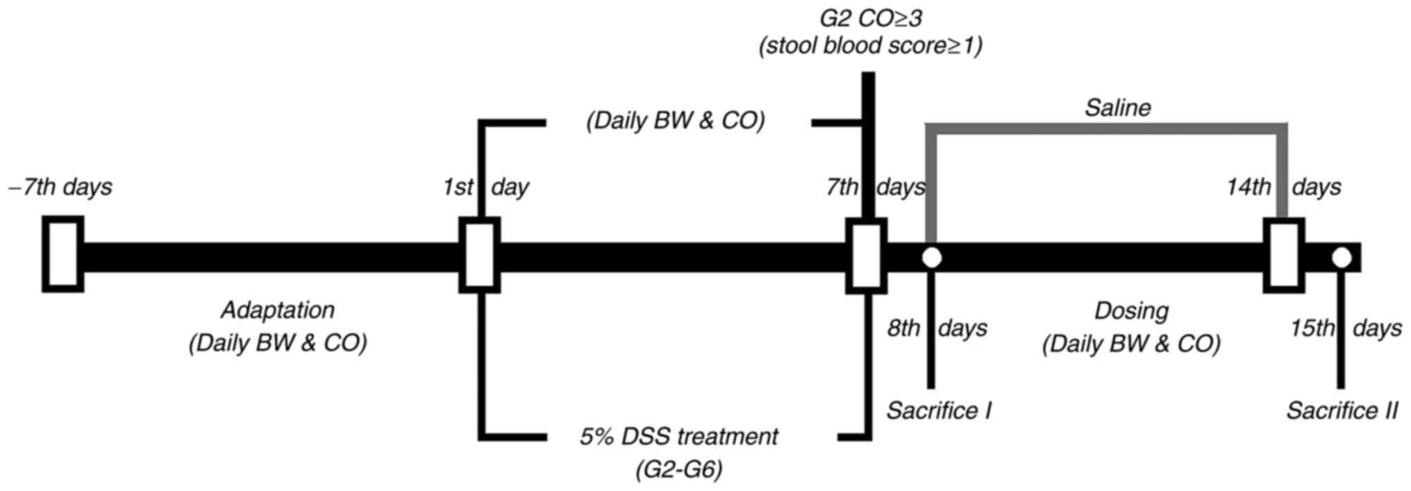 | Figure 1Schematic diagram of the experimental
process and study design. DSS, dextran sodium sulfate. Saline,
G1-G6 were administrated with saline from day 7 to day 14;
Sacrifice I, half of the rats in G1 and G2; Sacrifice II, all of
the remaining rats; CO, clinical observation; BW, body weight; G1,
control group; G2, 5% DSS group; G3, prednisolone group; G4,
vitamin D group; G5a-c, vitamin E (low, medium and high) groups;
G6, vitamin D + vitamin E group; DSS, dextran sodium sulfate. |
 | Table IGroups of rats analyzed in the
present study. |
Table I
Groups of rats analyzed in the
present study.
| Group name | Treatment | Number of rats | Route of
administration | Dosage of
administration |
|---|
| G1 | Control
(vehicle) | 24 | Drinking water | |
| G2 | DSS (Day 1-7) | 24 | Dissolved in
drinking water | 5% |
| G3 | DSSa (Day 1-7) + prednisolone (Day
8-14) | 12 | Orally administered
(Day 8-14) | 1.0 mg/kg |
| G4 | DSSa (Day 1-7) + vitamin D (Day
8-14) | 12 | Orally administered
(Day 8-14) | 50 ng |
| G5a | DSSa (Day 1-7) + vitamin E (low dose)
(Day 8-14) | 12 | Orally administered
(Day 8-14) | 6 IU/kg |
| G5b | DSSa (Day 1-7) + vitamin E (medium
dose) (Day 8-14) | 12 | Orally administered
(Day 8-14) | 30 IU/kg |
| G5c | DSSa (Day 1-7) + vitamin E (high
dose) (Day 8-14) | 12 | Orally administered
(Day 8-14) | 150 IU/kg |
| G6 | DSSa (Day 1-7) + vitamin D + vitamin
E (Day 8-14) | 12 | Orally administered
(Day 8-14) | 50 ng + 30
IU/kg |
Study design and clinical observation
(CO) score
For each rat in the six groups, BW and CO score were
recorded daily. Half of the rats in both the G1 and G2 groups were
sacrificed on day 8 to collect the blood and tissue samples for
histopathological examination, and cytokine measurement. The
remaining rats also underwent these tests, when they were culled on
day 15. The experimental design is shown in Fig. 1.
For the CO score, both stool score and stool blood
score were recorded separately and combined to generate a total CO
score with a maximum score of 5. Stool scoring was performed as
follows: 0, normal; 1, moist/sticky stool; 2, stool in or around
anus; 3, diarrhea. Stool blood scoring was carried out as follows:
0, no blood; 1, evidence of blood in stool or around anus; 2,
severe bleeding. For CO scoring, the experiment was conducted
according to a GLP standard. No stool images are provided in this
study.
For the colon weight/length ratio (%), half of the
rats in groups G1 and G2 were sacrificed on day 8 and the colon
weight/length ratio (%) was measured. The remaining rats in groups
G1-G6 were sacrificed on day 15 and the colon weight/length ratio
(%) was measured.
Gross and histopathological evaluation
of hematoxylin and eosin (H&E)-stained colonic tissue
For the histopathological examination, the proximal
colon (1 cm) was fixed in 10% formaldehyde for 24 h at room
temperature. The tissues were then dehydrated in a graded series of
alcohol [70% (2 h), 80% (2 h), 90% (2 h), 95% alcohol (2 h),
anhydrous alcohol (1 h)]; then cleared with xylene for 1 h. The
tissues were then infiltrated with paraffin at 58-60˚C for 2 h and
the paraffin blocks were sliced into ~5-µm sections at 45˚C and
mounted onto glass slides. The sections were dewaxed with xylene
for 15 min and were incubated with alcohol of different
concentrations (100, 95, 80 and 70% alcohol) to remove paraffin.
H&E staining was performed using a standard staining procedure;
the sections were incubated in hematoxylin solution for 5 min, then
incubated with eosin for 2-3 min. Eight randomly selected fields
(magnification, x100) in each slide were observed under a light
microscope (Olympus BX43; Olympus Corporation) as described
previously (54).
Enzyme-linked immunosorbent assay
(ELISA) of colonic cytokines
Briefly, colon tissues were homogenized in 1 ml
ice-cold RIPA assay lysis buffer, containing 1% protease inhibitor
cocktail and 1% phosphatase inhibitor cocktail. The lysate was
centrifuged at 15,000 x g for 15 min at 4˚C, and the supernatant
was transferred to 96-well ELISA plates before measurement of the
following inflammatory markers: IL-6 (cat. no. SEKR-0005), IL-12
(cat. no. SEKR-0057), IL-18 (cat. no. SEKR-0054), TNF-α (cat. no.
SEKR-0009) and IFN-γ (cat. no. SEKR-0008) using kits according to
the manufacturer's protocols.
Statistical analysis
Continuous data are presented as the mean ± standard
error of the mean, whereas CO score data are expressed as the
median and interquartile range. Comparisons between two groups were
performed using the unpaired Student's t-test. Multiple comparisons
were performed by one- or two-way analysis of variance and
Bonferroni's post hoc test. For the CO scores, Kruskal-Wallis was
conducted followed by Dunn's post hoc test for statistical
analysis. GraphPad Prism 6.0 software (GraphPad Software, Inc.) was
used to perform the statistical analysis. P<0.05 was considered
to indicate a statistically significant difference.
Results
Both vitamin D and vitamin E prevent
BW loss in a rat model of DSS-induced UC
DSS can cause damage to the colonic mucosal barrier,
leading to gut inflammation and BW loss (40). The present study demonstrated that
rats that received drinking water with 5% DSS had significantly
more BW loss than those in the control group (G1) from day 4 to day
8 (Fig. 2, F=7.43, P=0.01). Rats
in the vitamin D-treated group (G4), vitamin E-treated groups
(G5a-c), vitamin D + vitamin E group (G6) and prednisolone group
(G3) had a similar degree of BW loss as rats in G2 from day 4 to
day 8 (F=2.83, P=0.998). However, rats in G4, G5a-c and G6
experienced a rapid BW recovery from day 11 to day 15 compared with
those in G2 (F=6.13, P=0.01), whereas no significant difference was
found in BW between G3 and G6 (F=5.97, P=0.995).
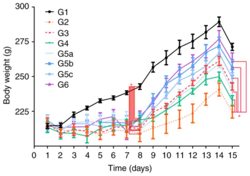 | Figure 2Measurements of body weight in G1,
G2, G3, G4, G5a-c and G6. The body weight in each group was
measured from day 1 to day 15. G1, control group; G2, 5% DSS group;
G3, prednisolone group; G4, vitamin D group; G5a-c, vitamin E (low,
medium and high) groups; G6, vitamin D + vitamin E group; DSS,
dextran sodium sulfate. *P<0.05. |
Vitamin D and vitamin E ameliorate the
clinical symptoms of UC in rats
In the experiment, CO scoring was performed daily
(Fig. 3). Compared with rats in
G1, rats treated with DSS (G2, G3, G4, G5a-c and G6) showed obvious
symptoms of diarrhea with noticeably higher CO scores from days 5
to 8 (χ2=19.28, P=0.007; Fig. 3). Rats in G3, G4, G5a-c and G6 had
a lower blood score and firmer stools than those in G2 from day 9
to day 15. On day 15, CO scores in the drug-treated groups (G3, G4,
G5a-c and G6) were significantly lower than those in G2
(χ2=12.77, P=0.04; Fig.
3).
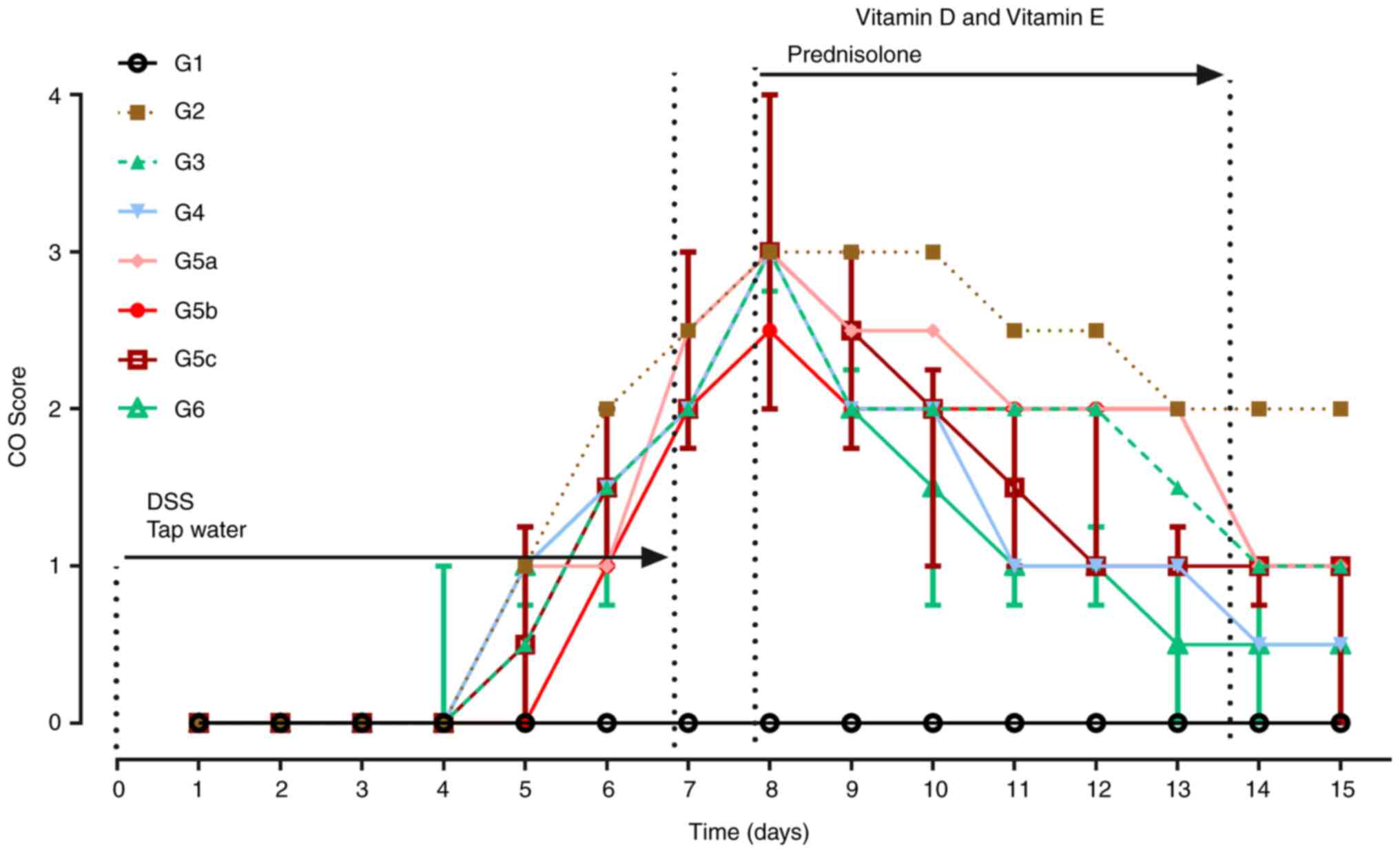 | Figure 3Changes in CO scores across the whole
experiment. From day 1 to day 7, rats in G1 received drinking
water, whereas rats in the other groups received water containing
DSS (5%, w/v). Compared with those in G1, CO scores were
significantly higher in the other groups (G2-G6) on day 8. On day
15, CO scores in G3, G4, G5c, G5b and G6 were significantly lower
than those in G2, whereas there was no statistically significant
difference between G2 and G5a. Data are presented as the median and
interquartile. n=12 for each group. G1, control group; G2, 5% DSS
group; G3, prednisolone group; G4, vitamin D group; G5a-c, vitamin
E (low, medium and high) groups; G6, vitamin D + vitamin E group;
CO, clinical observation; DSS, dextran sodium sulfate. |
Both vitamin D and vitamin E inhibit
DSS-induced colonic inflammation
To verify whether the rat model of DSS-induced UC
was successfully established, half of the rats in G1 and G2 were
sacrificed for histopathological examinations on day 8. Rats in G2
had more severe macroscopic inflammation than those in G1 (Fig. 4A). Consistent with macroscopic
observations, the colon length in G2 on day 8 was shorter than that
in G1 (Fig. 4A). On day 15, the
colon length in G3 and G6 was longer than that in G2 (Fig. 4B). Compared with G2, there was no
difference in colon length in G4 and G5, but there was a marked
difference in colon length between G6 and G2. Although colon length
in G6 was not as long as that in G3, the experimental result
provided novel evidence for the treatment of UC. These findings
indicated that the combination of vitamin D and vitamin E may be
better than individual use.
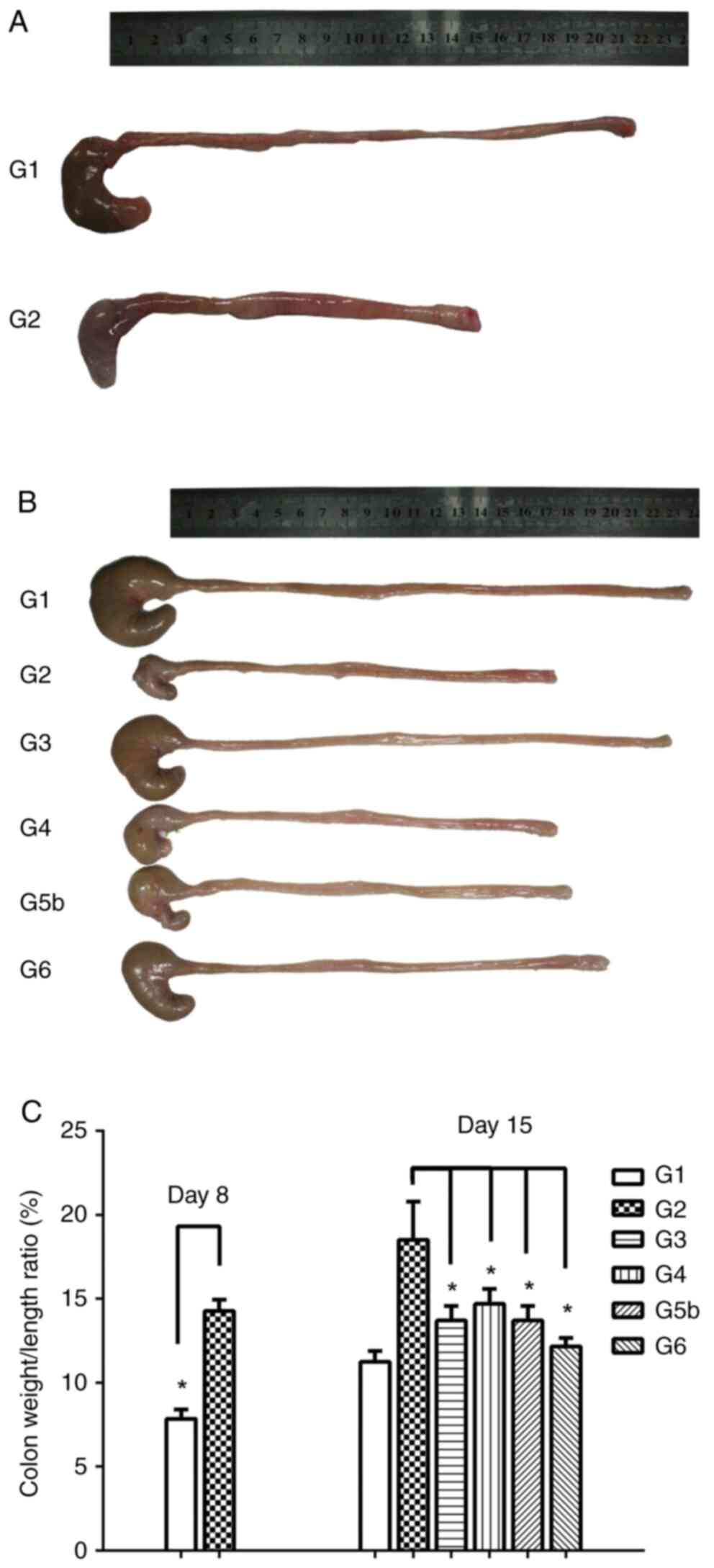 | Figure 4Vitamin D and vitamin E inhibit
DSS-induced colonic inflammation. (A) Macroscopic inflammation was
assessed, and the length of colons was measured for rats in G1 and
G2 on day 8. Colon length was shorter in G2 than that in G1. (B)
Colon length was measured for rats in the six groups on day 15.
Compared with that in G2, the colon length of rats in G3 and G6 was
markedly longer. (C) Colon weight to length ratio (%) in G1 and G2
on day 8, and in G1, G2, G3, G4, G5 and G6 on day 15. Compared with
that in G2, the colon weight/length ratio in G3, G4, G5b and G6 was
significantly decreased. Data are presented as the mean ± SEM. n=6.
*P<0.05. G1, control group; G2, 5% DSS group; G3,
prednisolone group; G4, vitamin D group; G5a-c, vitamin E (low,
medium and high) groups; G6, vitamin D + vitamin E group; DSS,
dextran sodium sulfate. |
In addition, the colon weight/length ratio (%) in G1
and G2 on day 8 was calculated. Compared with that in G1, the colon
weight/length ratio on day 8 in G2 was significantly increased
(F=-5.38, P=0.01), which suggested that the rat model of
DSS-induced UC was successfully established (Fig. 4C). Compared with that in G2, the
colon weight/length ratio on day 15 in G3, G4, G5b and G6 was
significantly lower (F3=5.67, P=0.03;
F4=3.83, P=0.049; F5b=4.08, P=0.048;
F6=4.97, P=0.04), indicating that both vitamin D and
vitamin E inhibited DSS-induced colonic inflammation.
Vitamin D and vitamin E promote the
recovery of DSS-induced UC in rats
One-half of the rats in G1 and G2 were sacrificed
after anesthesia, and their colon tissues were collected for
histopathological examination on day 8. Compared with in G1
(Fig. 5A), rats in G2 exhibited
edema, extensive ulceration of the epithelial layer, crypt damage
to the bowel wall, and infiltration of granulocytes and mononuclear
cells into the mucosa (Fig. 5B),
which revealed that DSS successfully induced UC in the rat
model.
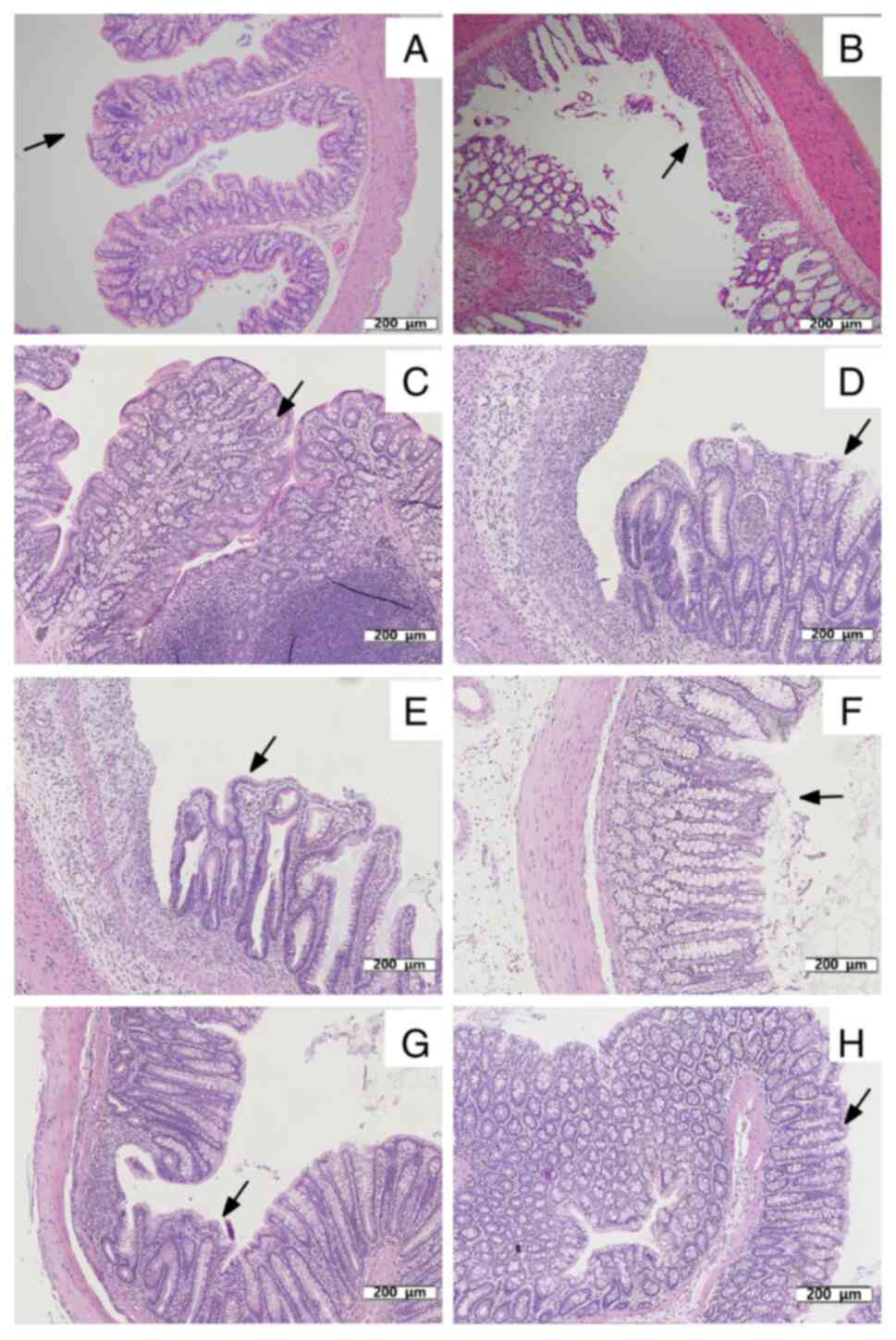 | Figure 5Histopathological examination of
colon tissues of rats. (A) Colon tissue of rats in G1 on day 8. The
mucosa is intact and free of inflammatory cell infiltration, as
indicated by the arrow. (B) Acute colitis induced by DSS on day 8.
Mucosal injury was shown as focal ulceration, epithelial necrosis
and infiltration of inflammatory cells, as indicated by the arrow.
Colon tissue of rats in (C) G1, (D) G2, (E) G3, (F) G4 (G) G5b and
(H) G6 on day 15. (C) Colon tissue of rats in G1 on day 15. Intact
mucosa and no inflammatory cell infiltration was indicated by
arrows. (D) Colon tissue of rats in G2 on day 15. The mucosal
injury was characterized by focal ulceration, epithelial necrosis
and infiltration of inflammatory cells, as indicated by arrows. (E)
Colon tissue of rats in G3 treated with prednisolone on day 15.
Colon tissue exhibited a relatively intact mucosa with only a small
infiltration of inflammatory cells, as indicated by arrows. (F)
Colon tissue of rats in G4 treated with vitamin D on day 15. The
colonic mucosa was intact but with some epithelial cell necrosis
and infiltration of inflammatory cells, as indicated by the arrows.
(G) Colon tissue of rats in G5b treated with vitamin E on day 15.
The colonic mucosa was intact and infiltrated by a few inflammatory
cells, as indicated by the arrows. (H) Colon tissue of rats in G6
treated with vitamin D and vitamin E on day 15. The colonic tissue
mucosa was more intact than that in G4 and G5b, as indicated by the
arrows. Treatment with vitamin D, vitamin E, vitamin D + vitamin E
and prednisolone reduced the morphological alterations associated
with DSS administration and protected the mucosal architecture.
Colon tissues in the figures were all stained with hematoxylin and
eosin. Scale bar, 200 µm; magnification, x110. G1, control group;
G2, 5% DSS group; G3, prednisolone group; G4, vitamin D group;
G5a-c, vitamin E (low, medium and high) groups; G6, vitamin D +
vitamin E group; DSS, dextran sodium sulfate. |
On day 15, the remaining rats were sacrificed after
anesthesia, and their colon tissues were also collected for
histopathological examination and detection of cytokines. The colon
tissues from rats in G1 did not exhibit inflammation (Fig. 5C). Compared with rats in G2
(Fig. 5D), treatment with
prednisolone (Fig. 5E), vitamin D
(Fig. 5F), vitamin E (medium)
(Fig. 5G), and vitamin D + vitamin
E (Fig. 5H) reduced DSS-induced
UC, including the severity of inflammation, extent of injury and
crypt damage. The histopathological examinations of colon tissues
suggested that vitamin D, vitamin E and their combination
ameliorated DSS-induced UC and promoted recovery.
Vitamin D and vitamin E decrease the
levels of inflammatory mediators
To determine the anti-inflammatory effects of
vitamin D and vitamin E on the rat model of DSS-induced UC, five
inflammatory markers (IL-6, IL-12, IL-18, TNF-α and IFN-γ) were
assessed. Compared with those in G1, the levels of the five
cytokines in G2 were significantly elevated on day 8
(FTNF-α=-2.99, P=0.03;
FIFN-γ=-172.53, P=0.04;
FIL-18=16.39, P=0.01; FIL-12=33.76, P=0.03;
FIL-6=2.88, P=0.04; Fig.
6).
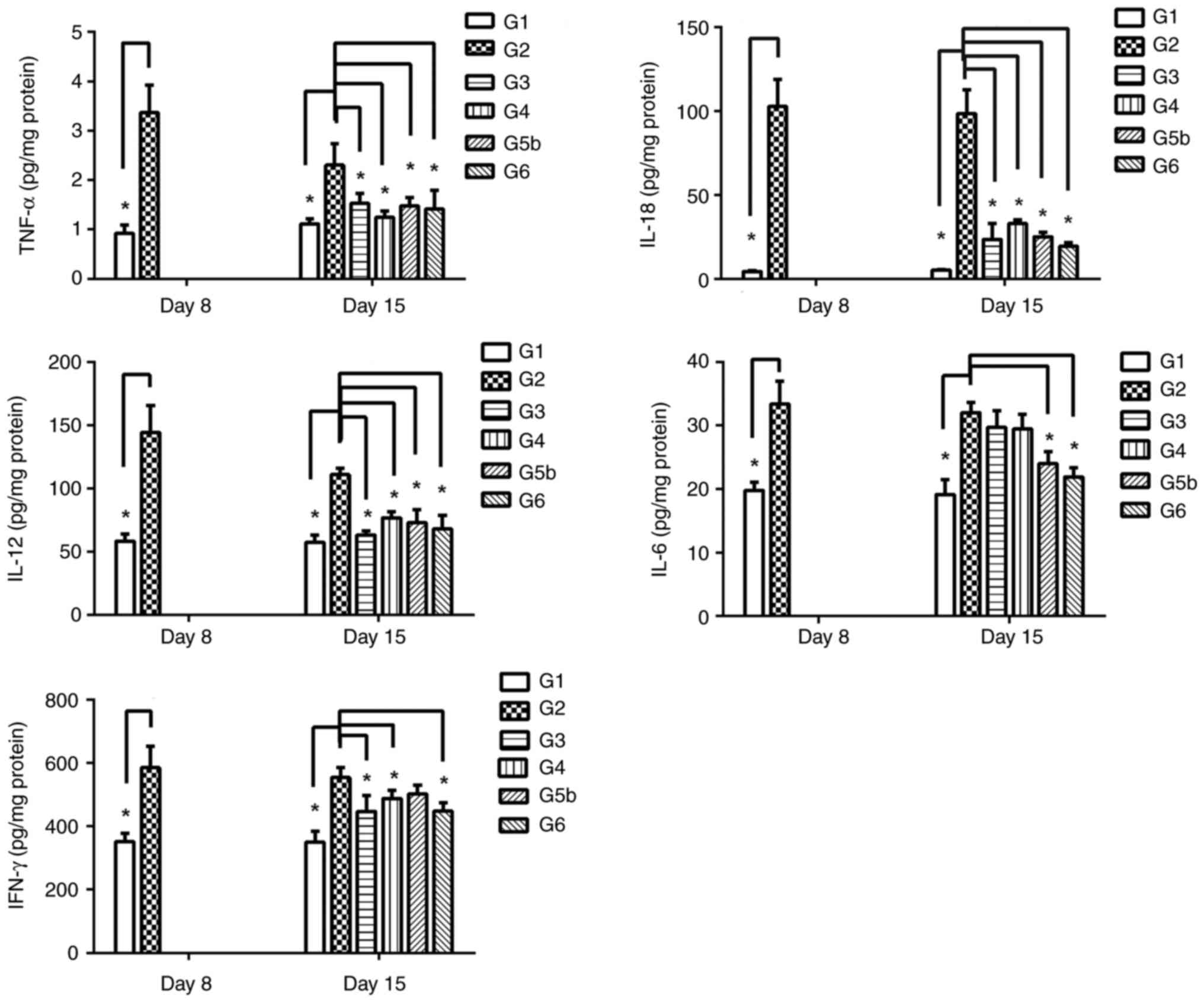 | Figure 6Effects of prednisolone, and vitamin
D and vitamin E, alone and combined, on the levels of IL-6, IL-12,
IL-18, TNF-α and IFN-γ in colon tissues at various time points. The
levels of these five cytokines were analyzed on day 8 in colon
tissues from G1 and G2. The levels of the five cytokines in G2 were
significantly elevated compared with those in G1. On day 15, the
levels of the five cytokines were analyzed in G1, G2, G3, G4, G5b
and G6. Data are presented as the mean ± SEM. n=6.
*P<0.05. G1, control group; G2, 5% DSS group; G3,
prednisolone group; G4, vitamin D group; G5a-c, vitamin E (low,
medium and high) groups; G6, vitamin D + vitamin E group; DSS,
dextran sodium sulfate; IFN-γ, interferon-γ; IL, interleukin;
TNF-α, tumor necrosis factor-α. |
On day 15, the remaining rats were sacrificed, and
their colon tissues were collected for the analysis of cytokines.
The results showed that the levels of four of the inflammatory
cytokines (TNF-α, IL-18, IL-12 and IFN-γ) in both G3 and G4 were
significantly decreased compared with those in G2 (TNF-α: G3 vs. G2
group: F=-3.82, P=0.01; G4 vs. G2 group, F=-8.81, P=0.01. IFN-γ:G3
vs. G2 group: F=138.62, P=0.01; G4 vs. G2 group, F=122.99, P=0.02.
IL-18:G3 vs. G2 group: F=51.04, P=0.01; G4 vs. G2 group, F=22.17,
P=0.01. IL-12: G3 vs. G2 group: F=42.09, P=0.01; G4 vs. G2 group,
F=13.78, P=0.04.), whereas there was no obvious change in IL-6
levels (G3 vs. G2 group: F=0.66, P=0.65; G4 vs. G2 group, F=0.49,
P=0.81) (Fig. 6). In addition, in
the colon tissues, the levels of four of the inflammatory cytokines
(TNF-α, IL-18, IL-12 and IL-6) were decreased in G5b compared with
in G2 (FIL-12=-34.97, P=0.03;
FIFN-γ=2.86, P=0.049;
FTNF-α=-3.91, P=0.03; FIL-6=-8.54,
P=0.04; FIL-18=46.19, P=0.01), whereas there was no
obvious change in the IFN-γ levels (F=1.75, P=0.14) (Fig. 6). Notably, the levels of five of
the inflammatory cytokines (IL-6, IL-12, IL-18, TNF-α and IFN-γ)
were significantly reduced in G6 compared with those in G2
(FTNF-α=-4.77, P=0.03;
FIFN-γ=138.66, P=0.04;
FIL-12=42.18, P=0.03; FIL-6=8.04, P=0.01;
FIL-18=17.39, P=0.01; Fig.
6). Collectively, these data showed that both vitamin D and
vitamin E could partly reduce the levels of inflammatory cytokines
in rats with DSS-induced UC, whereas their combined application
exhibited more noticeable inhibitory effects.
Discussion
IBD is commonly associated with immune
dysregulation; however, the precise physiological mechanisms
underlying this pathological state require further investigation,
so that therapeutic countermeasures can be improved. Prednisolone
is currently a commonly used drug for the treatment of severe cases
of IBD; therefore, in the present study, vitamins E and D were
compared with it to explore whether these vitamins are more
effective and thus provide a better treatment option. In the
present study, CO scoring, histopathological examination and ELISA
were performed on a rat model of DSS-induced UC; the results
revealed that both vitamin E and vitamin D positively promoted
recovery from DSS-induced UC, although their effects on the
regulation of inflammatory cytokines may differ. Hahn et al
(33) noted that the combination
of omega-3 fatty acids and vitamin D was more beneficial for
decreasing the risk of autoimmune diseases, whereas further
research is required to determine whether this combination is
advantageous for attenuating the incidence of IBD. The present
study also indicated that the combined use of vitamin E and vitamin
D may be associated with more noticeable anti-inflammatory effects
on UC than their separate application.
In the majority of patients with IBD, immune
pathogenesis is associated with the increased production of
proinflammatory cytokines, such as IL-6, IL-12, IL-18, TNF-α and
IFN-γ. Drugs targeting these cytokines or antibodies have exhibited
therapeutic effects on IBD (7,12,55);
thus, these cytokines are widely accepted as markers of UC.
DSS-induced inflammation has previously been shown to be associated
with the elevated production of IL-6, IL-18 and IFN-γ, and it could
be attenuated in the absence of IL-6, IL-18 or IFN-γ (56). Therefore, this model is widely
accepted for the analysis of the influence of any given drug or
compound on the promotion of epithelial cell repair and attenuation
of production of inflammatory mediators in animals with UC
(50,57). Studies have shown that even though
the absorption capacity and benefits of vitamin D vary by age and
category, optimal maintenance concentrations can reduce the risk of
inflammation and autoimmune diseases, as well as partly providing
innate autoimmunity, especially for COVID-19 (34,36).
From the perspective of clinical symptoms and data
from autopsies, the administration of DSS to rats in drinking water
resulted in BW loss, shortening of the colon, mucosal inflammation
and epithelial damage, which indicated that this rat model of IBD
may be a good model for studying and evaluating human colitis.
Vitamin E (30 and 150 IU/kg), vitamin D (50 ng), and
their combination prevented BW loss, relieved the symptoms of UC
and promoted the recovery of DSS-induced UC in the present study.
These data strongly indicated that both vitamin E and vitamin D had
obvious anti-inflammatory effects on UC and may have positive
therapeutic effects on patients with IBD. As vitamin E and vitamin
D are relatively safe drugs that have long been used clinically,
they could be appropriate for the treatment of IBD. In addition,
vitamin D has been shown to prevent osteoporosis in the elderly,
while it increases resistance to COVID-19 in appropriate
concentrations (35). In women and
children, vitamin D deficiency can lead to placental dysgenesis,
affecting the innate development and immunity of infants and
children (32,35,37).
Notably, as an antioxidant, the anti-inflammatory
mechanism of vitamin E (30 IU/kg) may differ slightly from that of
prednisolone (1 mg/kg) and vitamin D (50 ng), as indicated by the
different levels of inflammatory cytokines detected in the present
study. In addition, vitamin E plays an important role in immunity.
According to recent studies, vitamin E, which has been detected in
higher concentrations in immune cells compared with other blood
cells, affects the development and functional regulation of DCs,
macrophages, natural killer cells, T cells and B cells (58,59).
According to the results of the present study, vitamin E
administration (30 IU/kg) reduced the levels of TNF-α, IL-12, IL-18
and IL-6 compared with those in the DSS group, whereas it had no
significant effect on IFN-γ levels. By contrast, prednisolone and
vitamin D decreased the levels of TNF-α, IL-12, IL-18 and IFN-γ,
but not IL-6. Acute inflammation in DSS-induced UC could be
predominantly activated through the T helper (Th)2-mediated
inflammatory response in the chronic state (lower levels of TNF-α,
and elevated levels of IL-6, IFN-γ, IL-4 and IL-10) (50,57,60,61).
Thus, the anti-inflammatory effects of vitamin E may mainly target
Th2 cytokines, enabling us to find new drugs based on the Th2
cytokine mechanism, as the side effects of vitamin E are more
tolerable than those of prednisolone. However, compared with that
in the DSS group in the present study, colon length in the vitamin
D- and vitamin E-treated groups was not significantly increased;
however, there were some significant differences between these
groups regarding colon weight/length ratio and inflammatory
cytokine levels, especially TNF-α, IL-18 and IL-12, which may
indicate that colon length is not a very sensitive indicator
compared with cytokine levels. Therefore, although vitamin D and
vitamin E are effective, they are not as significant in changing
colon length as their combination.
In the present study, the rat model of DSS-induced
UC could not thoroughly reflect the symptoms of patients with IBD.
Thus, further studies are required to examine the effects of
vitamin E, vitamin D and their combination on UC.
As the rat model of DSS-induced UC could not fully
represent the complexity of human models of IBD, additional
research on the protective and therapeutic effects of vitamin E and
vitamin D for human UC should be performed. Additionally, in order
to provide more valuable information for combination therapy,
further studies need to be conducted to evaluate their effects in
combination with other anti-inflammatory drugs. In conclusion, the
combination of vitamin E and vitamin D proved to be feasible and
effective in the present study. This suggests that the addition of
vitamin E and vitamin D oral therapy may be an effective treatment
for IBD in the future.
Acknowledgements
The authors would like to thank Dr Trevor Smith
(Wolfson Centre for Age-Related Diseases, King's College London)
for his technical assistance and editing of the manuscript.
Funding
Funding: This study was financially supported by the National
Key Technologies R&D Program for New Drugs (grant nos.
2013ZX09302303 and 2012ZX09301003-001-008), the National Natural
Science Foundation of China (grant no. 82073833), the Beijing
Municipal Natural Science Foundation (grant nos. 7142123 and
Z131100006513010) and the State Key Laboratory of Proteomics
Foundation (grant no. SKLP-YB201403).
Availability of data and material
The datasets used and/or analyzed during the current
study are available from the corresponding author on reasonable
request.
Authors' contributions
XF performed multiple experiments, data acquisition
and data analysis. JieY collected experimental data and
participated in revising the manuscript. JiyeY contributed to the
experimental design and participated in completing the relevant
experiments. XW and RD contributed to the design of the study,
wrote the original manuscript, confirmed the authenticity of all
raw data, and agreed to be accountable for all aspects of the study
in ensuring that questions related to the accuracy or integrity of
any part of the study are appropriately investigated and resolved.
All authors have read and approved the final manuscript.
Ethics approval and consent to
participate
Experiments were performed in compliance with the
National Institutes of Health Guide for the Care and Use of
Laboratory Animals (62). The
experimental procedures were approved by the National Beijing
Center for Drug Safety Evaluation and Research Laboratory Animal
Welfare Ethics Committee (Beijing, China; approval no.
IACUC-2011-002).
Patient consent for publication
Not applicable.
Competing interests
The authors declare that they have no competing
interests.
References
|
1
|
Rufo PA, Denson LA, Sylvester FA, Szigethy
E, Sathya P, Lu Y, Wahbeh GT, Sena LM and Faubion WA: Health
supervision in the management of children and adolescents with IBD:
NASPGHAN recommendations. J Pediatr Gastroenterol Nutr. 55:93–108.
2012.PubMed/NCBI View Article : Google Scholar
|
|
2
|
Lucendo AJ, Hervías D, Roncero Ó, Lorente
R, Bouhmidi A, Angueira T, Verdejo C, Salueña I, González-Castillo
S and Arias Á: Epidemiology and temporal trends (2000-2012) of
inflammatory bowel disease in adult patients in a central region of
Spain. Eur J Gastroenterol Hepatol. 26:1399–1407. 2014.PubMed/NCBI View Article : Google Scholar
|
|
3
|
Rescigno M: The pathogenic role of
intestinal flora in IBD and colon cancer. Curr Drug Targets.
9:395–403. 2008.PubMed/NCBI View Article : Google Scholar
|
|
4
|
Ng SC, Tang W, Ching JY, Wong M, Chow CM,
Hui AJ, Wong TC, Leung VK, Tsang SW, Yu HH, et al: Incidence and
phenotype of inflammatory bowel disease based on results from the
Asia-pacific Crohn's and colitis epidemiology study.
Gastroenterology. 145:158–165.e2. 2013.PubMed/NCBI View Article : Google Scholar
|
|
5
|
Kuenzig ME, Fung SG, Marderfeld L, Mak
JWY, Kaplan GG, Ng SC, Wilson DC, Cameron F, Henderson P, Kotze PG,
et al: Twenty-first century trends in the global epidemiology of
pediatric-onset inflammatory bowel disease: Systematic review.
Gastroenterology. 162:1147–1159.e4. 2022.PubMed/NCBI View Article : Google Scholar
|
|
6
|
Bauer C, Duewell P, Mayer C, Lehr HA,
Fitzgerald KA, Dauer M, Tschopp J, Endres S, Latz E and Schnurr M:
Colitis induced in mice with dextran sulfate sodium (DSS) is
mediated by the NLRP3 inflammasome. Gut. 59:1192–1199.
2010.PubMed/NCBI View Article : Google Scholar
|
|
7
|
Kojouharoff G, Hans W, Obermeier F, Männel
DN, Andus T, Schölmerich J, Gross V and Falk W: Neutralization of
tumour necrosis factor (TNF) but not of IL-1 reduces inflammation
in chronic dextran sulphate sodium-induced colitis in mice. Clin
Exp Immunol. 107:353–358. 1997.PubMed/NCBI View Article : Google Scholar
|
|
8
|
Matsunaga H, Hokari R, Ueda T, Kurihara C,
Hozumi H, Higashiyama M, Okada Y, Watanabe C, Komoto S, Nakamura M,
et al: Physiological stress exacerbates murine colitis by enhancing
proinflammatory cytokine expression that is dependent on IL-18. Am
J Physiol Gastrointest Liver Physiol. 301:G555–G564.
2011.PubMed/NCBI View Article : Google Scholar
|
|
9
|
Takagi H, Kanai T, Okazawa A, Kishi Y,
Sato T, Takaishi H, Inoue N, Ogata H, Iwao Y, Hoshino K, et al:
Contrasting action of IL-12 and IL-18 in the development of dextran
sodium sulphate colitis in mice. Scand J Gastroenterol. 38:837–844.
2003.PubMed/NCBI View Article : Google Scholar
|
|
10
|
Hans W, Schölmerich J, Gross V and Falk W:
Interleukin-12 induced interferon-gamma increases inflammation in
acute dextran sulfate sodium induced colitis in mice. Eur Cytokine
Netw. 11:67–74. 2000.PubMed/NCBI
|
|
11
|
Wang Y, Tong J, Chang B, Wang BF, Zhang D
and Wang BY: Genetic polymorphisms in the IL-18 gene and ulcerative
colitis risk: A meta-analysis. DNA Cell Biol. 33:438–447.
2014.PubMed/NCBI View Article : Google Scholar
|
|
12
|
Zhu Y, Mahon BD, Froicu M and Cantorna MT:
Calcium and 1 alpha,25-dihydroxyvitamin D3 target the TNF-alpha
pathway to suppress experimental inflammatory bowel disease. Eur J
Immunol. 35:217–224. 2005.PubMed/NCBI View Article : Google Scholar
|
|
13
|
Stio M, Martinesi M, Bruni S, Treves C,
d'Albasio G, Bagnoli S and Bonanomi AG: Interaction among vitamin
D(3) analogue KH 1060, TNF-alpha, and vitamin D receptor protein in
peripheral blood mononuclear cells of inflammatory bowel disease
patients. Int Immunopharmacol. 6:1083–1092. 2006.PubMed/NCBI View Article : Google Scholar
|
|
14
|
Magro F, Cordeiro G, Dias AM and Estevinho
MM: Inflammatory bowel disease-non-biological treatment. Pharmacol
Res. 160(105075)2020.PubMed/NCBI View Article : Google Scholar
|
|
15
|
Jeong DY, Kim S, Son MJ, Son CY, Kim JY,
Kronbichler A, Lee KH and Shin JI: Induction and maintenance
treatment of inflammatory bowel disease: A comprehensive review.
Autoimmun Rev. 18:439–454. 2019.PubMed/NCBI View Article : Google Scholar
|
|
16
|
Jiang Y, Xia X, Wang W, Lin L, Xu C, Cai
Z, Zheng B, Pei J, Shen S and Xia B: Hyperhomocysteinemia and
related genetic polymorphisms correlate with ulcerative colitis in
Chinese Han population in Central China [corrected]. Cell Biochem
Biophys. 62:203–210. 2012.PubMed/NCBI View Article : Google Scholar
|
|
17
|
Hart AL: Vitamin D and inflammatory bowel
disease: Chicken or egg? Inflamm Bowel Dis. 19:459–460.
2013.PubMed/NCBI View Article : Google Scholar
|
|
18
|
Levin AD, Wadhera V, Leach ST, Woodhead
HJ, Lemberg DA, Mendoza-Cruz AC and Day AS: Vitamin D deficiency in
children with inflammatory bowel disease. Dig Dis Sci. 56:830–836.
2011.PubMed/NCBI View Article : Google Scholar
|
|
19
|
Blanck S and Aberra F: Vitamin d
deficiency is associated with ulcerative colitis disease activity.
Dig Dis Sci. 58:1698–1702. 2013.PubMed/NCBI View Article : Google Scholar
|
|
20
|
Hassan V, Hassan S, Seyed-Javad P, Ahmad
K, Asieh H, Maryam S, Farid F and Siavash A: Association between
serum 25 (OH) vitamin D concentrations and inflammatory bowel
diseases (IBDs) activity. Med J Malaysia. 68:34–38. 2013.PubMed/NCBI
|
|
21
|
Selhub J, Byun A, Liu Z, Mason JB, Bronson
RT and Crott JW: Dietary vitamin B6 intake modulates colonic
inflammation in the IL10-/- model of inflammatory bowel disease. J
Nutr Biochem. 24:2138–2143. 2013.PubMed/NCBI View Article : Google Scholar
|
|
22
|
Li YC: Investigating the role of vitamin D
in IBD pathophysiology and treatment. Gastroenterol Hepatol (N Y).
4:20–21. 2008.PubMed/NCBI
|
|
23
|
Jankowska M, Trzonkowski P, Dębska-Ślizień
A, Marszałł M and Rutkowski B: Vitamin B6 status, immune response
and inflammation markers in kidney transplant recipients treated
with polyclonal anti-thymocyte globulin. Transplant Proc.
46:2631–2635. 2014.PubMed/NCBI View Article : Google Scholar
|
|
24
|
De Silva P and Ananthakrishnan AN: Vitamin
D and IBD: More pieces to the puzzle, still no complete picture.
Inflamm Bowel Dis. 18:1391–1393. 2012.PubMed/NCBI View Article : Google Scholar
|
|
25
|
Xue LN, Xu KQ, Zhang W, Wang Q, Wu J and
Wang XY: Associations between vitamin D receptor polymorphisms and
susceptibility to ulcerative colitis and Crohn's disease: A
meta-analysis. Inflamm Bowel Dis. 19:54–60. 2013.PubMed/NCBI View Article : Google Scholar
|
|
26
|
Zator ZA, Cantu SM, Konijeti GG, Nguyen
DD, Sauk J, Yajnik V and Ananthakrishnan AN: Pretreatment
25-hydroxyvitamin D levels and durability of anti-tumor necrosis
factor-α therapy in inflammatory bowel diseases. JPEN J Parenter
Enteral Nutr. 38:385–391. 2014.PubMed/NCBI View Article : Google Scholar
|
|
27
|
Ooi JH, Li Y, Rogers CJ and Cantorna MT:
Vitamin D regulates the gut microbiome and protects mice from
dextran sodium sulfate-induced colitis. J Nutr. 143:1679–1686.
2013.PubMed/NCBI View Article : Google Scholar
|
|
28
|
Mackawy AMH and Badawi MEH: Association of
vitamin D and vitamin D receptor gene polymorphisms with chronic
inflammation, insulin resistance and metabolic syndrome components
in type 2 diabetic Egyptian patients. Meta Gene. 2:540–556.
2014.PubMed/NCBI View Article : Google Scholar
|
|
29
|
Li YC, Chen Y and Du J: Critical roles of
intestinal epithelial vitamin D receptor signaling in controlling
gut mucosal inflammation. J Steroid Biochem Mol Biol. 148:179–183.
2015.PubMed/NCBI View Article : Google Scholar
|
|
30
|
Leslie WD, Miller N, Rogala L and
Bernstein CN: Vitamin D status and bone density in recently
diagnosed inflammatory bowel disease: The manitoba IBD cohort
study. Am J Gastroenterol. 103:1451–1459. 2008.PubMed/NCBI View Article : Google Scholar
|
|
31
|
Ghaly S and Lawrance I: The role of
vitamin D in gastrointestinal inflammation. Expert Rev
Gastroenterol Hepatol. 8:909–923. 2014.PubMed/NCBI View Article : Google Scholar
|
|
32
|
Phillips EA, Hendricks N, Bucher M and
Maloyan A: Vitamin D supplementation improves mitochondrial
function and reduces inflammation in placentae of obese women.
Front Endocrinol (Lausanne). 13(893848)2022.PubMed/NCBI View Article : Google Scholar
|
|
33
|
Hahn J, Cook NR, Alexander EK, Friedman S,
Walter J, Bubes V, Kotler G, Lee IM, Manson JE and Costenbader KH:
Vitamin D and marine omega 3 fatty acid supplementation and
incident autoimmune disease: VITAL randomized controlled trial.
BMJ. 376(e066452)2022.PubMed/NCBI View Article : Google Scholar
|
|
34
|
Bui L, Zhu Z, Hawkins S, Cortez-Resendiz A
and Bellon A: Vitamin D regulation of the immune system and its
implications for COVID-19: A mini review. SAGE Open Med.
9(20503121211014073)2021.PubMed/NCBI View Article : Google Scholar
|
|
35
|
Hribar CA, Cobbold PH and Church FC:
Potential role of vitamin D in the elderly to resist COVID-19 and
to slow progression of Parkinson's disease. Brain Sci.
10(284)2020.PubMed/NCBI View Article : Google Scholar
|
|
36
|
Charoenngam N and Holick MF: Immunologic
effects of vitamin D on human health and disease. Nutrients.
12(2097)2020.PubMed/NCBI View Article : Google Scholar
|
|
37
|
Mailhot G and White JH: Vitamin D and
immunity in infants and children. Nutrients.
12(1233)2020.PubMed/NCBI View Article : Google Scholar
|
|
38
|
Zhao H, Zhang H, Wu H, Li H, Liu L, Guo J,
Li C, Shih DQ and Zhang X: Protective role of 1,25(OH)2 vitamin D3
in the mucosal injury and epithelial barrier disruption in
DSS-induced acute colitis in mice. BMC Gastroenterol.
12(57)2012.PubMed/NCBI View Article : Google Scholar
|
|
39
|
Vaghari-Tabari M, Mohammadzadeh I, Qujeq
D, Majidinia M, Alemi F, Younesi S, Mahmoodpoor A, Maleki M,
Yousefi B and Asemi Z: Vitamin D in respiratory viral infections: A
key immune modulator? Crit Rev Food Sci Nutr. 1–16. 2021.PubMed/NCBI View Article : Google Scholar : (Epub ahead of
print).
|
|
40
|
Colón AL, Madrigal JL, Menchén LA, Moro
MA, Lizasoain I, Lorenzo P and Leza JC: Stress increases
susceptibility to oxidative/nitrosative mucosal damage in an
experimental model of colitis in rats. Dig Dis Sci. 49:1713–1721.
2004.PubMed/NCBI View Article : Google Scholar
|
|
41
|
Narushima S, Spitz DR, Oberley LW,
Toyokuni S, Miyata T, Gunnett CA, Buettner GR, Zhang J, Ismail H,
Lynch RG and Berg DJ: Evidence for oxidative stress in
NSAID-induced colitis in IL10-/- mice. Free Radic Biol Med.
34:1153–1166. 2003.PubMed/NCBI View Article : Google Scholar
|
|
42
|
Seven A, Seymen O, Inci F, Oz B, Yiğit G
and Burçak G: Evaluation of oxidative stress in experimental
colitis: Effects of L-arginine-nitric oxide pathway manipulation. J
Toxicol Environ Health A. 61:167–176. 2000.PubMed/NCBI View Article : Google Scholar
|
|
43
|
Rana SV, Sharma S, Kaur J, Prasad KK,
Sinha SK, Kochhar R, Malik A and Morya RK: Relationship of
cytokines, oxidative stress and GI motility with bacterial
overgrowth in ulcerative colitis patients. J Crohns Colitis.
8:859–865. 2014.PubMed/NCBI View Article : Google Scholar
|
|
44
|
Rana SV, Sharma S, Prasad KK, Sinha SK and
Singh K: Role of oxidative stress & antioxidant defence in
ulcerative colitis patients from north India. Indian J Med Res.
139:568–571. 2014.PubMed/NCBI
|
|
45
|
Korkina L, Suprun M, Petrova A,
Mikhal'chik E, Luci A and De Luca C: The protective and healing
effects of a natural antioxidant formulation based on ubiquinol and
Aloe vera against dextran sulfate-induced ulcerative colitis in
rats. Biofactors. 18:255–264. 2003.PubMed/NCBI View Article : Google Scholar
|
|
46
|
Abdala-Valencia H, Berdnikovs S and
Cook-Mills JM: Vitamin E isoforms as modulators of lung
inflammation. Nutrients. 5:4347–4363. 2013.PubMed/NCBI View Article : Google Scholar
|
|
47
|
Almohanna HM, Ahmed AA, Tsatalis JP and
Tosti A: The role of vitamins and minerals in hair loss: A review.
Dermatol Ther (Heidelb). 9:51–70. 2019.PubMed/NCBI View Article : Google Scholar
|
|
48
|
Carrier J, Aghdassi E, Cullen J and Allard
JP: Iron supplementation increases disease activity and vitamin E
ameliorates the effect in rats with dextran sulfate sodium-induced
colitis. J Nutr. 132:3146–3150. 2002.PubMed/NCBI View Article : Google Scholar
|
|
49
|
Tahan G, Aytac E, Aytekin H, Gunduz F,
Dogusoy G, Aydin S, Tahan V and Uzun H: Vitamin E has a dual effect
of anti-inflammatory and antioxidant activities in acetic
acid-induced ulcerative colitis in rats. Can J Surg. 54:333–338.
2011.PubMed/NCBI View Article : Google Scholar
|
|
50
|
Parmar AR, Trivedi PP and Jena GB: Dextran
sulfate sodium-induced ulcerative colitis leads to testicular
toxicity in mice: Role of inflammation, oxidative stress and DNA
damage. Reprod Toxicol. 49:171–184. 2014.PubMed/NCBI View Article : Google Scholar
|
|
51
|
Hu D, Wu CQ, Li ZJ, Liu Y, Fan X, Wang QJ
and Ding RG: Characterizing the mechanism of
thiazolidinedione-induced hepatotoxicity: An in vitro model in
mitochondria. Toxicol Appl Pharmacol. 284:134–141. 2015.PubMed/NCBI View Article : Google Scholar
|
|
52
|
Meng G, Zhao J, Wang HM, Ding RG, Zhang
XC, Huang CQ and Ruan JX: Injury of cell tight junctions and
changes of actin level in acute lung injury caused by the
perfluoroisobutylene exposure and the role of Myosin light chain
kinase. J Occup Health. 53:250–257. 2011.PubMed/NCBI View Article : Google Scholar
|
|
53
|
Weng XC, Fan X, Wang QX, Shi C, Li LN,
Ouyang ZH, Kong Q, Wang QJ, Guan YB and Ding RG: Research on the
methods of active systemic anaphylaxis on guinea pig in drug safety
evaluation. Chin J Comp Med. 22:51–55. 2012.
|
|
54
|
Cooper HS, Murthy SN, Shah RS and
Sedergran DJ: Clinicopathologic study of dextran sulfate sodium
experimental murine colitis. Lab Invest. 69:238–249.
1993.PubMed/NCBI
|
|
55
|
Ten Hove T, Corbaz A, Amitai H, Aloni S,
Belzer I, Graber P, Drillenburg P, van Deventer SJ, Chvatchko Y and
Te Velde AA: Blockade of endogenous IL-18 ameliorates TNBS-induced
colitis by decreasing local TNF-alpha production in mice.
Gastroenterology. 121:1372–1379. 2001.PubMed/NCBI View Article : Google Scholar
|
|
56
|
Wang L, Feng Y and Wang J, Luo T, Wang X,
Wu M, Wang R, Chen D, Li J and Wang J: Arbutin ameliorates murine
colitis by inhibiting JAK2 signaling pathway. Front Pharmacol.
12(683818)2021.PubMed/NCBI View Article : Google Scholar
|
|
57
|
Chassaing B, Aitken JD, Malleshappa M and
Vijay-Kumar M: Dextran sulfate sodium (DSS)-induced colitis in
mice. Curr Protoc Immunol. 104:15.25.1–15.25.14. 2014.PubMed/NCBI View Article : Google Scholar
|
|
58
|
Sun M, Yan Z, Sun R, Tian W, Yi W and
Zhang J: Dynamic monitoring and a clinical correlation analysis of
the serum vitamin A, D, and E levels in children with recurrent
respiratory tract infections. Am J Transl Res. 14:3533–3538.
2022.PubMed/NCBI
|
|
59
|
Lewis ED, Meydani SN and Wu D: Regulatory
role of vitamin E in the immune system and inflammation. IUBMB
Life. 71:487–494. 2019.PubMed/NCBI View Article : Google Scholar
|
|
60
|
Perše M and Cerar A: Dextran sodium
sulphate colitis mouse model: Traps and tricks. J Biomed
Biotechnol. 2012(718617)2012.PubMed/NCBI View Article : Google Scholar
|
|
61
|
Schwager J, Seifert N, Bompard A,
Raederstorff D and Bendik I: Resveratrol, EGCG and vitamins
modulate activated T lymphocytes. Molecules.
26(5600)2021.PubMed/NCBI View Article : Google Scholar
|
|
62
|
National Research Council (US) Committee
for the Update of the Guide for the Care and Use of Laboratory
Animals: The national academies collection: Reports funded by
national institutes of health. In: Guide for the care and use of
laboratory animals. National Academies Press (US) Copyright ©.
2011, National Academy of Sciences, Washington (DC), 2011.
|


