Spandidos Publications Logo
  • About
    • About Spandidos
    • Aims and Scopes
    • Abstracting and Indexing
    • Editorial Policies
    • Reprints and Permissions
    • Job Opportunities
    • Terms and Conditions
    • Contact
  • Journals
    • All Journals
    • Oncology Letters
      • Oncology Letters
      • Information for Authors
      • Editorial Policies
      • Editorial Board
      • Aims and Scope
      • Abstracting and Indexing
      • Bibliographic Information
      • Archive
    • International Journal of Oncology
      • International Journal of Oncology
      • Information for Authors
      • Editorial Policies
      • Editorial Board
      • Aims and Scope
      • Abstracting and Indexing
      • Bibliographic Information
      • Archive
    • Molecular and Clinical Oncology
      • Molecular and Clinical Oncology
      • Information for Authors
      • Editorial Policies
      • Editorial Board
      • Aims and Scope
      • Abstracting and Indexing
      • Bibliographic Information
      • Archive
    • Experimental and Therapeutic Medicine
      • Experimental and Therapeutic Medicine
      • Information for Authors
      • Editorial Policies
      • Editorial Board
      • Aims and Scope
      • Abstracting and Indexing
      • Bibliographic Information
      • Archive
    • International Journal of Molecular Medicine
      • International Journal of Molecular Medicine
      • Information for Authors
      • Editorial Policies
      • Editorial Board
      • Aims and Scope
      • Abstracting and Indexing
      • Bibliographic Information
      • Archive
    • Biomedical Reports
      • Biomedical Reports
      • Information for Authors
      • Editorial Policies
      • Editorial Board
      • Aims and Scope
      • Abstracting and Indexing
      • Bibliographic Information
      • Archive
    • Oncology Reports
      • Oncology Reports
      • Information for Authors
      • Editorial Policies
      • Editorial Board
      • Aims and Scope
      • Abstracting and Indexing
      • Bibliographic Information
      • Archive
    • Molecular Medicine Reports
      • Molecular Medicine Reports
      • Information for Authors
      • Editorial Policies
      • Editorial Board
      • Aims and Scope
      • Abstracting and Indexing
      • Bibliographic Information
      • Archive
    • World Academy of Sciences Journal
      • World Academy of Sciences Journal
      • Information for Authors
      • Editorial Policies
      • Editorial Board
      • Aims and Scope
      • Abstracting and Indexing
      • Bibliographic Information
      • Archive
    • International Journal of Functional Nutrition
      • International Journal of Functional Nutrition
      • Information for Authors
      • Editorial Policies
      • Editorial Board
      • Aims and Scope
      • Abstracting and Indexing
      • Bibliographic Information
      • Archive
    • International Journal of Epigenetics
      • International Journal of Epigenetics
      • Information for Authors
      • Editorial Policies
      • Editorial Board
      • Aims and Scope
      • Abstracting and Indexing
      • Bibliographic Information
      • Archive
    • Medicine International
      • Medicine International
      • Information for Authors
      • Editorial Policies
      • Editorial Board
      • Aims and Scope
      • Abstracting and Indexing
      • Bibliographic Information
      • Archive
  • Articles
  • Information
    • Information for Authors
    • Information for Reviewers
    • Information for Librarians
    • Information for Advertisers
    • Conferences
  • Language Editing
Spandidos Publications Logo
  • About
    • About Spandidos
    • Aims and Scopes
    • Abstracting and Indexing
    • Editorial Policies
    • Reprints and Permissions
    • Job Opportunities
    • Terms and Conditions
    • Contact
  • Journals
    • All Journals
    • Biomedical Reports
      • Information for Authors
      • Editorial Policies
      • Editorial Board
      • Aims and Scope
      • Abstracting and Indexing
      • Bibliographic Information
      • Archive
    • Experimental and Therapeutic Medicine
      • Information for Authors
      • Editorial Policies
      • Editorial Board
      • Aims and Scope
      • Abstracting and Indexing
      • Bibliographic Information
      • Archive
    • International Journal of Epigenetics
      • Information for Authors
      • Editorial Policies
      • Editorial Board
      • Aims and Scope
      • Abstracting and Indexing
      • Bibliographic Information
      • Archive
    • International Journal of Functional Nutrition
      • Information for Authors
      • Editorial Policies
      • Editorial Board
      • Aims and Scope
      • Abstracting and Indexing
      • Bibliographic Information
      • Archive
    • International Journal of Molecular Medicine
      • Information for Authors
      • Editorial Policies
      • Editorial Board
      • Aims and Scope
      • Abstracting and Indexing
      • Bibliographic Information
      • Archive
    • International Journal of Oncology
      • Information for Authors
      • Editorial Policies
      • Editorial Board
      • Aims and Scope
      • Abstracting and Indexing
      • Bibliographic Information
      • Archive
    • Medicine International
      • Information for Authors
      • Editorial Policies
      • Editorial Board
      • Aims and Scope
      • Abstracting and Indexing
      • Bibliographic Information
      • Archive
    • Molecular and Clinical Oncology
      • Information for Authors
      • Editorial Policies
      • Editorial Board
      • Aims and Scope
      • Abstracting and Indexing
      • Bibliographic Information
      • Archive
    • Molecular Medicine Reports
      • Information for Authors
      • Editorial Policies
      • Editorial Board
      • Aims and Scope
      • Abstracting and Indexing
      • Bibliographic Information
      • Archive
    • Oncology Letters
      • Information for Authors
      • Editorial Policies
      • Editorial Board
      • Aims and Scope
      • Abstracting and Indexing
      • Bibliographic Information
      • Archive
    • Oncology Reports
      • Information for Authors
      • Editorial Policies
      • Editorial Board
      • Aims and Scope
      • Abstracting and Indexing
      • Bibliographic Information
      • Archive
    • World Academy of Sciences Journal
      • Information for Authors
      • Editorial Policies
      • Editorial Board
      • Aims and Scope
      • Abstracting and Indexing
      • Bibliographic Information
      • Archive
  • Articles
  • Information
    • For Authors
    • For Reviewers
    • For Librarians
    • For Advertisers
    • Conferences
  • Language Editing
Login Register Submit
  • This site uses cookies
  • You can change your cookie settings at any time by following the instructions in our Cookie Policy. To find out more, you may read our Privacy Policy.

    I agree
Search articles by DOI, keyword, author or affiliation
Search
Advanced Search
presentation
Experimental and Therapeutic Medicine
Join Editorial Board Propose a Special Issue
Print ISSN: 1792-0981 Online ISSN: 1792-1015
Journal Cover
March-2023 Volume 25 Issue 3

Full Size Image

Sign up for eToc alerts
Recommend to Library

Journals

International Journal of Molecular Medicine

International Journal of Molecular Medicine

International Journal of Molecular Medicine is an international journal devoted to molecular mechanisms of human disease.

International Journal of Oncology

International Journal of Oncology

International Journal of Oncology is an international journal devoted to oncology research and cancer treatment.

Molecular Medicine Reports

Molecular Medicine Reports

Covers molecular medicine topics such as pharmacology, pathology, genetics, neuroscience, infectious diseases, molecular cardiology, and molecular surgery.

Oncology Reports

Oncology Reports

Oncology Reports is an international journal devoted to fundamental and applied research in Oncology.

Experimental and Therapeutic Medicine

Experimental and Therapeutic Medicine

Experimental and Therapeutic Medicine is an international journal devoted to laboratory and clinical medicine.

Oncology Letters

Oncology Letters

Oncology Letters is an international journal devoted to Experimental and Clinical Oncology.

Biomedical Reports

Biomedical Reports

Explores a wide range of biological and medical fields, including pharmacology, genetics, microbiology, neuroscience, and molecular cardiology.

Molecular and Clinical Oncology

Molecular and Clinical Oncology

International journal addressing all aspects of oncology research, from tumorigenesis and oncogenes to chemotherapy and metastasis.

World Academy of Sciences Journal

World Academy of Sciences Journal

Multidisciplinary open-access journal spanning biochemistry, genetics, neuroscience, environmental health, and synthetic biology.

International Journal of Functional Nutrition

International Journal of Functional Nutrition

Open-access journal combining biochemistry, pharmacology, immunology, and genetics to advance health through functional nutrition.

International Journal of Epigenetics

International Journal of Epigenetics

Publishes open-access research on using epigenetics to advance understanding and treatment of human disease.

Medicine International

Medicine International

An International Open Access Journal Devoted to General Medicine.

Journal Cover
March-2023 Volume 25 Issue 3

Full Size Image

Sign up for eToc alerts
Recommend to Library

  • Article
  • Citations
    • Cite This Article
    • Download Citation
    • Create Citation Alert
    • Remove Citation Alert
    • Cited By
  • Similar Articles
    • Related Articles (in Spandidos Publications)
    • Similar Articles (Google Scholar)
    • Similar Articles (PubMed)
  • Download PDF
  • Download XML
  • View XML
Case Report Open Access

Odontogenic keratocyst in the mandibular condyle base region: A case report

  • Authors:
    • Sho Miyamoto
    • Takahiro Goto
    • Jumpei Shirakawa
    • Toshihiro Kawano
    • Makoto Murahashi
    • Kentaro Ide
    • Nobuyuki Maruyama
    • Akira Matayoshi
    • Kazuhide Nishihara
    • Hiroyuki Nakamura
  • View Affiliations / Copyright

    Affiliations: Department of Oral and Maxillofacial Functional Rehabilitation, Graduate School of Medicine, University of The Ryukyus, Nishihara, Okinawa 903‑0215, Japan, Department of Oral and Maxillofacial Surgery, Okinawa Red Cross Hospital, Naha, Okinawa 902‑8588, Japan
    Copyright: © Miyamoto et al. This is an open access article distributed under the terms of Creative Commons Attribution License.
  • Article Number: 141
    |
    Published online on: February 13, 2023
       https://doi.org/10.3892/etm.2023.11840
  • Expand metrics +
Metrics: Total Views: 0 (Spandidos Publications: | PMC Statistics: )
Metrics: Total PDF Downloads: 0 (Spandidos Publications: | PMC Statistics: )
Cited By (CrossRef): 0 citations Loading Articles...

This article is mentioned in:


Abstract

Odontogenic keratocysts (OKCs) often occur in the molars in the mandibular ramus; they often progress asymptomatically and are discovered only after widespread development. Some cases of OKC progress to the mandibular condyle; however, very few cases exist only in the condyle. To the best of our knowledge, in all of the previously reported cases, OKCs occurred in the mandibular ramus, which underwent resection. The present study reports the case of a 31‑year‑old man in whom an OKC (13x12x6 mm) occurred discretely in the base of the condyle, in which the condylar head was successfully preserved. The tumor was removed under general anesthesia using the approach of shaving the anterior surface of the mandible. The extraction cavity was managed using the packed open technique and with an obturator. Approximately 20 months post‑operation, the patient remained recurrence‑free. This report presents a rare case of an OKC in the mandibular condyle base region. Resection was performed under general anesthesia and the condylar process was successfully preserved.

Introduction

Odontogenic keratocysts (OKCs) account for 11.7% of all jaw cysts worldwide, making them the third most common type of cyst in the jaws after radicular and dentigerous cysts (1). OKCs are characterized by aggressive behavior and have a relatively high recurrence rate (2). Similar to other entities with an odontogenic origin, OKCs originate in tooth-bearing regions, and they occur twice as often in the mandible as in the maxilla (3). When OKCs originate in the mandible, the most common location is the posterior sextant, the angle or the ramus (4,5). Cases of OKCs often progress to the condyle because of their aggressive nature (6,7); however, to the best of our knowledge, only two reports of OKCs in the condyloid process that are not continuous with the surrounding areas have been reported (8,9). In both cases, OKCs occurred in the condylar head, which was resected. The present study reports on the case of an OKC that occurred discretely in the base of the condyle, which was treated successfully to preserve the condylar process.

Case report

Patient information

A 31-year-old man was referred to the University of the Ryukyus Hospital (Nishihara, Japan) in November 2020 with a complaint of repeated inflammatory symptoms around the condylar process. Cysts were first detected from the right mandibular body to the base of the condyle when the patient visited a dental clinic for scaling in January 2019. The patient was referred to another hospital, where a cystectomy and wisdom tooth extraction were performed in March 2019; however, the cyst at the base of the condyle was rediscovered. In January 2020, inflammatory symptoms around the condylar process appeared. The patient was then referred to the University of the Ryukyus Hospital. The patient had used antibiotics repeatedly and for an extended period at the time of his first visit to the University of the Ryukyus Hospital. The patient had no medical history and no family history of congenital anomalies. The patient had a smoking habit (one pack a day for 12 years) and he denied having any allergies. A general physical examination revealed normal physical and nutritional status.

Investigations

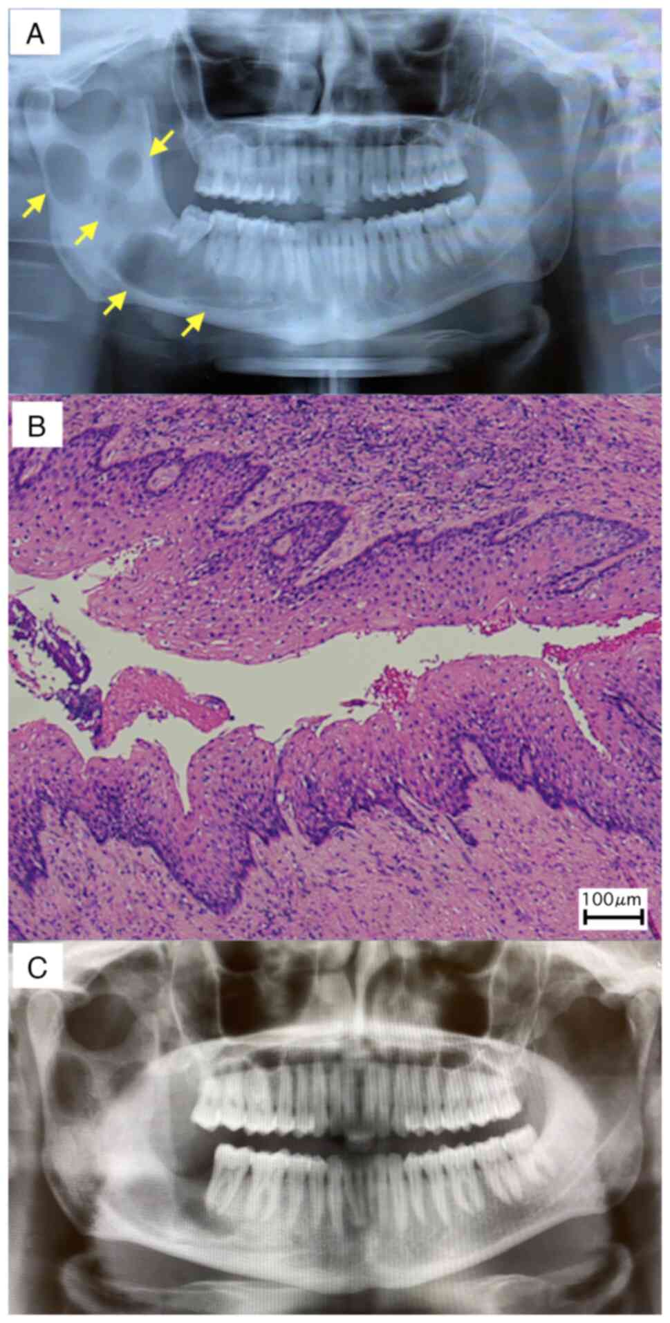
On the first visit to the referral hospital, a panoramic radiographic examination revealed a multivesicular cyst formation extending from the right mandibular body to the base of the condyle (Fig. 1A).

Figure 1

Referral hospital data. (A) Panoramic radiographic examination findings at the first visit to the referral hospital. Multivesicular cyst formation (yellow arrows) extended from the right mandibular body to the base of the condyle. (B) Histopathological image. The cyst wall was lined by a uniform stratified squamous epithelium with a corrugated surface of parakeratin, and palisaded and hyperchromatic basal cells. The histopathological diagnosis was OKC. (C) Postoperative panoramic radiographic examination findings at the referral hospital. All cysts appeared to have been removed, including the cyst at the base of the condyle.

The histopathological result of the cyst excised during surgery was OKC (Fig. 1B). Furthermore, the postoperative panoramic radiographic examination showed that all cysts were removed (Fig. 1C). At the time of their first visit 
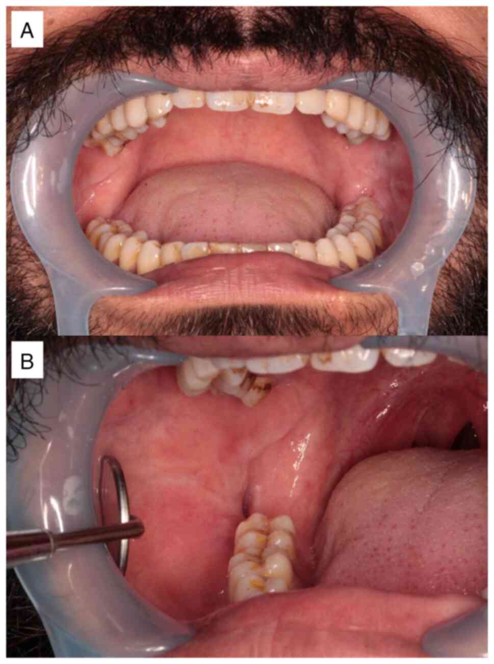
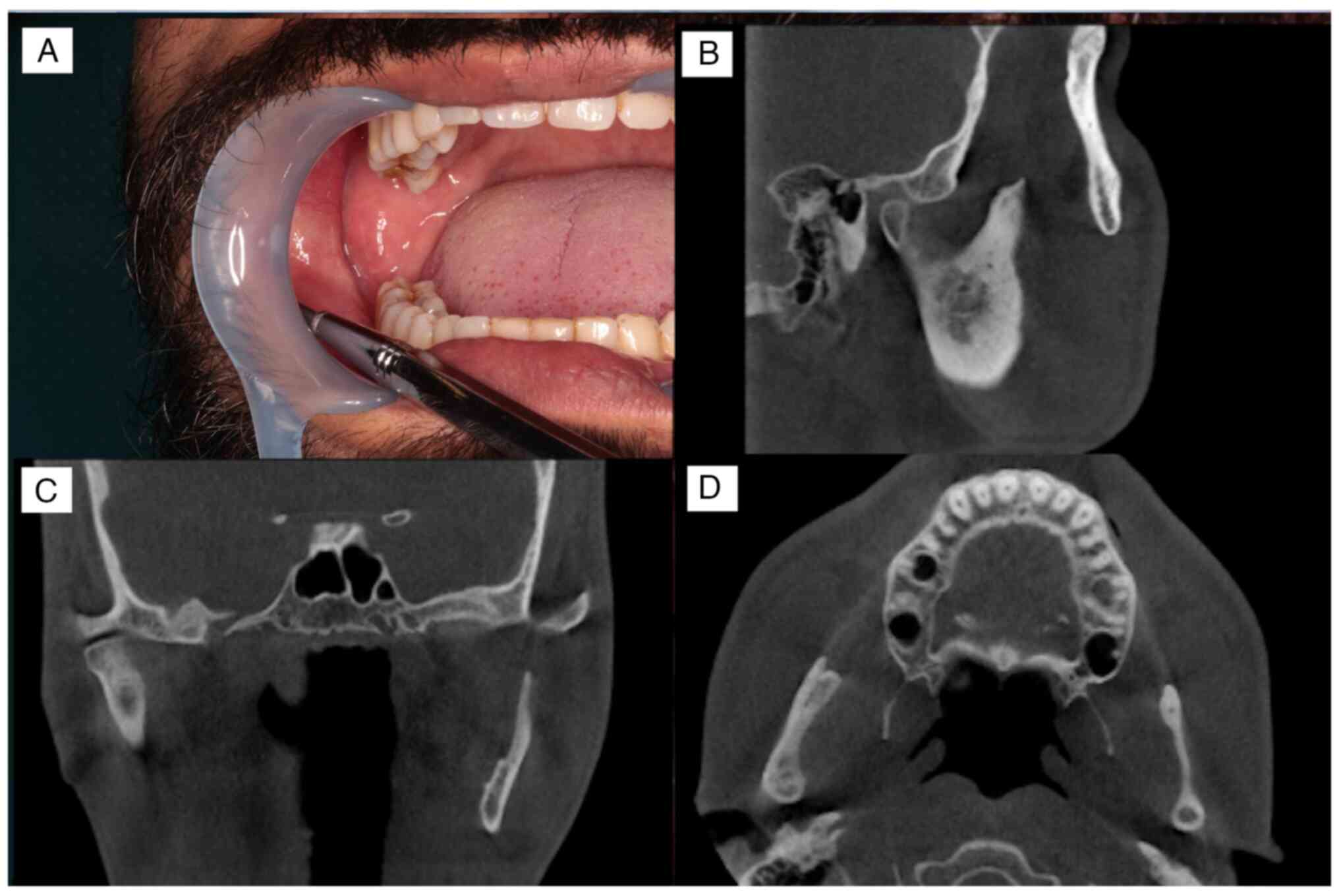
to the University of the Ryukyus Hospital, an intraoral examination showed that the buccal gingiva corresponding to the lower right 
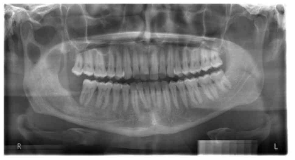
wisdom tooth was depressed owing to previous surgery (Fig. 2A and B). Panoramic radiographic examination showed an isolated cyst-like transmission in the base of the condyle and the anterior margin of the right mandibular ramus 
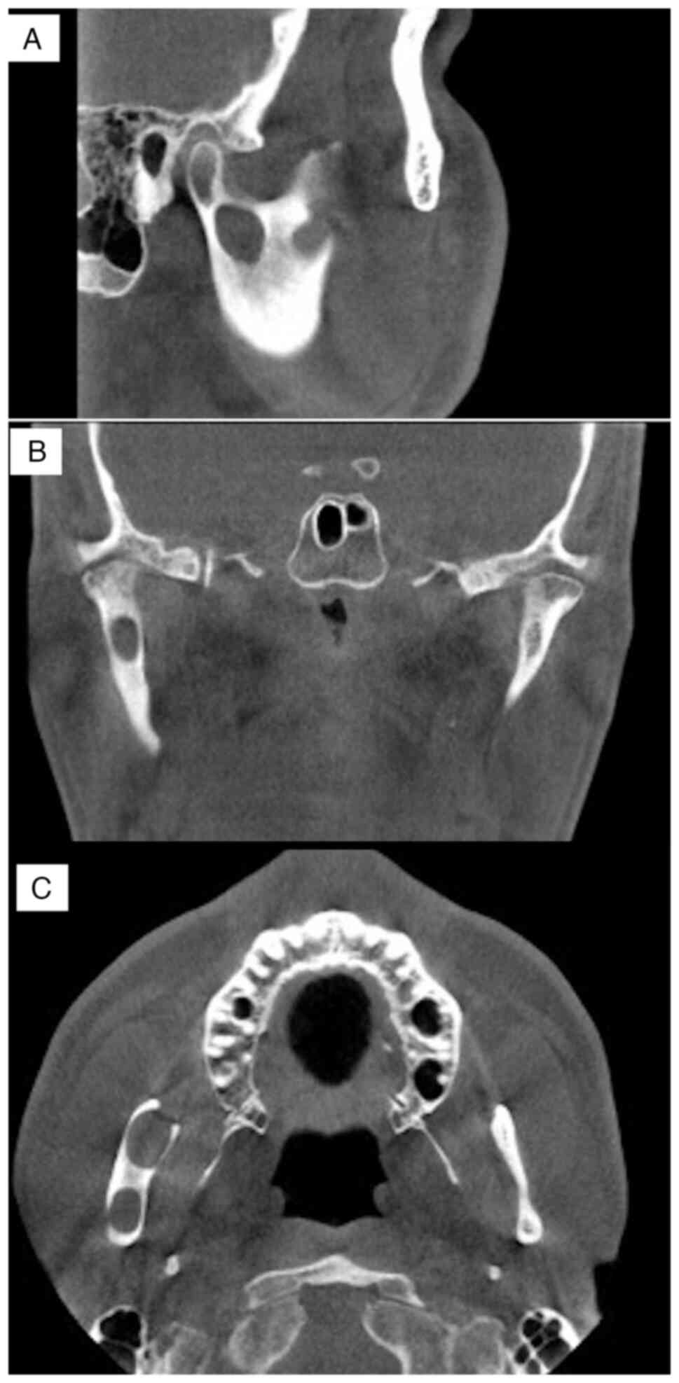
(Fig. 3). Cone-beam computed tomography examination disclosed a cyst-like transmission image measuring 13x12x6 mm in the base of the condyle. A suspected residual cyst was detected in the anterior margin of the right mandibular ramus. The cysts were not connected and both were not in contact with the inferior alveolar nerve (Fig. 4A-C). The long-term, continuous use of antibiotics was terminated because no inflammatory symptoms were observed. Although the patient was scheduled to undergo surgery in the General Anesthesia Department, symptoms of swelling and pain 
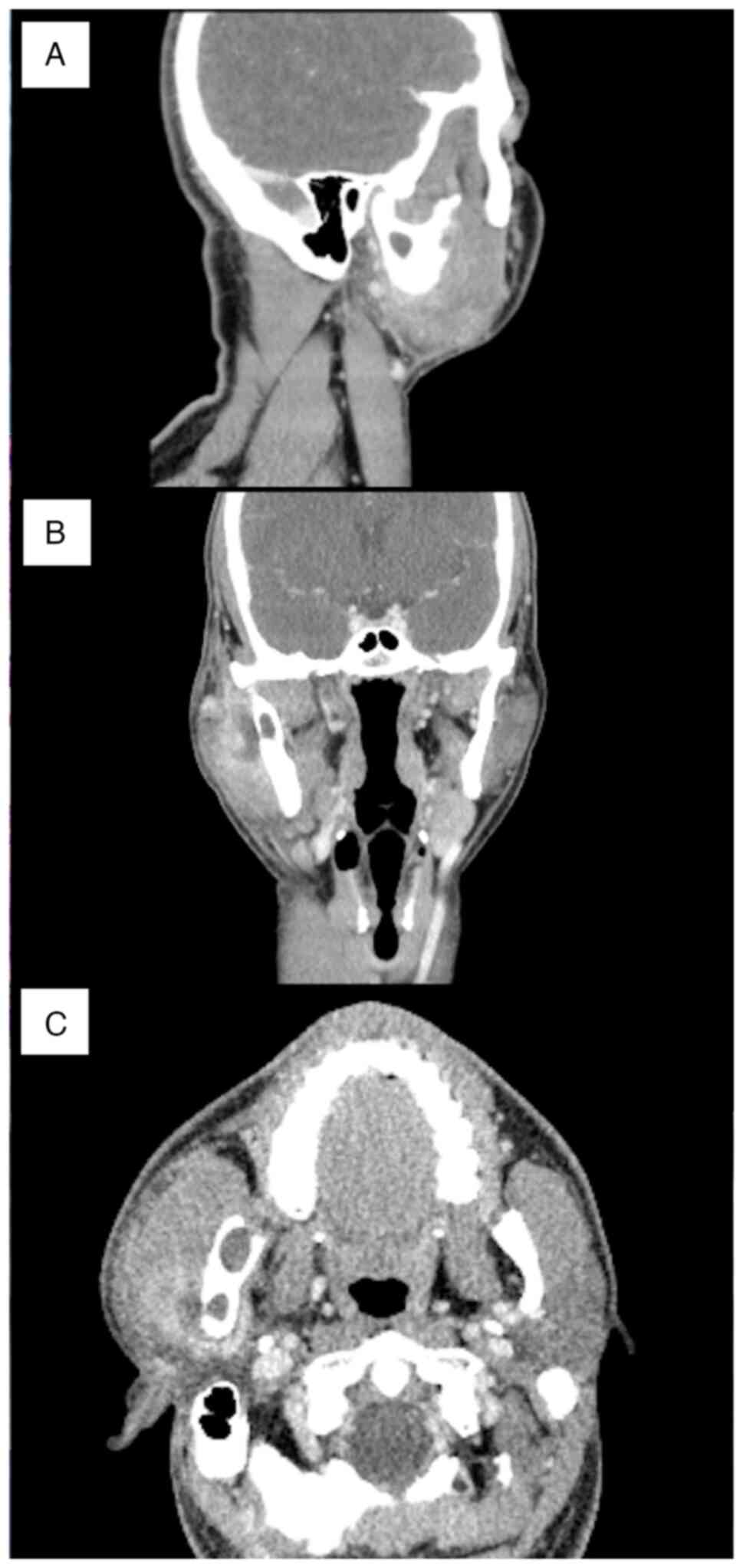
occurred around the condylar process in December 2020. A contrast computed tomography examination showed the formation of an abscess at the cyst at the base of the condyle. Swelling of the surrounding tissues, including the parotid gland, was also observed (Fig. 5A-C). Anti-inflammatory treatment included incision, drainage and antibiotics.

Figure 2

Intraoral view of the mandible. The buccal gingiva corresponding to the lower right wisdom tooth was depressed owing to a previous surgery. (A) Frontal view and (B) view focusing on the operated area.

Figure 3

Panoramic radiographic examination findings 1 year and 8 months after surgery at the referred hospital. Cyst-like transmission images in the base of the condyle and the anterior margin of the right mandibular ramus. Cyst-like transmission images of the mandibular molars seen before surgery have disappeared, thus indicating that the cyst in the mandibular molars was completely excised at the referral hospital and there has been no recurrence.

Figure 4

Cone-beam computed tomography images. (A) Sagittal, (B) coronal and (C) horizontal views of a cyst-like transmission image measuring 13x12x6 mm in the base of the condyle. A suspected residual cyst was detected on the anterior margin of the right mandibular ramus. The cysts were not connected.

Figure 5

Contrast computed tomography images. (A) Sagittal, (B) coronal and (C) horizontal views of abscess formation at the cyst at the base of the condyle and swelling of the surrounding tissues, including the parotid gland.

Treatment and follow-up

In January 2021, the patient underwent surgery under general anesthesia. The packed open technique (10,11) was selected to preserve the condyle process. To avoid damaging the mandibular nerve as it entered the mandibular foramen before approaching the cyst, the lingual side of the mandible was 
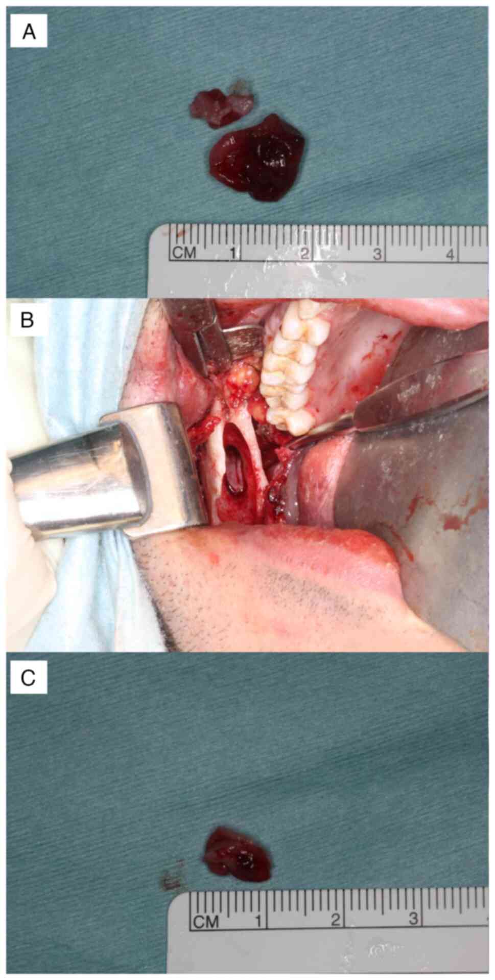
carefully detached. First, the anterior cyst left behind from the anterior margin of the mandibular ramus was removed (Fig. 6A). After removing the anterior cyst, an osteotomy was performed at the height between the mandibular notch and the mandibular foramen. Care was taken to protect the buccal and lingual sides of the base of the condyle and to ensure that only the bone was scraped and the burs did not penetrate to prevent damage to the jaw artery around the mandibular head. An ultrasonic bone scalpel was used as needed. The posterior cyst was confirmed and excised (Fig. 6B and C) after further grinding of the surrounding bone to remove the daughter cyst, which caused OKC recurrence. The method of enucleation was chosen followed by open packing. Because the open cavity width was so small, it might close prematurely, creating a dead space. The oral mucosa and bone were sutured with absorbent thread. To reduce pain when changing the packing gauze, a bovine collagen sheet (TERUDERMIS®, Terumo Co., Ltd.) was attached to the cyst cavity and tetracycline hydrochloride ointment gauze was inserted. Hospitalization was managed with attention paid to postoperative fractures as the bone at the articular process was thinned. After surgery, the patient followed an oral liquid diet for 6 days. During hospitalization, the patient's mouth opening was restricted, with only a minimum opening allowed when eating or brushing teeth. A total of 7 days after surgery, the tetracycline hydrochloride ointment gauze was replaced and the thread was removed. The patient was discharged 14 days after surgery owing to good progress. From February to May 2021, an obturator was attached to prevent the cavity from closing. A total of 20 months post-operation, the patient showed no signs of local recurrence (Fig. 7A-D). In addition, there was no reappearance of inflammation after surgery.

Figure 6

Intraoperative findings. (A) Anterior cyst left behind from the anterior margin of the mandibular ramus. (B) Posterior cyst after osteotomy at the height between the mandibular notch and the mandibular foramen. (C) Excised posterior cyst.

Figure 7

Postoperative findings. (A) Intraoral view of the mandible. (B) Sagittal, (C) coronal and (D) horizontal views of postoperative cone-beam computed tomography image findings. There were no signs of local recurrence.

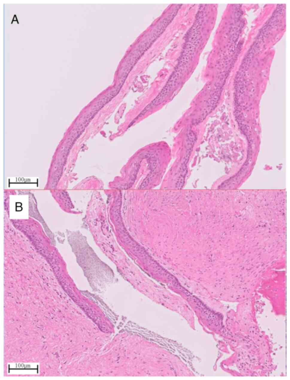
Histopathological examination of the removed specimen showed cyst walls with a well-ordered squamous epithelial lining in both the anterior (Fig. 8A) and posterior (Fig. 8B) cysts. These were typical findings of OKCs showing a palisade arrangement of basal cells and a wavy structure on the surface.

Figure 8

Histopathological images. The specimen showed cyst walls with a well-ordered squamous epithelial lining in both the (A) anterior and (B) posterior cysts. It was a typical odontogenic keratocyst finding showing a palisade arrangement of basal cells and a wavy structure on the surface.

Discussion

OKCs were initially described in 1876 and were named in 1956 by Phillipsen (12) to describe an odontogenic cyst lined with keratinized stratified squamous epithelium (13). In 1992, the term ‘odontogenic keratocyst’, which is synonymous with ‘primordial cyst’, was introduced by the World Health Organization (WHO) to denote benign cysts of odontogenic origin with a specific histological appearance. However, in 2005, this pathology was reclassified as a benign keratocystic odontogenic tumor (KCOT) in the third edition of the WHO classification of head and neck tumors because of the high risk of recurrence, aggressive clinical course, mutations in the protein patched homolog tumor suppressor gene, the occurrence of satellite cysts and the association with Gorlin-Goltz syndrome (14,15). However, due to insufficient evidence to categorize this pathology as a neoplastic lesion, in the fourth edition of the WHO classification of head and neck tumors, KCOT was moved back into the cyst category under the name OKC (16) and it is still categorized as a cyst in the current classification (fifth edition) (17).

The recurrence rate of OKC ranges between 7 and 28% (2). Since the 1980s, it has been suggested that to reduce the high recurrence rate of OKCs, surgery should include marginal resection, including a rim of uninvolved bone, which is similar to the treatment for unicystic ameloblastoma (18). Various suggestions have been made regarding surgical methods for OKCs. The main suggestions are as follows: Enucleation; enucleation plus adjunctive therapy, such as enucleation plus curettage, enucleation with Carnoy's solution, enucleation with cryotherapy and enucleation with peripheral ostectomy and Carnoy's solution; marsupialization; resection; or combinations of these techniques (2). Although the difference in recurrence rates due to the surgical method has been examined, several factors other than the surgical method contribute to the recurrence rate, such as multilocular cysts or unilocular cysts and perforated or nonperforated bony walls; therefore, there is no one-size-fits-all surgical procedure for OKC (2,19). There is another important consideration when choosing jawbone cyst surgery, which is the size of the cyst. Enucleation and primary closure are often selected. This technique is accepted for lesions <2 cm, in which angiogenic and osteogenic cells from the adjacent bone and periosteum can reach the remaining blood clot and could promote ossification (20). It was predicted that the OKC described in the present case report would create a cavity of >2 cm, including a reduction of the bone. Enucleation, followed by open packing, has been reported to be a less invasive method for the treatment of OKCs with a lower recurrence rate and this method was thus used in the present study (10,11). In the present case, after the packing became open, the tetracycline hydrochloride ointment gauze was replaced once a week and an impression was taken 1 month after the operation to create an obturator to prevent a dead space from collecting food or debris (21). As a result, a good healing course was observed without recurrence.

To the best of our knowledge, there have only been two reports of OKCs in the condyloid process that are not continuous with the surrounding areas (8,9). In both cases, OKCs occurred in the head of the condyle, which was resected. Although there is a difference between the condylar heads and the base of the condyle, the present case is the first in which the condylar process was successfully preserved. Although the details of the subsequent course were not described in the previous two cases, the excision of the articular process without reconstruction may result in malocclusion and facial deformity. In the present case, because the bone at the articular process was thinned, a hospitalization plan was made to avoid fractures. The patient received an oral liquid diet in the first 6 days after surgery, and during hospitalization, the patient was allowed only a minimum amount of mouth opening while eating or when brushing their teeth. In addition, from February to May 2021, an obturator was attached. A total of 20 months post-operation, the patient showed no signs of local recurrence.

In conclusion, this report presents a rare case of an OKC in the mandibular condyle base region of a 31-year-old man. Resection was performed under general anesthesia and the condylar process was successfully preserved. The patient has remained disease-free for 20 months post-operation.

Acknowledgements

Not applicable.

Funding

Funding: No funding was received.

Availability of data and materials

The datasets used and/or analyzed during the current study are available from the corresponding author on reasonable request.

Author contributions

SM, TG, JS, TK, MM, KI, NM, AM and KN examined and treated the patient and collected the data. SM, TG, JS, KN and HN analyzed and discussed the case and data. SM, TK, AM and KN confirm the authenticity of all the raw data. SM wrote the manuscript. All authors read and approved the final manuscript.

Ethics approval and consent to participate

Not applicable.

Patient consent for publication

Written informed consent was obtained from the patient for both the surgical treatment and publication of any accompanying images.

Competing interests

The authors declare that they have no competing interests.

References

1 

Johnson NR, Gannon OM, Savage NW and Batstone MD: Frequency of odontogenic cysts and tumors: A systematic review. J Investig Clin Dent. 5:9–14. 2014.PubMed/NCBI View Article : Google Scholar

2 

Antonoglou GN, Sándor GK, Koidou VP and Papageorgiou SN: Non-syndromic and syndromic keratocystic odontogenic tumors: Systematic review and meta-analysis of recurrences. J Craniomaxillofac Surg. 42:e364–e371. 2014.PubMed/NCBI View Article : Google Scholar

3 

Harmon M, Arrigan M, Toner M and O'Keeffe SA: A radiological approach to benign and malignant lesions of the mandible. Clin Radiol. 70:335–350. 2015.PubMed/NCBI View Article : Google Scholar

4 

MacDonald D: Lesions of the jaws presenting as radiolucencies on cone-beam CT. Clin Radiol. 71:972–985. 2016.PubMed/NCBI View Article : Google Scholar

5 

Kaneda T, Minami M and Kurabayashi T: Benign odontogenic tumors of the mandible and maxilla. Neuroimaging Clin N Am. 13:495–507. 2003.PubMed/NCBI View Article : Google Scholar

6 

Farzad P: A case of an extensive keratocystic odontogenic tumor in the mandible reconstructed with a custom-made total joint prosthesis. Craniomaxillofac Trauma Reconstr. 11:131–137. 2018.PubMed/NCBI View Article : Google Scholar

7 

Fidele NB, Bing L, Sun Y, Wu T, Zheng Y and Zhao Y: Management of mandibular odontogenic keratocyst through radical resection: Report of 35 cases. Oncol Lett. 18:733–741. 2019.PubMed/NCBI View Article : Google Scholar

8 

Kallalli B, Rawson K, Telkar S and Penumatcha M: Keratocystic odontogenic tumor of the right mandibular condyle: A rare case. J Indian Acad Oral Med Radiol. 26(103)2014.

9 

Managutti A, Managutti S, Patel H and Menat S: Orthokeratinized odontogenic cyst (OOC) of condylar head: A rare entity. J Maxillofac Oral Surg. 15 (Suppl 2):S315–S319. 2016.PubMed/NCBI View Article : Google Scholar

10 

Kshirsagar RA, Bhende RC, Raut PH, Mahajan V, Tapadiya VJ and Singh V: Odontogenic Keratocyst: Developing a protocol for surgical intervention. Ann Maxillofac Surg. 9:152–157. 2019.PubMed/NCBI View Article : Google Scholar

11 

Yildirim G, Ataoglu H, Kalayci A, Özkan BT, Kucuk K and Esen A: Conservative treatment protocol for keratocystic odontogenic tumour: A follow-up study of 3 cases. J Oral Maxillofac Res. 1(e7)2010.PubMed/NCBI View Article : Google Scholar

12 

Philipsen HP: OM Keratocysts (kole steatoma). Tandlaegebladet. 60:963–980. 1956.(In Danish).

13 

Chi AC, Owings JR Jr and Muller S: Peripheral odontogenic keratocyst: Report of two cases and review of the literature. Oral Surg Oral Med Oral Pathol Oral Radiol Endod. 99:71–78. 2005.PubMed/NCBI View Article : Google Scholar

14 

Tomasz K, Jadwiga S and Romana T: Update of the WHO classification of odontogenic and maxillofacial bone tumours. Journal of Stomatology. 23:484–506. 2017.

15 

Soluk-Tekkeşin M and Wright JM: The World Health Organization Classification of Odontogenic Lesions: A summary of the changes of the 2017 (4th) Edition. Turk Patoloji Derg 34, 2018.

16 

Speight PM and Takata T: New tumour entities in the 4th edition of the World Health Organization Classification of Head and Neck tumours: Odontogenic and maxillofacial bone tumours. Virchows Arch. 472:331–339. 2018.PubMed/NCBI View Article : Google Scholar

17 

Soluk-Tekkesin M and Wright JM: The World Health Organization Classification of Odontogenic Lesions: A summary of the changes of the 2022 (5th) Edition. Turk Patoloji Derg. 38:168–184. 2022.PubMed/NCBI View Article : Google Scholar

18 

Ahlfors E, Larsson A and Sjögren S: The odontogenic keratocyst: A benign cystic tumor? J Oral Maxillofac Surg. 42:10–19. 1984.PubMed/NCBI View Article : Google Scholar

19 

Forssell K, Forssell H and Kahnberg KE: Recurrence of keratocysts. A long-term follow-up study. Int J Oral Maxillofac Surg. 17:25–28. 1988.PubMed/NCBI View Article : Google Scholar

20 

Carbonell-Asins P and Sánchez Aniceto G: A retrospective risk factor analysis of infection and wound dehiscence following guided bone regeneration in cystectomy defects of the jaws. J Craniomaxillofac Surg. 50:657–663. 2022.PubMed/NCBI View Article : Google Scholar

21 

Troiano A, Lo Giudice G, De Luca R, Lo Giudice F, D'Amato S, Tartaro G and Colella G: Salvage of dental implant located in mandibular odontogenic Cyst. A conservative surgical treatment proposal. Dent J (Basel). 8(E49)2020.PubMed/NCBI View Article : Google Scholar

Related Articles

  • Abstract
  • View
  • Download
  • Twitter
Copy and paste a formatted citation
Spandidos Publications style
Miyamoto S, Goto T, Shirakawa J, Kawano T, Murahashi M, Ide K, Maruyama N, Matayoshi A, Nishihara K, Nakamura H, Nakamura H, et al: Odontogenic keratocyst in the mandibular condyle base region: A case report. Exp Ther Med 25: 141, 2023.
APA
Miyamoto, S., Goto, T., Shirakawa, J., Kawano, T., Murahashi, M., Ide, K. ... Nakamura, H. (2023). Odontogenic keratocyst in the mandibular condyle base region: A case report. Experimental and Therapeutic Medicine, 25, 141. https://doi.org/10.3892/etm.2023.11840
MLA
Miyamoto, S., Goto, T., Shirakawa, J., Kawano, T., Murahashi, M., Ide, K., Maruyama, N., Matayoshi, A., Nishihara, K., Nakamura, H."Odontogenic keratocyst in the mandibular condyle base region: A case report". Experimental and Therapeutic Medicine 25.3 (2023): 141.
Chicago
Miyamoto, S., Goto, T., Shirakawa, J., Kawano, T., Murahashi, M., Ide, K., Maruyama, N., Matayoshi, A., Nishihara, K., Nakamura, H."Odontogenic keratocyst in the mandibular condyle base region: A case report". Experimental and Therapeutic Medicine 25, no. 3 (2023): 141. https://doi.org/10.3892/etm.2023.11840
Copy and paste a formatted citation
x
Spandidos Publications style
Miyamoto S, Goto T, Shirakawa J, Kawano T, Murahashi M, Ide K, Maruyama N, Matayoshi A, Nishihara K, Nakamura H, Nakamura H, et al: Odontogenic keratocyst in the mandibular condyle base region: A case report. Exp Ther Med 25: 141, 2023.
APA
Miyamoto, S., Goto, T., Shirakawa, J., Kawano, T., Murahashi, M., Ide, K. ... Nakamura, H. (2023). Odontogenic keratocyst in the mandibular condyle base region: A case report. Experimental and Therapeutic Medicine, 25, 141. https://doi.org/10.3892/etm.2023.11840
MLA
Miyamoto, S., Goto, T., Shirakawa, J., Kawano, T., Murahashi, M., Ide, K., Maruyama, N., Matayoshi, A., Nishihara, K., Nakamura, H."Odontogenic keratocyst in the mandibular condyle base region: A case report". Experimental and Therapeutic Medicine 25.3 (2023): 141.
Chicago
Miyamoto, S., Goto, T., Shirakawa, J., Kawano, T., Murahashi, M., Ide, K., Maruyama, N., Matayoshi, A., Nishihara, K., Nakamura, H."Odontogenic keratocyst in the mandibular condyle base region: A case report". Experimental and Therapeutic Medicine 25, no. 3 (2023): 141. https://doi.org/10.3892/etm.2023.11840
Follow us
  • Twitter
  • LinkedIn
  • Facebook
About
  • Spandidos Publications
  • Careers
  • Cookie Policy
  • Privacy Policy
How can we help?
  • Help
  • Live Chat
  • Contact
  • Email to our Support Team