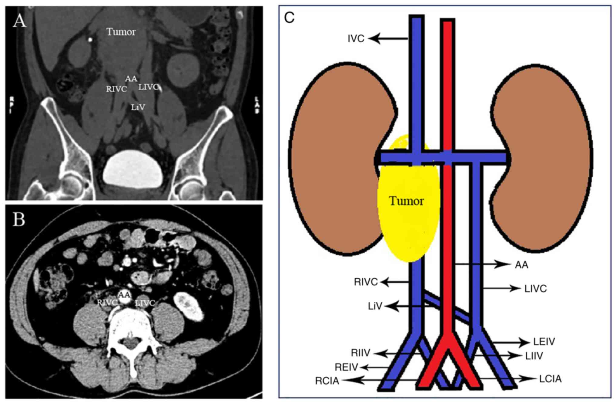
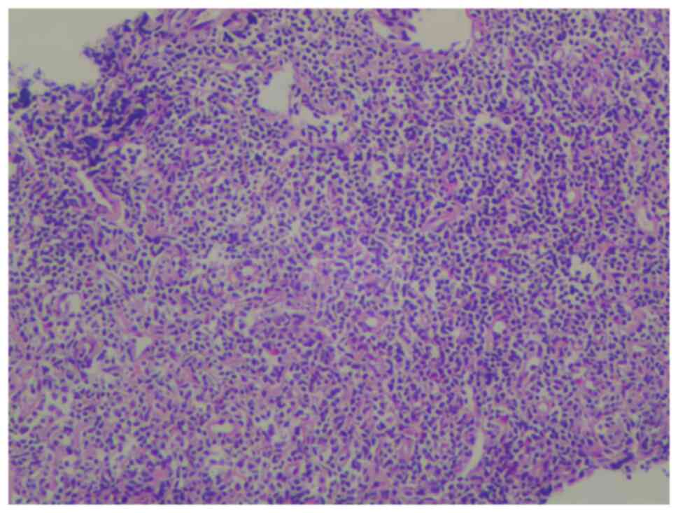
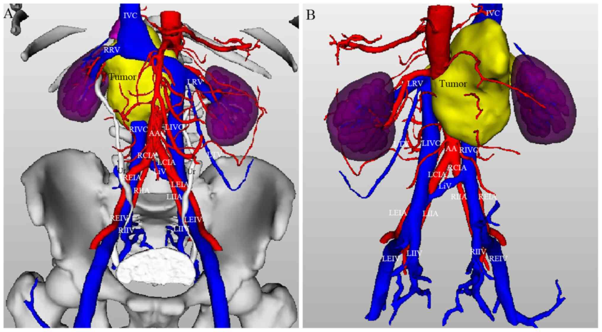
|
1
|
Shaheen S, Alyahya KI, Fouhil AFE, Salama
EEA, Atteya M, Elshaer F and Darwish H: An extremely rare complete
bilateral duplication of inferior vena cava in a male cadaver:
Anatomy, embryology and clinical relevance. Folia Morphol (Warsz).
81:247–253. 2022.PubMed/NCBI View Article : Google Scholar
|
|
2
|
Banerjee A, Maharana S, Kumar IA and
Jhansi P: Duplication of the inferior vena cava-report of a rare
congenital variation. IJAV. 5:141–143. 2012.
|
|
3
|
Petik B: Inferior vena cava anomalies and
variations: Imaging and rare clinical findings. Insights Imaging.
6:613–639. 2015.PubMed/NCBI View Article : Google Scholar
|
|
4
|
Babaian RJ and Johnson DE: Major venous
anomalies complicating retroperitoneal surgery. South Med J.
72:1254–1258. 1979.PubMed/NCBI View Article : Google Scholar
|
|
5
|
Yoshimura S, Yamamoto K, Fujimura S,
Kawata S, Shimada K, Omotehara T and Itoh M: A case of double
inferior vena cava with the connection to sacral venous plexus.
Anat Sci Int. 97:143–146. 2022.PubMed/NCBI View Article : Google Scholar
|
|
6
|
Waśniewska A, Ruzik K, Olewnik Ł,
Stefańczyk L and Polguj M: Unusual coexistence of double inferior
vena cava with nutcracker syndrome-a case report and review of the
literature. J Int Med Res. 48(300060520904520)2020.PubMed/NCBI View Article : Google Scholar
|
|
7
|
Klinkhachorn PS, Ritz BK, Umstot SI,
Skrzat J and Zdilla MJ: Duplication of the inferior vena cava:
Evidence of a novel type IV. Folia Cracov. 60:5–13. 2020.PubMed/NCBI View Article : Google Scholar
|
|
8
|
Chen HY, Emura S, Nagasaki S and Kubo K:
Double inferior vena cava with interiliac vein: A case report and
literature review. Okajimas Folia Anat Jpn. 88:147–151.
2012.PubMed/NCBI View Article : Google Scholar
|
|
9
|
Matsuoka A, Tate S, Nishikimi K and Shozu
M: Retroperitoneal lymphadenectomy for ovarian cancer with double
inferior vena cava. Gynecol Oncol. 148:632–633. 2018.PubMed/NCBI View Article : Google Scholar
|
|
10
|
Onoda K, Shomura Y and Komada T: Double
inferior vena cava with azygos continuation and retroaortic left
renal vein associated with juxtarenal abdominal aortic aneurysm
surgery. Ann Vasc Dis. 11:123–126. 2018.PubMed/NCBI View Article : Google Scholar
|
|
11
|
Wang X, Chen Z and Cai Q:
Catheter-directed thrombolysis for double inferior vena cava with
deep venous thrombosis: A case report and literature review.
Phlebology. 29:480–483. 2014.PubMed/NCBI View Article : Google Scholar
|
|
12
|
Pilichowska E, Ostrowski P, Kotowski MJ,
Tejchman K, Ostrowska-Clark K, Ostrowski M and Sieńko J:
Transplantation of a kidney with duplicated ureter harvested from a
donor with vascular anomaly in the form of double inferior vena
cava: A Case Report. Transplant Proc. 52:2533–2535. 2020.PubMed/NCBI View Article : Google Scholar
|
|
13
|
Ito T and Ikeda Y: A case of double
inferior vena cava with renal, ovarian and iliac vein variation.
Anat Sci Int. 93:139–143. 2018.PubMed/NCBI View Article : Google Scholar
|
|
14
|
Coco D, Cecchini S, Leanza S, Viola M,
Ricci S and Campagnacci R: Inferior vena cava duplication:
Incidental case in a young woman. Case Rep Radiol.
2016(3071873)2016.PubMed/NCBI View Article : Google Scholar
|
|
15
|
Chaijaroonkhanarak W, Pannangrong W,
Welbat JU, Namking M, Khamanarong K and Prachaney P: Double
inferior vena cava with three shunts: A rare anomaly with important
implications for surgeons. Folia Morphol (Warsz). 76:307–311.
2017.PubMed/NCBI View Article : Google Scholar
|
|
16
|
Jiang Y, Duan L, Lu L, Zhao WG, Zeng ZP,
Li HZ and Zhang XB: Rare case of reninoma with double inferior vena
cava. Clin Exp Hypertens. 33:325–327. 2011.PubMed/NCBI View Article : Google Scholar
|
|
17
|
Nakatani T, Kim T, Naganuma T, Uchida J,
Takemoto Y and Sugimura K: Kidney transplants from living related
donors having double inferior vena cava. Urol Int. 72:358–360.
2004.PubMed/NCBI View Article : Google Scholar
|
|
18
|
Kumar S, Panigrahy B, Ravimohan SM, Pandya
S, Mandal AK and Singh SK: Rare case of renal cell carcinoma with
double inferior vena cava with venous thrombosis. Urology.
72:461.e7–e10. 2008.PubMed/NCBI View Article : Google Scholar
|
|
19
|
Yano R, Hayakawa D, Emura S, Chen H, Ozawa
Y, Taguchi H and Shoumura S: Two cases of the double inferior venae
cavae. Okajimas Folia Anat Jpn. 77:133–136. 2000.PubMed/NCBI View Article : Google Scholar
|
|
20
|
Fronek JP, Morsy MA, Singh U, Chemla E and
Chang RW: Retroperitoneoscopic live donor nephrectomy in a patient
with a double inferior vena cava. J Laparoendosc Adv Surg Tech A.
16:378–380. 2006.PubMed/NCBI View Article : Google Scholar
|
|
21
|
Vasanth Kumar A, Anirudh Kumar A, Hussain
A and Sameeraja V: An uncommon encounter during temporary pacemaker
implantation-A double inferior vena cava. Indian Heart J. 68 (Suppl
2):S216–S217. 2016.PubMed/NCBI View Article : Google Scholar
|
|
22
|
Furutani A, Yoshida S, Yoshida T, Nishi M,
Yamagishi T, Goto H, Otsubo D, Yamane H, Matsumoto T, Fujino Y and
Tominaga M: A case of laparoscopic anterior resection for rectal
cancer with duplication of the inferior vena cava using
preoperative 3D computed tomography angiography. J Surg Case Rep.
2020(rjaa223)2020.PubMed/NCBI View Article : Google Scholar
|
|
23
|
Habuchi T, Okagaki T, Arai K and Miyakawa
M: Renal cell carcinoma extending into left side of double inferior
vena cava. Urology. 41:181–184. 1993.PubMed/NCBI View Article : Google Scholar
|
|
24
|
Mao YQ, Zhu SX and Zhang W: The iatrogenic
injury of double vena cava due to misdiagnosis during the radical
nephroureterectomy and cystectomy. World J Surg Oncol.
13(41)2015.PubMed/NCBI View Article : Google Scholar
|
|
25
|
Yamaguchi A, Negoro H, Kojo K, Ikeda A,
Kimura T, Kandori S, Hoshi A, Kojima T, Kawai K and Nishiyama H:
Retroperitoneal lymph node dissection for testicular cancer in a
patient with a double inferior vena cava. IJU Case Rep. 4:86–88.
2021.PubMed/NCBI View Article : Google Scholar
|
|
26
|
Sousa Gomes M, Pardal C, Monteiro C and
Serrano P: Double inferior vena cava in gynaecological oncology
surgery. BMJ Case Rep. 13(e240361)2020.PubMed/NCBI View Article : Google Scholar
|
|
27
|
Aljabri B, MacDonald PS, Satin R, Stein
LS, Obrand DI and Steinmetz OK: Incidence of major venous and renal
anomalies relevant to aortoiliac surgery as demonstrated by
computed tomography. Ann Vasc Surg. 15:615–618. 2001.PubMed/NCBI View Article : Google Scholar
|
|
28
|
Shammas NW, Rachwan RJ, Daher G and
Dargham BB: Double inferior vena cava and its implications during
endovascular and surgical interventions: A word of caution. J
Invasive Cardiol. 29:51–53. 2017.PubMed/NCBI
|
|
29
|
Eldefrawy A, Arianayagam M, Kanagarajah P,
Acosta K and Manoharan M: Anomalies of the inferior vena cava and
renal veins and implications for renal surgery. Cent European J
Urol. 64:4–8. 2011.PubMed/NCBI View Article : Google Scholar
|
|
30
|
Sitwala PS, Ladia VM, Brahmbhatt PB, Jain
V and Bajaj K: Inferior vena cava anomaly: A risk for deep vein
thrombosis. N Am J Med Sci. 6:601–603. 2014.PubMed/NCBI View Article : Google Scholar
|
|
31
|
Sartori MT, Zampieri P, Andres AL,
Prandoni P, Motta R and Miotto D: Double vena cava filter insertion
in congenital duplicated inferior vena cava: A case report and
literature review. Haematologica. 91 (Suppl
6)(ECR30)2006.PubMed/NCBI
|
|
32
|
Vo NJ, Wieseler KW, Burdick TR, Goswami
GK, Vaidya SS and Andrews RT: The use of paired optionally
retrievable günther tulip filters in trauma patients with
anatomical variants. Semin Intervent Radiol. 24:20–28.
2007.PubMed/NCBI View Article : Google Scholar
|
|
33
|
Hoppe RT, Advani RH, Ai WZ, Ambinder RF,
Armand P, Bello CM, Benitez CM, Bierman PJ, Boughan KM, Dabaja B,
et al: Hodgkin lymphoma, version 2.2020, NCCN clinical practice
guidelines in oncology. J Natl Compr Canc Netw. 18:755–781.
2020.PubMed/NCBI View Article : Google Scholar
|
|
34
|
Lucas MF: A case of double inferior vena
cava. J Anat. 51(Pt 1). 69–70. 1916.PubMed/NCBI
|
|
35
|
Yagel S, Kivilevitch Z, Cohen SM, Valsky
DV, Messing B, Shen O and Achiron R: The fetal venous system, part
I: Normal embryology, anatomy, hemodynamics, ultrasound evaluation
and Doppler investigation. Ultrasound Obstet Gynecol. 35:741–750.
2010.PubMed/NCBI View
Article : Google Scholar
|
|
36
|
Mayo J, Gray R, St Louis E, Grosman H,
McLoughlin M and Wise D: Anomalies of the inferior vena cava. AJR
Am J Roentgenol. 140:339–345. 1983.PubMed/NCBI View Article : Google Scholar
|
|
37
|
Mathews R, Smith PA, Fishman EK and
Marshall FF: Anomalies of the inferior vena cava and renal veins:
Embryologic and surgical considerations. Urology. 53:873–880.
1999.PubMed/NCBI View Article : Google Scholar
|
|
38
|
Bass JE, Redwine MD, Kramer LA, Huynh PT
and Harris JH Jr: Spectrum of congenital anomalies of the inferior
vena cava: Cross-sectional imaging findings. Radiographics.
20:639–652. 2000.PubMed/NCBI View Article : Google Scholar
|
|
39
|
Tisnado J, Amendola MA, Vines FS and
Beachley MC: Computed tomography of double inferior vena cava: The
‘double cava’ sign. Comput Tomogr. 3:195–199. 1979.PubMed/NCBI View Article : Google Scholar
|
|
40
|
Inamasu J and Guiot BH: Laparoscopic
anterior lumbar interbody fusion: A review of outcome studies.
Minim Invasive Neurosurg. 48:340–347. 2005.PubMed/NCBI View Article : Google Scholar
|