Introduction
Lacrimal canaliculitis is an infection of the
lacrimal canaliculus, which is the proximal part of the tear
drainage system of the eye. It is a relatively rare condition and
typically occurs in people aged >40. Although it is most
commonly caused by a bacterial pathogen, it can also be caused by a
fungal or viral infection (1,2). The
infection may lead to the formation of small dacryoliths. Multiple
dacryoliths can be present and obstruct the lacrimal outflow system
with time; finally, the infection and obstruction of lacrimal
canaliculus are combined to form a vicious circle. Lacrimal
canaliculitis can be divided into primary canaliculitis and
secondary canaliculitis according to the cause of the disease
(3). Secondary canaliculitis is
most commonly associated with punctal or intracanalicular plug
placement, while there is often no history of surgery related to
the lacrimal canaliculus in primary canaliculitis.
Primary canaliculitis is an uncommon disease,
comprising 2% of all patients with lacrimal disease worldwide
(4), typically manifesting as
pouting erythematous punctum, punctal or canalicular swelling, and
expressible punctal discharge. It is caused by various
microorganisms, with Actinomyces infection reported to be
the most common pathogen (5).
However, owing to its rarity and lack of typical presentation in
the clinics, primary canaliculitis is often misdiagnosed as chronic
conjunctivitis, chronic dacryocystitis, chalazion or hordeolum
(4,6-8),
which causes a delay in effective treatment. Therefore, there is
often a prolonged symptomatic period until diagnosis; this can have
serious impacts on work and life and even lead to adverse
psychological effects.
The present study aimed to summarize the pathogenic
characteristics, diagnosis and treatment of patients with primary
canaliculitis admitted to Affiliated Wuxi Clinical College of
Nantong University (Wuxi, China) between May 2018 and April 2021.
This information may serve as a basis for developing guidelines for
the clinical diagnosis and treatment of canaliculitis. To the best
of our knowledge, the current study is the first to report this
information for the specific region analyzed.
Materials and methods
Participants
Patients diagnosed with primary canaliculitis
according to the clinical manifestations and examinations (such as
pouting erythematous punctum, punctal or canalicular swelling, as
well as expressible punctal discharge) at the Department of
Ophthalmology, Affiliated Wuxi Clinical College of Nantong
University (Wuxi, China), between May 2018 and April 2021, were
enrolled in the present prospective study (Table I). Patients with canaliculitis
secondary to trauma or punctal and canalicular plugs were excluded.
All patients underwent slit-lamp examination and lacrimal duct
irrigation and exploration. Ultrasound biomicroscopy (UBM) (Quantel
Medical, Cournon d'Auvergne Cedex, France) was performed with a 50
MHz probe as an ancillary examination only in patients with severe
lacrimal duct dilatation. The protocol of the current prospective
study was approved by the Institutional Review Board of the
Affiliated Wuxi Clinical College of Nantong University (approval
no. 2022-Y-98). Prior to inclusion, written informed consent was
obtained from all patients. The present study adhered to the tenets
of The Declaration of Helsinki.
 | Table IPatient clinicopathological
characteristics (n=50). |
Table I
Patient clinicopathological
characteristics (n=50).
| Characteristic | Number of patients, n
(%) |
|---|
| Sex | |
|
Male | 11(22) |
|
Female | 39(78) |
| Age, years | |
|
38-63 | 27(54) |
|
63-87 | 23(46) |
| Lacrimal duct
position | |
|
Superior | 17(34) |
|
Inferior | 29(58) |
|
Superior +
inferior | 4(8) |
| Eye position | |
|
Left
eye | 30(60) |
|
Right
eye | 20(40) |
Specimen collection and smear staining
examination
Care was taken in specimen collection since this can
directly impact the results of smears and cultures. Following the
application of one drop of topical anesthesia (Oxybuprocaine
Hydrochloride Eye Drops; Santen Pharmaceutical), the eyes were
cleaned and disinfected. The affected lacrimal punctum was dilated.
Thereafter, the mucopurulent discharge and concretions were
manually expressed from the punctum using two sterile cotton buds
to compress the affected canaliculus. Possibly contaminated
discharge was removed from the anterior segment. A sterile spatula
was subsequently used to obtain a discharge from the middle or
posterior segment. The dacryoliths from discharge were crushed
evenly and then smears were immediately prepared for Gram staining
according to the manufacturer's protocols and microscopic
examination under a fluorescence microscope (Olympus BX53; Olympus
Corporation).
Bacterial culture and
identification
The collected specimens (both mucopurulent discharge
and dacryoliths) were used for bacterial, fungal and anaerobic
cultures. Specimens for aerobic culture were inoculated onto
Columbia blood agar plates, nutrient broth and chocolate agar
plates without antibiotics (All of the above plates were purchased
from Shanghai Comagal Microbial Technology Co., Ltd.). Plates were
incubated for 24-48 h at 35˚C with 5% CO2. Subsequently,
bacteria from culture-positive specimens were isolated to obtain
single colonies. Specimens for anaerobic culture were plated into
CDC blood agar plates (Qingdao Hi-Tech Industrial Park Hope
Bio-Technology Co., Ltd.). The plates were placed in an AnaeroPack
system (bioMérieux, Inc.) at 35˚C for 2-7 days. Subsequently,
bacteria from culture-positive specimens were streaked for
isolation on the same medium.
The VITEK® MS automated rapid mass
spectrometry microbial identification system (bioMérieux, Inc.) was
used to identify the isolated colonies. The analysis was performed
according to the manufacturer's instructions.
Treatments
Initially, an intracanalicular ointment infiltration
(IOI) treatment with an ophthalmic corticosteroid/antibiotic
combination (tobramycin and dexamethasone eye ointment; TobraDex;
Alcon) was adopted for all patients, which was a safe and
non-invasive approach that was initially introduced by Xu et
al (9). Specifically, the
discharges and concretions were expressed thoroughly, followed by
lacrimal duct irrigation with physiological saline. Subsequently,
~0.2 ml antibiotic/corticosteroid eye ointment was injected into
the lacrimal duct weekly for 2-8 weeks and topical antibiotic eye
drops were administered four times a day.
A total of five patients who responded poorly to IOI
underwent routine surgical treatment. First, a silicone Crawford
tube (Shandong Bausch & Lomb Freda Pharmaceutical Co., Ltd.,)
was intubated into the lacrimal passage through the upper and lower
lacrimal puncta under topical anesthesia. Using probe guidance, an
incision was made on the canaliculus, parallel to the eyelid margin
and at a distance of 2 mm from the punctum. The canalicular lumen
was exposed to remove intracanalicular dacryoliths and hyperplastic
granulation tissue. After that, the canalicular incision was closed
with an 8-0 absorbable thread. Subsequently, the lacrimal duct was
irrigated and injected with 0.2 ml antibiotic/corticosteroid eye
ointment. Irrigation and injection with eye ointment were performed
weekly for 2-8 weeks after surgery. In addition, the patients were
treated with topical antibiotic eye drops four times daily. The
silicone drainage tube remained in place for 3-6 months.
Follow-up
Follow-up was performed monthly for 3-12 months
after the end of conservative treatment or for 3-12 months after
silicone drainage tube removal in patients who underwent surgical
treatment. During follow-up, conjunctival hyperemia, discharge,
epiphora, canalicular swelling and lacrimal duct irrigation were
observed.
Data analysis
Graphs were plotted using GraphPad Prism version 9
(GraphPad Software; Dotmatics). Count data are presented as
frequency (%).
Results
Patient characteristics
A total of 50 patients diagnosed with primary
canaliculitis were recruited to the present study. The age of the
participants ranged from 38-87 years (average age, 63 years), and
there were 11 males (22%) and 39 females (78%) (Table I). There were 17 cases (34%) of
superior canaliculitis, 29 cases (58%) of inferior canaliculitis
and 4 cases (8%) of superior and inferior canaliculitis. A total of
30 cases (60%) were affected on the left eye, whereas 20 cases
(40%) were affected on the right eye. All patients had a history of
chronic red eye and increased discharge, occasionally accompanied
by epiphora (10%).
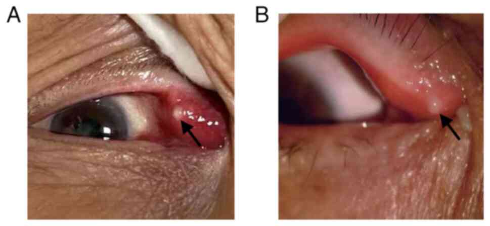
Clinical manifestations
Slit-lamp examination revealed conjunctival
hyperemia. The inflamed lacrimal canaliculus was hyperemic and
edematous. A yellowish-white mucopurulent discharge attached to the
punctum was observed (Fig. 1).
Upon lacrimal duct irrigation the discharges always regurgitated
from the affected punctum; however, the inflamed lacrimal
canaliculus was patent in most patients.
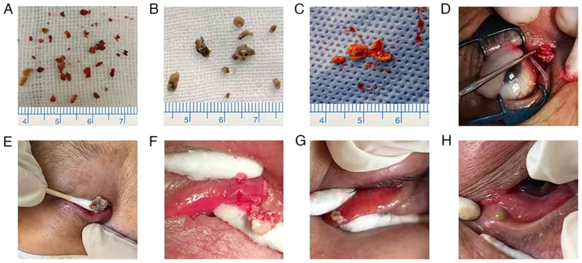
Lacrimal duct exploration revealed a dilated
canalicular lumen that was not smooth, narrowed or obstructed, with
a palpable gritty feel on the canalicular wall. Canalicular
compression produced a sulfurous, cottage cheese-like discharge,
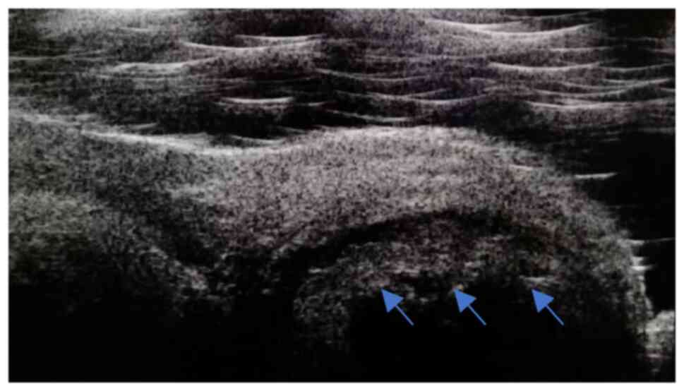
dacryoliths, which varied in color (Fig. 2). UBM revealed canalicular dilation
in patients with several dacryoliths, with uneven, moderate-to-high
intensity signals within the lumen (Fig. 3).
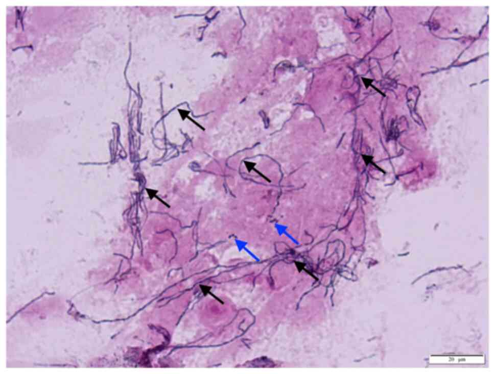
Discharge smears
Among the 50 cases with canaliculitis, 48 showed
dacryoliths in their discharge specimens. The dacryoliths were
crushed to prepare smears for Gram staining and microscopic
examination; all smears of dacryoliths revealed the presence of
Actinomyces species (spp.) (Fig. 4). The discharge in the two cases
without dacryoliths was negative for Actinomyces. All smears
were also positive for cocci or bacilli, with more cases testing
positive for cocci. None of the smears was positive for fungi.
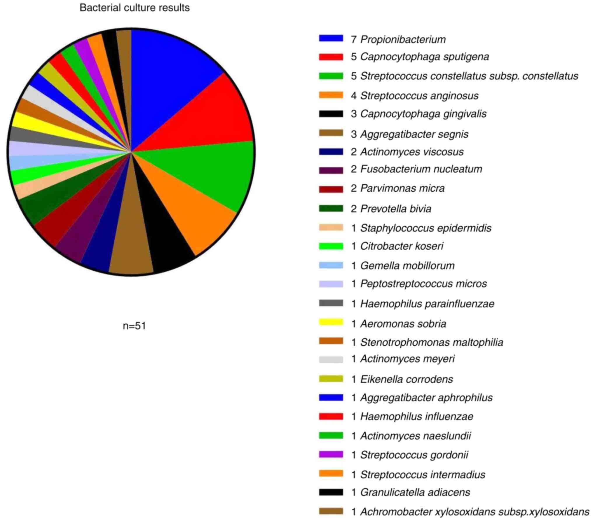
Culture
Bacterial, fungal and anaerobic cultures were
prepared. Among the 50 cases with canaliculitis, 41 were positive
for bacteria, with a culture positivity rate of 82%. A total of 51
relatively heterogeneous bacterial strains were identified in these
cultures. The top three most frequently identified bacteria were
Streptococcus spp. (11 strains), Capnocytophaga spp.
(eight strains) and Propionibacterium (seven strains;
(Fig. 5). Aerobes accounted for
27.5% (14/51), anaerobes for 35.3% (18/51) and facultative
anaerobes for 37.2% (19/51) of the identified strains. Among the 51
strains, 56.9% (29/51) were gram-positive and 43.1% (22/51) were
gram-negative (Table II). All
patient cultures were negative for fungal infection.
| Table IIDistribution of the 51 strains
isolated from bacterial cultures. |
Table II
Distribution of the 51 strains
isolated from bacterial cultures.
| A, Aerobes
(n=14) |
|---|
| Strain | Total, n (%) |
|---|
| Gram-positive
bacteria | |
|
Streptococcus
anginosus | 4 (7.84) |
|
Staphylococcus
epidermidis | 1 (1.96) |
|
Streptococcus
constellatus subsp. constellatus | 5 (9.80) |
|
Streptococcus
gordonii | 1 (1.96) |
|
Streptococcus
intermedius | 1 (1.96) |
| Gram-negative
bacteria | |
|
Stenotrophomonas
maltophilia | 1 (1.96) |
|
Achromobacter
xylosoxidans subsp. xylosoxidans | 1 (1.96) |
| B, Obligate
anaerobes (n=18) |
| Strain | Total, n (%) |
| Gram-positive
bacteria | |
|
Propionibacterium | 7 (13.73) |
|
Actinomyces
viscosus | 2 (3.92) |
|
Peptostreptococcus
micros | 1 (1.96) |
|
Actinomyces
meyeri | 1 (1.96) |
|
Parvimonas
micra | 2 (3.92) |
|
Actinomyces
naeslundii | 1 (1.96) |
| Gram-negative
bacteria | |
|
Fusobacterium
nucleatum | 2 (3.92) |
|
Prevotella
bivia | 2 (3.92) |
| C, Facultative
anaerobes (n=19) |
| Strain | Total, n (%) |
| Gram-positive
bacteria | |
|
Citrobacter
koseri | 1 (1.96) |
|
Gemella
morbillorum | 1 (1.96) |
|
Granulicatella
adiacens | 1 (1.96) |
| Gram-negative
bacteria | |
|
Capnocytophaga
sputigena | 5 (9.80) |
|
Capnocytophaga
gingivalis | 3 (5.88) |
|
Haemophilus
parainfluenzae | 1 (1.96) |
|
Aeromonas
sobria | 1 (1.96) |
|
Aggregatibacter
segnis | 3 (5.88) |
|
Eikenella
corrodens | 1 (1.96) |
|
Aggregatibacter
aphrophilus | 1 (1.96) |
|
Haemophilus
influenzae | 1 (1.96) |
Treatment outcomes
Among the 50 cases with canaliculitis, 45 showed
improvements in eye redness, discharge, epiphora and canalicular
oedema following IOI treatment. Clinically meaningful improvements
were generally achieved 3-4 days after treatment initiation, and
most patients were cured within 2-8 weeks. No relapse occurred at
the follow-up appointments, and the success rate was 90%. A total
of 5 patients who responded poorly to IOI treatment or experienced
relapse underwent surgical treatment. Canalicular dacryoliths and
hyperplastic granulation tissues were completely removed during
surgery, and bicanalicular lacrimal drainage tube placement was
performed. Ocular symptoms were alleviated in all cases after
surgery, without complications (such as canalicular luminal
narrowing or scarring, lacrimal pump dysfunction or canalicular
fistula formation) during follow-up. All patients with
canaliculitis achieved satisfactory treatment outcomes. The total
cure rate was 100%.
Discussion
The results of the present study are similar to
previous reports, stating that middle-aged and older women were
more commonly affected by primary canaliculitis and that the lower
eyelid was involved more often than the upper eyelid (6-8).
There were also similarities in the clinical presentations and the
prolonged symptomatic period until diagnosis, including pouting
erythematous punctum, punctal or canalicular swelling, and
expressible punctal discharge (7-14).
UBM and dacryoendoscopy may be used as diagnostic tools for
canaliculitis (13-15);
however, these require special equipment and techniques. In most
cases, a thorough clinical examination is sufficient for making a
correct diagnosis (8,12).
To the best of our knowledge, there are few detailed
descriptions of laboratory findings in canaliculitis in the
literature. The present study comprised 50 cases of canaliculitis,
of which 48 had canalicular dacryolith. Discharge smear staining
examination revealed a 96% (48/50) positivity rate of
Actinomyces, which is similar to previous reports (10,16,17).
Most smears were also positive for cocci or bacilli, with more
positive smears for cocci. Canaliculitis is typically associated
with mixed bacterial growth (5,16).
The culture positivity rate of canaliculitis
reportedly ranges from 11.1-91.0% (7,8,10,14,17,18).
The present study found a bacterial culture positivity rate of 82%,
which was moderately high. The anaerobic culture may have increased
the bacterial detection rate. The bacterial strains were relatively
heterogeneous (mostly anaerobes or facultative anaerobes and
gram-positive bacteria). The three commonest strains identified
through culture were Streptococcus spp.,
Capnocytophaga spp. and Propionibacterium. In
previous studies (8-10,12,13,17,18),
Streptococcus and Staphylococcus were isolated as the commonest
causative agents. This difference could have occurred as a result
of specimen contamination and differences in culture conditions or
due to changes in the microbiological profile of canaliculitis.
Specimens should be collected aseptically to reduce contamination
and increase culture accuracy. In the current study, anaerobic
culture was also performed to increase the detection rate for
anaerobic bacteria. Only three cases (6%) were positive for
Actinomyces, which is similar to the culture results of
other studies (7,10,13,17).
The low positivity rate for Actinomyces found in the current
study may be related to the small sample size, difficulty in
isolating Actinomyces and stringent culture requirements.
Perumal et al (16)
reported that the success of bacterial culture could be variable
but that smear examination analysis can assist in determining the
etiology. Conducting both smear and culture examinations will help
improve the pathogen detection rate in canaliculitis and guide
clinical treatment. All smears and cultures in the present study
were negative for fungal infection, which is consistent with the
findings of a previous report (17).
Various treatments have been described for
canaliculitis. Conservative measures include topical antibiotics
and intracanalicular antibiotic irrigation, punctal dilatation and
canalicular expression (8,9,12).
Surgical measures include punctoplasty and canalicular curettage,
canaliculotomy with canalicular curettage, or canaliculostomy
(4,11,13,14,18).
Conservative management not only has a high failure rate of
65-100%, but also a high recurrence rate of 33% (3). Failure of antibiotic therapy is
attributed to the inability of antibiotics to penetrate canalicular
concretions. The concretions and inflamed tissue interfere with
tear flow, causing a cycle of canalicular stasis and infection
(19). Surgical treatment has
proven to be effective (4,7,11,14,18);
however, it could lead to canalicular luminal narrowing or
scarring, lacrimal pump dysfunction and canalicular fistula
formation (6,7,9,11,14).
IOI was described in 2015 by Xu et al
(9) as a minimally invasive
technique. The technique is particularly effective when the
ointment is retained for a longer duration inside the canaliculus,
thereby increasing its bioavailability around the focus of
infection. The resolution rate of canaliculitis after
intracanalicular ointment single injection was 72.7% in the study
by Xu et al (9). One
patient had a canalicular laceration during ointment infiltration,
but no canalicular block was noted at the 2-month follow-up. In
2021, Alam et al (20) used
the same technique; however, their protocol involved multiple
sessions of ointment loading, which achieved a complete cure in all
cases. Three patients developed a canalicular block, but the
authors could not determine if this resulted from the primary
disease or a cannula-related injury. The same treatment was
selected for canaliculitis in the present study, where canalicular
dacryoliths were thoroughly removed through the lacrimal punctum
followed by intracanalicular injection of tobramycin/dexamethasone
eye ointment. The treatment was performed weekly for 2-8 weeks,
resulting in a cure rate of 90%. Five patients relapsed at
subsequent follow-up sessions. These patients underwent
canaliculotomy with canalicular curettage. Canalicular dacryoliths
and hyperplastic granulation tissues were completely removed during
surgery. The present study preserved the integrity of the punctum
and performed bicanalicular lacrimal drainage placement. All five
cases were cured without any complications. In the present study,
all patients with canaliculitis achieved satisfactory treatment
outcomes, and the resolution rate was 100%. During surgery,
residual dacryoliths are observed in the lacrimal tubule (11,18).
It is difficult to completely remove dacryoliths through the
lacrimal punctal without canaliculotomy and residual dacryoliths
may lead to relapse (5,19). Placement of the lacrimal drainage
tube is beneficial for the drainage of tears through the inflamed
canalicular system and for preventing lacrimal duct adhesion,
narrowing or obstruction (21).
According to a previous study, the ointment can incite a foreign
body reaction or lead to lipogranuloma formation (22). No paracanalicular lipogranulomata
were noted in the present study. If penetration of the canalicular
walls occurs creating a false passage, the ointment injected may be
deposited in the eyelid tissue, inducing lipogranulomatous
inflammation. Therefore, the ointment administration should be
performed carefully and gently. Eye ointment should only be
injected by an experienced specialist.
The present study was limited by its relatively
small sample size; thus, the present findings require further
verification to be considered representative.
In conclusion, although primary canaliculitis is an
uncommon clinical condition and may have no specific features, it
is not difficult to be diagnosed using detailed smear staining
examinations and laboratory testing. Surgery has a high resolution
rate, and for a number of patients, it is the treatment of choice.
By contrast, IOI is an effective and less invasive procedure with
fewer complications and could be used as the gold standard for
canaliculitis treatment. Thorough removal of the dacryoliths is the
key to successful treatment and prevention of relapse. In the
future, the present authors will endeavor to continue collecting
data on this condition, hoping that clinical research results based
on a larger sample will provide more evidence regarding the
etiology of canaliculitis, which can further guide its treatment
and diagnosis.
Acknowledgements
Not applicable.
Funding
Funding: The present study was supported by The Fund of Top
Talent Support Program for Young and Middle-aged People of Wuxi
Health Committee (grant no. HB2020030), The Research Project of
Wuxi Commission of Health (grant no. M202131), The Elderly Health
Research Project of Jiangsu Province (grant no. 2022043) and The
Social Development Project of Jiangsu Provincial Department of
Science and Technology (grant no. BE2022699).
Availability of data and materials
The datasets used and/or analyzed during the current
study are available from the corresponding author on reasonable
request.
Authors' contributions
QW, SS and ZZ conceived the study. QW, SL, RH, HS
and YG collected the clinical information of the patients. QW, SS,
SL, RH, HS and ZZ analyzed and interpreted the clinical data. QW
and SL wrote the draft of the manuscript. SS, YG and ZZ reviewed
and edited the manuscript. ZZ was responsible for study
supervision. QW and ZZ confirm the authenticity of all the raw
data. All authors agreed to be accountable for all aspects of the
work. All authors have read and approved the final manuscript.
Ethics approval and consent to
participate
The protocol of the present prospective study was
approved by the Institutional Review Board of the Affiliated Wuxi
Clinical College of Nantong University (Wuxi, China; approval no.
2022-Y-98). Prior to inclusion, written informed consent was
obtained from all patients. The present study adhered to the tenets
of the Declaration of Helsinki.
Patient consent for publication
Written informed consent was obtained from the
patient whose images are presented.
Competing interests
The authors declare that they have no competing
interests.
References
|
1
|
Anand AR, Harinee R, Jeyalatha MV, Poonam
NS, Therese KL, Rajeshwari H, Narasimhan L and Gopinath R:
Microbiological profile of canaliculitis and their antibiotic
susceptibility patterns: A 11-year review at a referral eye care
centre. Indian J Med Microbiol. 40:378–383. 2022.PubMed/NCBI View Article : Google Scholar
|
|
2
|
Ali MJ: Metagenomics of infective
canaliculitis: The Lacriome paper 3. Eur J Ophthalmol.
32:3346–3352. 2022.PubMed/NCBI View Article : Google Scholar
|
|
3
|
Freedman JR, Markert MS and Cohen AJ:
Primary and secondary lacrimal canaliculitis: A review of
literature. Surv Ophthalmol. 56:336–347. 2011.PubMed/NCBI View Article : Google Scholar
|
|
4
|
Baldursdóttir E, Sigurdsson H, Jónasson L
and Gottfredsson M: Actinomycotic canaliculitis: Resolution
following surgery and short topical antibiotic treatment. Acta
Ophthalmol. 88:367–370. 2010.PubMed/NCBI View Article : Google Scholar
|
|
5
|
Hussain I, Bonshek RE, Loudon K, Armstrong
M and Tullo AB: Canalicular infection caused by Actinomyces. Eye
(Lond). 7 (Pt 4):542–544. 1993.PubMed/NCBI View Article : Google Scholar
|
|
6
|
Fulmer NL, Neal JG, Bussard GM and Edlich
RF: Lacrimal canaliculitis. Am J Emerg Med. 17:385–386.
1999.PubMed/NCBI View Article : Google Scholar
|
|
7
|
Anand S, Hollingworth K, Kumar V and
Sandramouli S: Canaliculitis: The incidence of long-term epiphora
following canaliculotomy. Orbit. 23:19–26. 2004.PubMed/NCBI View Article : Google Scholar
|
|
8
|
Kaliki S, Ali MJ, Honavar SG,
Chandrasekhar G and Naik MN: Primary canaliculitis: Clinical
features, microbiological profile, and management outcome. Ophthal
Plast Reconstr Surg. 28:355–360. 2012.PubMed/NCBI View Article : Google Scholar
|
|
9
|
Xu J, Liu Z, Mashaghi A, Sun X, Lu Y, Li
Y, Wu D, Yang Y, Wei A, Zhao Y, et al: Novel therapy for primary
canaliculitis: A pilot study of intracanalicular ophthalmic
corticosteroid/antibiotic combination ointment infiltration.
Medicine (Baltimore). 94(e1611)2015.PubMed/NCBI View Article : Google Scholar
|
|
10
|
Zaldívar RA and Bradley EA: Primary
canaliculitis. Ophthal Plast Reconstr Surg. 25:481–484.
2009.PubMed/NCBI View Article : Google Scholar
|
|
11
|
Lee MJ, Choung HK, Kim NJ and Khwarg SI:
One-snip punctoplasty and canalicular curettage through the
punctum: A minimally invasive surgical procedure for primary
canaliculitis. Ophthalmology. 116:2027–2030.e2. 2009.PubMed/NCBI View Article : Google Scholar
|
|
12
|
Gogandy M, Al-Sheikh O and Chaudhry I:
Clinical features and bacteriology of lacrimal canaliculitis in
patients presenting to a tertiary eye care center in the Middle
East. Saudi J Ophthalmol. 28:31–35. 2014.PubMed/NCBI View Article : Google Scholar
|
|
13
|
Xiang S, Lin B, Pan Q, Zheng M, Qin X,
Wang Y and Zhang Z: Clinical features and surgical outcomes of
primary canaliculitis with concretions. Medicine (Baltimore).
96(e6188)2017.PubMed/NCBI View Article : Google Scholar
|
|
14
|
Su Y, Zhang L, Li L, Fan X and Xiao C:
Surgical procedure of canaliculoplasty in the treatment of primary
canaliculitis associated with canalicular dilatation. BMC
Ophthalmol. 20(245)2020.PubMed/NCBI View Article : Google Scholar
|
|
15
|
Luo B and Qi X: Utility of 80-MHz
ultrasound biomicroscopy and lacrimal endoscopy in chronic lacrimal
canaliculitis. J Ultrasound Med. 40:2513–2520. 2021.PubMed/NCBI View Article : Google Scholar
|
|
16
|
Perumal B, Carlson JA and Meyer DR: A
pathological analysis of canaliculitis concretions: More than just
Actinomyces. Scientifica (Cairo). 2016(6313070)2016.PubMed/NCBI View Article : Google Scholar
|
|
17
|
Zhang Y, Deng SJ, Wang ZQ, Ding JW and Sun
XG: Etiological and drug sensitivity analysis of lacrimal
canaliculitis. Zhonghua Yan Ke Za Zhi. 54:111–114. 2018.PubMed/NCBI View Article : Google Scholar : (In Chinese).
|
|
18
|
Kim UR, Wadwekar B and Prajna L: Primary
canaliculitis: The incidence, clinical features, outcome and
long-term epiphora after snip-punctoplasty and curettage. Saudi J
Ophthalmol. 29:274–277. 2015.PubMed/NCBI View Article : Google Scholar
|
|
19
|
Pavilack MA and Frueh BR: Through
curettage in the treatment of chronic canaliculitis. Arch
Ophthalmol. 110:200–202. 1992.PubMed/NCBI View Article : Google Scholar
|
|
20
|
Alam MS, Poonam NS, Koka K, Vijay V and
Ganesh S: Intracanalicular antibiotic ointment loading as a
management option for canaliculitis. Orbit. 40:295–300.
2021.PubMed/NCBI View Article : Google Scholar
|
|
21
|
Jin X, Zhao Y, Tong N and Xu W: Use of
crawford tube for chronic suppurative lacrimal canaliculitis.
Ophthal Plast Reconstr Surg. 30:229–232. 2014.PubMed/NCBI View Article : Google Scholar
|
|
22
|
Wang Y, Xiao C, Bi X, Zhou H, Ge S and Fan
X: Palpebral lipogranuloma caused by transcanalicular ointment
injection after laser canaliculoplasty. Ophthal Plast Reconstr
Surg. 27:333–337. 2011.PubMed/NCBI View Article : Google Scholar
|