Introduction
An abdominal aortic aneurysm (AAA) is an abnormal
dilation of the abdominal aorta and is associated with a mortality
rate of >50% in the event it ruptures. AAAs are characterized by
an increase of ³50% in aortic diameter compared with the normal
diameter or a maximum infrarenal aortic diameter ≥30 mm (1). The prevalence of AAA increases with
age and ~4% of men >55 years have an AAA (1) and AAAs cause ~1% of all deaths in
developed countries (2).
Therapeutic options for AAA are currently limited to
surgical or interventional repair to prevent rupture which can be
effective for several decades; however, no pharmacologic therapy
has been reported to have a significant effect in humans (1,3,4). A
delay in the development of AAA treatments is partly due to the
absence of an AAA animal model of human AAA (3) and because human AAA tissue can only
be obtained during surgical repair, investigation of AAA with human
tissue is limited (5). Previous
studies have proposed AAA animal models (2,6);
however, more evidence of histological features and similarities in
shape is needed to establish an effective AAA animal model for the
study of human clinical applications. A common animal model for the
aneurysmal aortic lesions consists of the transient perfusion of
the abdominal aorta with porcine pancreatic elastase (PPE)
(2,7-10).
Another common model of AAA induction is CaCl2 perfusion
of the aorta (2,4). Other AAA induction methods, including
mechanical induction through aortic stretching, patching or injury,
have also been reported (2). In
certain cases, a combination of the aforementioned methods have
been reported (11-13).
The present model causes mechanical injury to the aortic intima,
making it similar to human AAA. The present study established a
novel and reproducible animal model of AAA using mechanical injury.
This model is based on the hypothesis that the mechanical injury
will initiate the inflammation process and destruction of the
elastin and collagen fibers at the abdominal aorta, as observed in
human AAA tissue.
Materials and methods
This experimental animal study was approved by the
Pusan National University Institutional Animal Care and Use
Committee (approval no. PNU-2018-1909; Republic of Korea). A total
of 22 adult Lewis rats (male; age, 8-12 weeks; Koatech Technology
Corporation) weighing ~330 g (range, 256-450 g) were used. Rats
were housed and maintained at 25˚C and 50% humidity, with a 12 h
light/dark cycle according to the institutional animal protocols.
All rats had regular food and water ad libitum with no
restrictions on movement. All rats were from a specific pathogen
free herd and evaluated as healthy by a veterinarian prior to
inclusion. The rats were randomly divided into 2 groups and
harvested after 42 (6-week group) and 63 (9-week group) days to
investigate the progression of the AAAs. All experimental
procedures were performed according to Pusan National University's
guidelines for animal care.
AAA induction
High blood pressure could be the cause of mechanical
injuries that initiate the inflammation process and destruction of
the elastin and collagen fibers of the abdominal aorta. Therefore,
the present study used a mechanical injury model to mimic the
physical effects that may lead to AAAs.
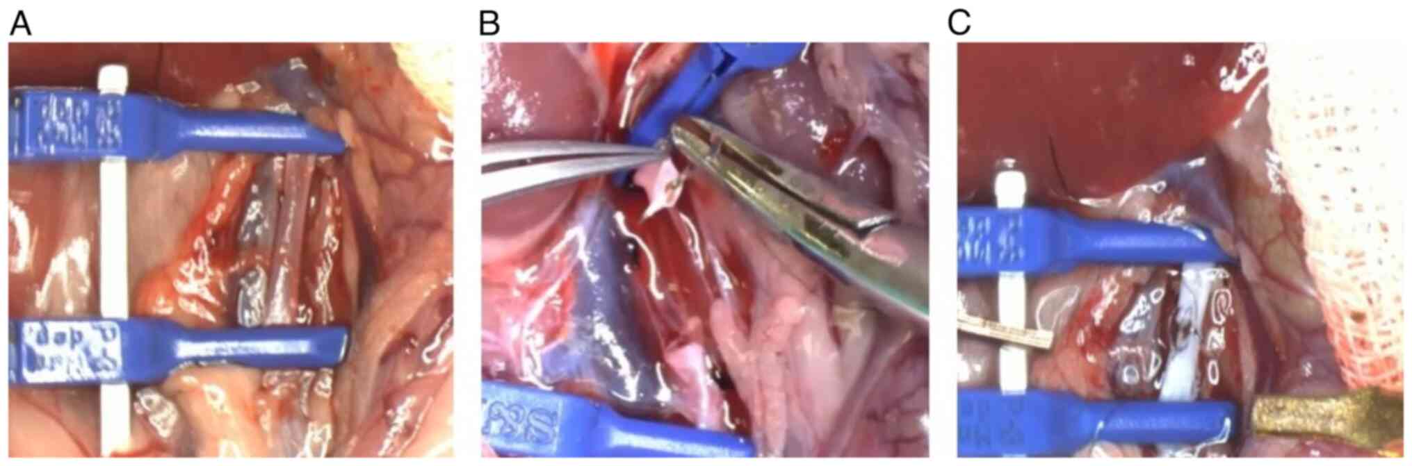
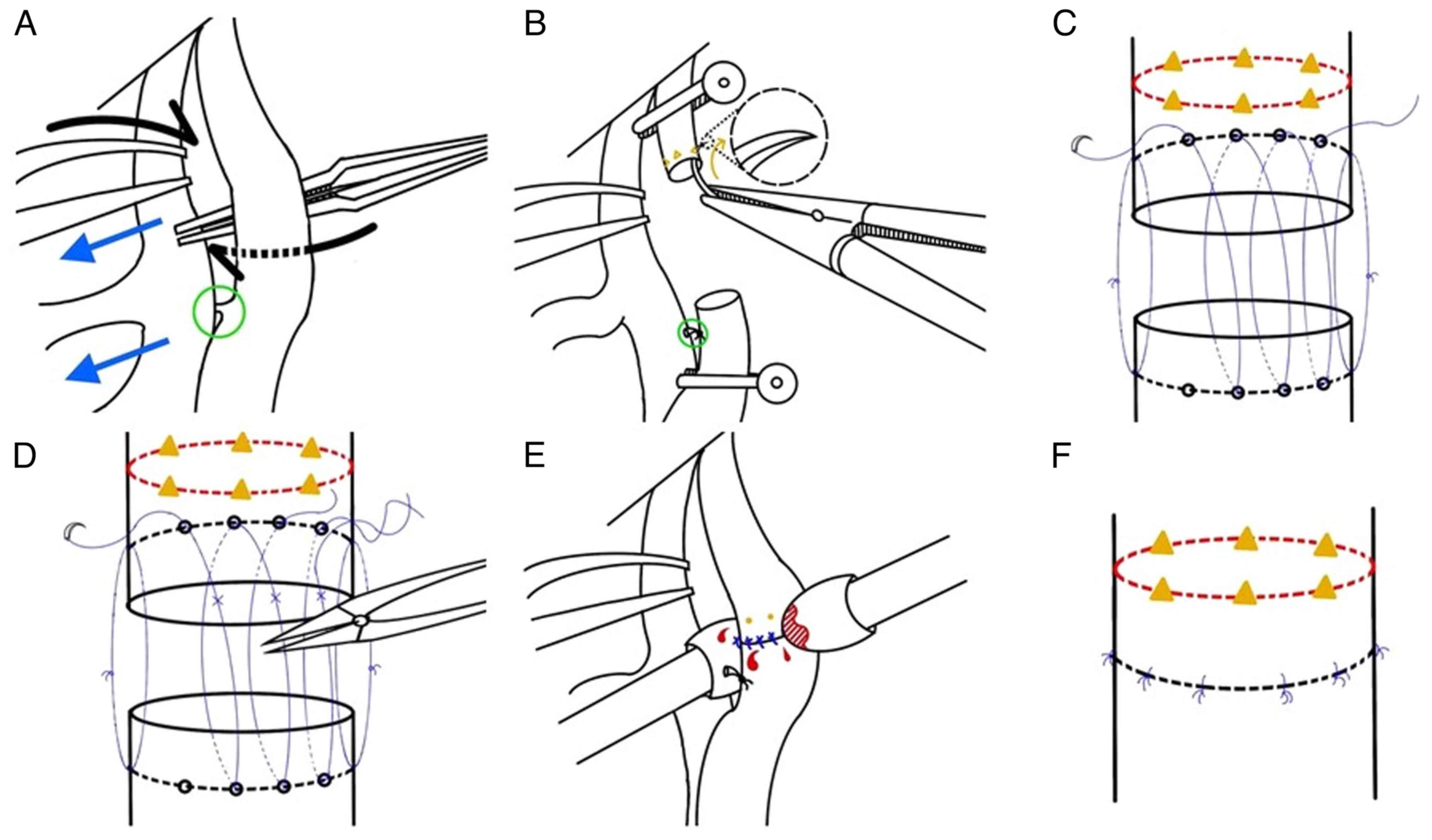
The rats were anesthetized with an intraperitoneal
injection of pentobarbital sodium (120 mg/kg). The skin incision
site was disinfected with a povidone-iodine swab (Q & Q Pham
Co. Ltd.). After midline laparotomy, the aorta and inferior vena
cava were carefully dissected from neighboring tissues. The
infrarenal aorta was then exposed by isolating it from the inferior
vena cava. The aorta was clamped distally and superiorly to the
renal artery and proximally and inferiorly to the gonadal artery.
After ligating the lumbar artery, the entire circumference of the
abdominal aorta was temporarily opened to apply the same level of
injury and tension in all directions. Six deep stabs from the
intima to the adventitia around the aortic wall were performed
using a 7-0 blue nylon cutting needle (25 mm; 3/8; 75 cm; AILEE
Co., Ltd.). The opened aortic wall was anastomosed using 9-0
Monosof™ sutures (5 mm; 3/8; 13 cm; Medtronic) using the 12-14
point open-loop technique. Hemostasis was performed using cotton
swabs. Finally, the retroperitoneum and laparotomy were closed with
5-0 or 6-0 Vicryl™ sutures (Ethicon, Inc.; Johnson & Johnson)
(Figs. 1 and 2).
Postoperative care
The rats were separated and raised in the same
environment as aforementioned. Rats were housed and maintained at
25˚C and 50% humidity, with a 12 h light/dark cycle according to
the institutional animal protocols. All rats had regular food and
water ad libitum with no restrictions on movement.
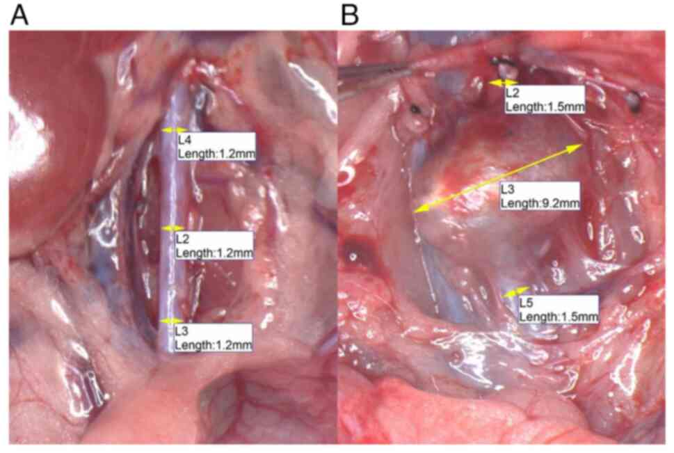
Harvest and data collection
On postoperative day 42 and 63, the respective
groups of rats were euthanized by intraperitoneal injection with a
lethal dose of pentobarbital (120 mg/kg). After a midline
laparotomy, the aorta from the level of the renal artery to the
gonadal artery, including the AAAs, was harvested. Microscopic AAA
were defined as cases that had a >50% increase in aorta diameter
at the time of harvest compared with the initially measured
diameter of the infrarenal aorta in each rat (Fig. 3). Representative 4-µm sections of
formalin-fixed, in 10% neutral-buffered formalin at room
temperature for 12 h, paraffin-embedded tissues were cut on charged
glass slides. Slides were placed in a 65˚C oven for 20 min to dry
the tissue. Immunostaining was performed using the Bond-Max
staining instrument (Leica Microsystems, Inc.) as described by the
manufacturer. Briefly, paraffin wax was removed with a Bond Dewax
Solution (cat. no. AR9222; Leica Biosystems Nussloch GmbH) at 72˚C
and the sections were rehydrated. Heat-induced epitope retrieval
was achieved using a Bond Epitope Retrieval Solution (EDTA-based
buffer; Leica Microsystems, Inc.) for 20 min at 100˚C. The section
was then incubated with antibodies SMA (cat. no. M0851; monoclonal;
1:800; DAKO; Agilent Technologies, Inc.) for 15 min at room
temperature. The secondary antibody was incubated for 8 min and
horseradish peroxidase (HRP) for 10 min at room temperature. Both
the secondary antibody and HRP were provided as ready-to-use kit
(Bond™ Polymer Refine Detection; cat. no. DS9800; Leica
Microsystems, Inc.). Finally, 3,3'-diaminobenzidine
tetrahydrochloride was used as the chromogen. Verhoeff-van Gieson
staining for elastin were also performed at room temperature for 30
min. The slides were examined using the Olympus BX53 light
microscope (Olympus Corporation) by a single pathologist.
Results
Summary data
In the 6-week group, 10/11 rats survived the study
period and in the 9-week group 11/11 rats survived the study period
(Table I). The mean weight of the
rats was 338.5±49.3 g (range, 256-450 g) and 452±21.3 g (range,
411-500 g) at the time of the procedure and the time of harvest,
respectively. The mean operative time was 33±3.3 min (range, 24-37
min). No immediate postoperative complications were noted. One rat
died during the follow-up period due to an unknown cause and its
data were omitted from the analysis.
 | Table IChanges in animal weight and maximum
diameter of the infrarenal aorta. |
Table I
Changes in animal weight and maximum
diameter of the infrarenal aorta.
| A, 6-week group |
|---|
| No. | Initial weight,g | Weight at harvest,
g | Preoperative maximum
diameter of infrarenal aorta, mm | Maximum diameter of
infrarenal aorta at harvest, mm | Increase in aorta
diameter, (%) |
|---|
| 1 | 320 | 500 | 1.3 | 2.5 | 92 |
| 2 | 293 | 454 | 1.1 | 1.1 | 0 |
| 3 | 256 | 423 | 1.2 | 1.1 | -8 |
| 4 | 260 | 443 | 1.3 | 1.3 | 0 |
| 5 | 364 | 452 | 1.3 | 7.2 | 454 |
| 6 | 364 | 444 | 1.4 | 4.8 | 243 |
| 7 | 363 | 463 | 1.2 | 9.2 | 667 |
| 8 | 364 | 470 | 1.3 | 3.2 | 146 |
| 9 | 364 | 443 | 1.4 | 3.8 | 171 |
| 10 | 357 | 411 | 1 | 1.5 | 50 |
| 11 | 450 | Died | 1.4 | Died | Died |
| Mean ± SD | 363±53.3 | 448±23.4 | 1.3±0.12 | 2.9±2.6 | 119±210 |
| B, 9-week group |
| No. | Initial weight,g | Weight at harvest,
g | Preoperative maximum
diameter of infrarenal aorta, mm | Maximum diameter of
infrarenal aorta at harvest, mm | Increase in aorta
diameter, (%) |
| 1 | 359 | 490 | 1.4 | 5.5 | 293 |
| 2 | 366 | 478 | 1.5 | 5 | 233 |
| 3 | 370 | 466 | 1.4 | 1.8 | 29 |
| 4 | 365 | 438 | 1.3 | 2.9 | 123 |
| 5 | 270 | 462 | 1.2 | 1.3 | 8 |
| 6 | 276 | 443 | 1.2 | 2.8 | 133 |
| 7 | 290 | 454 | 1.1 | 2.9 | 164 |
| 8 | 290 | 446 | 1.3 | 2.8 | 115 |
| 9 | 284 | 458 | 1.1 | 3.1 | 182 |
| 10 | 281 | 416 | 1.1 | 1.5 | 36 |
| 11 | 288 | 450 | 1.2 | 3.6 | 200 |
| Mean ± SD | 312±40 | 454±19.1 | 1.2±0.13 | 2.9±1.3 | 133±85 |
Macroscopic study
Preoperatively, the overall mean maximum diameter of
the infrarenal aorta was 1.3±0.13 mm (range, 1.0-1.5 mm). In the
6-week group, a marked increase in the diameter of the aneurysm was
observed in 6/10 (60%) of the surviving rats. The mean maximum
diameter of the infrarenal aorta was 2.85±2.63 mm which was a 119%
increase from the preoperative size. In the 9-week group, changes
in the diameter of the aneurysm were observed in 8/11 (88%) of the
rats. The mean maximum diameter of the infrarenal aorta was
2.9±1.25 mm, a 123% increase from the preoperative size (Table I).
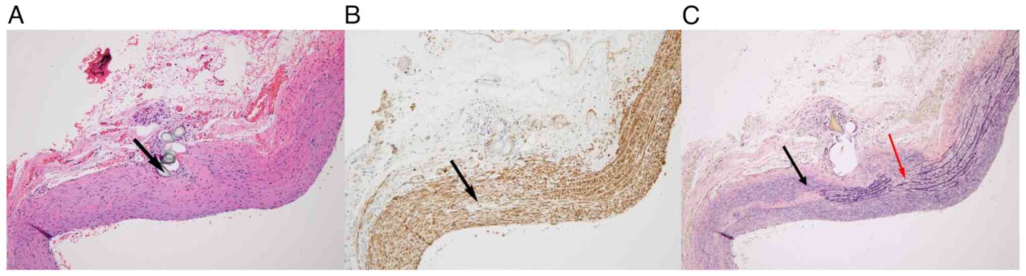
Microscopic study
In the transverse section of the aorta from all
induced samples in the 6- and 9-week groups, the difference in the
diameter compared with the control under the same magnification was
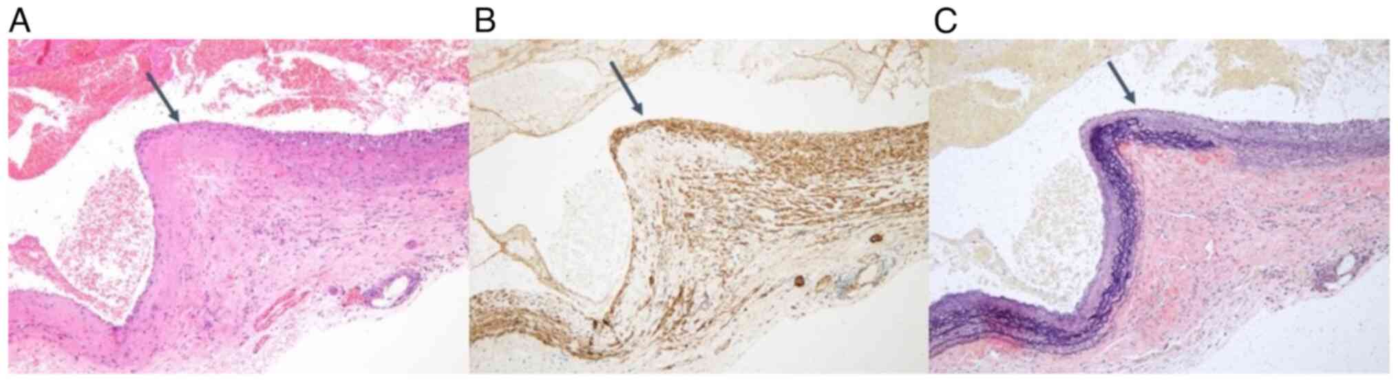
clear (Fig. 4). The decrease in
the cellular density was not apparent in the H&E slides. The
vascular smooth muscle cells were gone and were replaced by
hyalinizing material. The loss of smooth muscle cells was confirmed
by staining for smooth muscle actin (Fig. 5). The thickness of the protruding
vessel wall was irregular and elastin production was reduced
(Fig. 6). The elastic fibers
showed breaks and looked flat with no coiling or resilience, and
the number of elastic fibers was reduced (Fig. 4).
Discussion
The present study aimed to establish evidence for a
novel and reproducible animal model of AAA. It was hypothesized
that mechanical injury would initiate the inflammation process and
destruction of the elastin and collagen fibers at the abdominal
aorta, thus mimicking the formation of AAAs in humans. To evaluate
this hypothesis, changes in the AAA tissue at 6 and 9 weeks were
evaluated by observing macro- and microscopic. The results of the
present supported this hypothesis, as the animal models
demonstrated similar morphological and microscopic findings as
human cases of AAA. This can be seen by how induced AAAs in the
present model had a symmetric fusiform shape in the infrarenal
area, similar to human AAAs.
The development of new experimental techniques,
genetic analysis and robust molecular tools has contributed to the
understanding of the pathophysiology of AAAs. While certain
cellular mechanisms, such as inflammatory response mechanisms, have
been reported to be responsible for AAA formation (14), the application of physical force to
put pressure on the heartbeat and blood flow are not commonly used
techniques for AAA animal modeling. However, hypertension is
positively associated with the incidence of AAAs (15) and physical force, such as that from
high blood pressure, could be the cause of mechanical injuries that
initiate the inflammation process and the destruction of the
elastin and collagen fibers of the abdominal aorta (16). Therefore, a mechanical injury model
was used to mimic the physical effects that may lead to AAAs in the
present study. Several models were evaluated in pilot studies,
including end-to-end anastomosis, out-in-needle injury and
in-out-needle injury. Aneurysm was not formed in the end-to-end
anastomosis model, and bleeding was not controlled in the
out-in-needle injury model. Therefore, the in-out-needle injury
model was further evaluated in the present study as this had
demonstrated the best results in the pilot studies (data not
shown).
There are numerous AAA animal models in use, each of
which have drawbacks in the matching of the effects of human AAAs.
For example, the porcine pancreatic elastase model is commonly used
and AAAs have been previously induced via PPE perfusion in mice
(2,8). PPE modeling in rats has been reported
to demonstrate similar temporal and pathophysiological findings as
those found in humans, such as the correlation of aortic dilation
with the state of the inflammatory response, which leads to the
destruction of the medial elastic tissue (2,8). In
rat PPE models, the rates of successful induction have been high
but in larger animals, the success rates have been reported to be
lower (9) due to technical
difficulties with the model. Specifically, when infusing the PPE,
cannulation of the aorta is required and leakage of the PPE
solution during perfusion happens frequently, and this process can
result in the death of the animal (2).
The CaCl2 model is another frequently
used model (2). Exposing the aorta
to CaCl2 leads to the destruction of the elastin fibers
of the wall of the aorta, the infiltration of inflammatory cells
and AAA induction. However, periaortic CaCl2 application
does not reliably induce AAA formation, with success varying from
80-100% of the models developing aneurysms (2,10,17).
An angiotensin II model in mice is technically less
challenging than others mentioned here and there is no need for
major surgical manipulation. It has been previously reported that
AAA is encouraged in hyperlipidemic apolipoprotein E-deficient mice
with continuous subcutaneous angiotensin II infusion (5,18).
However, the induced AAAs in this model have certain differences
when compared with human AAAs such as a significantly higher
incidence of aortic dissection and rupture, and more frequently
observed suprarenal AAAs, while humans present a much higher
incidence of infrarenal AAAs (2).
The xenograft model uses implanted xenograft
arterial tissues to generate AAAs using the rejection of the
implanted tissue by the immune response (7,19).
Models which use knockout animals, which focus on the disruption of
one or more gene alleles resulting in the creation of
strain-deficient animals have also been previously reported
(2).
The mechanical injury model used in the present
study has three distinct advantages. First, the model had high
reproducibility. At 9 weeks, the size of the aorta increased by up
to 133% in 88% of the rats (8/11). Reproducibility is one of the
most important conditions to meet in animal models so that
additional studies can be performed reliably. Second, the gross and
microscopic findings of the model were very similar to findings
previously reported in human AAAs. The induced AAAs in this model
had a symmetric fusiform shape in the infrarenal area, similar to
human AAAs. However, in other animal models, saccular shaped AAAs
(20) or suprarenal AAAs (5) were reported to have been induced.
Because the different shapes could indicate different blood flow
and progression, gross similarities are important to consider. In
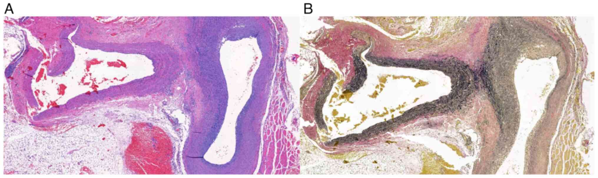
the present model, important microscopic findings were found such
as the destruction of the tunica media and the elastic fibers,
neovascularization, infiltration of lymphocytes and intraluminal
mural hematomas (19). In
particular, the histological characteristic of broken elastin in
the formation of an aneurysm is similar to that of human aneurysm.
Finally, the model described in the present study can be reproduced
at a very low cost as it did not require special drugs or
equipment. Only typical vascular surgical instruments were needed
to perform the surgical intervention and because the rats were
allowed to live without subsequent manipulation, there was no
additional cost. Furthermore, no paraplegia occurred in the short
operation time of 30-50 min.
The limitation of the model comes from the surgical
intervention which required microsurgical skills, which can be
difficult for inexperience staff to follow. Improper surgical
technique may result in animal deaths or unsuccessful induction of
AAAs, which could cause reproducibility to decrease and costs to
increase. Furthermore, the present study only performed a 9-week
investigation. Because the AAA diameters increased from 6- to
9-weeks, it could be hypothesized that the AAAs would grow larger
over time, however this and whether they eventually rupture like
human AAAs was not confirmed. Before starting to use this model, it
is recommended that researchers should be trained on animal
experiments and vascular management. Furthermore, the present study
did not yield clear results for the inflammatory response and
confirmation of the inflammatory response requires further
microscopic examination in future studies.
In the 6-week group, it was demonstrated that the
aortic diameter did not increase in 3/10 animals. They typically
had an initial weight of <300 g, which was 18% less than the
mean weight of 365 g. This suggested the need to investigate the
relationship between weight and hemodynamic parameters such as
blood pressure and cardiac output. Further studies are also
required to elucidate the timeframe for the occurrence of AAAs and
what the effect of proteins expressed by different genes on the
formation of aneurysms in mice with and without aneurysms are.
Whether the use of certain drugs can prevent the formation of
aneurysms, delay the continued increase of an aneurysm until
rupture or even make the aneurysm shrink also requires evaluation.
Future studies, are also required to assess the long-term results
of this model while measuring the AAAs with ultrasound. This model
should also be trialed in larger animals although there have been
many reports of failures when applied to larger animals (2,21).
However, a trial in larger animals is needed to confirm that the
AAAs in the present model are similar to human AAAs with infrarenal
symmetric fusiform shape.
In conclusion, the mechanical injury-induced AAA
animal model reported in the present study demonstrated very
similar morphological and microscopic findings to those of human
AAAs. The model showed high reproducibility, with 60% induction in
the 6-week group and 88% induction in the 9-week group. Though the
present study was performed in rodents, this method could provide
evidence for AAA treatment if it can be applied to middle and large
sized animals.
Acknowledgements
Not applicable.
Funding
Funding: The present study was supported by a 2022 research
grant from Pusan National University Yangsan Hospital.
Availability of data and materials
The datasets used and/or analyzed during the current
study are available from the corresponding author on reasonable
request.
Authors' contributions
SHK, JHP and SSL were responsible for study
conception and design. SHK, JHP, DHK, JHM and JHC were responsible
for data analysis and interpretation. SSL was responsible for data
collection. SHK, JHP and SSL confirm the authenticity of all the
raw data. SHK, JHL and SSL were responsible for writing and
revising the article. SHK, JHP, DHK, JHL and SSL were responsible
for final approval of the article. SSL was responsible for overall
supervision. All authors have read and approved the final
manuscript.
Ethics approval and consent to
participate
The present study was approved by Pusan National
University Institutional Animal Care and Use Committee (approval
no. PNU-2018-1909; Republic of Korea).
Patient consent for publication
Not applicable.
Author information
Dr Sang Su Lee ORCID: https://orcid.org/0000-0003-0648-976X.
Competing interests
The authors declare that they have no competing
interests.
References
|
1
|
Rughani G, Robertson L and Clarke M:
Medical treatment for small abdominal aortic aneurysms. Cochrane
Database Syst Rev. (CD009536)2012.PubMed/NCBI View Article : Google Scholar
|
|
2
|
Patelis N, Moris D, Schizas D, Damaskos C,
Perrea D, Bakoyiannis C, Liakakos T and Georgopoulos S: Animal
models in the research of abdominal aortic aneurysms development.
Physiol Res. 66:899–915. 2017.PubMed/NCBI View Article : Google Scholar
|
|
3
|
Yoshimura K, Morikage N, Nishino-Fujimoto
S, Furutani A, Shirasawa B and Hamano K: Current status and
perspectives on pharmacologic therapy for abdominal aortic
aneurysm. Curr Drug Targets. 19:1265–1275. 2018.PubMed/NCBI View Article : Google Scholar
|
|
4
|
Wang Y, Krishna S and Golledge J: The
calcium chloride-induced rodent model of abdominal aortic aneurysm.
Atherosclerosis. 226:29–39. 2013.PubMed/NCBI View Article : Google Scholar
|
|
5
|
Fashandi AZ, Hawkins RB, Salmon MD,
Spinosa MD, Montgomery WG, Cullen JM, Lu G, Su G, Ailawadi G and
Upchurch GR Jr: A novel reproducible model of aortic aneurysm
rupture. Surgery. 163:397–403. 2018.PubMed/NCBI View Article : Google Scholar
|
|
6
|
Mouton R, Pollock J, Soar J, Mitchell DC
and Rogers CA: Remote ischaemic preconditioning versus sham
procedure for abdominal aortic aneurysm repair: An external
feasibility randomized controlled trial. Trials.
16(377)2015.PubMed/NCBI View Article : Google Scholar
|
|
7
|
Allaire E, Muscatelli-Groux B, Mandet C,
Guinault AM, Bruneval P, Desgranges P, Clowes A, Méllière D and
Becquemin JP: Paracrine effect of vascular smooth muscle cells in
the prevention of aortic aneurysm formation. J Vasc Surg.
36:1018–1026. 2002.PubMed/NCBI View Article : Google Scholar
|
|
8
|
Halpern VJ, Nackman GB, Gandhi RH,
Irizarry E, Scholes JV, Ramey WG and Tilson MD: The elastase
infusion model of experimental aortic aneurysms: Synchrony of
induction of endogenous proteinases with matrix destruction and
inflammatory cell response. J Vasc Surg. 20:51–60. 1994.PubMed/NCBI View Article : Google Scholar
|
|
9
|
Marinov GR, Marois Y, Pâris E, Roby P,
Formichi M, Douville Y and Guidoin R: Can the infusion of elastase
in the abdominal aorta of the Yucatán miniature swine consistently
produce experimental aneurysms? J Invest Surg. 10:129–150.
1997.PubMed/NCBI View Article : Google Scholar
|
|
10
|
Zhu JX, Tang QQ, Zhou C, Shi XC, Yi SY and
Yang Y: Establishment of a new abdominal aortic aneurysm model in
rats by a retroperitoneal approach. Front Cardiovasc Med.
9(808732)2022.PubMed/NCBI View Article : Google Scholar
|
|
11
|
Li MW, Mian MO, Barhoumi T, Rehman A, Mann
K, Paradis P and Schiffrin EL: Endothelin-1 overexpression
exacerbates atherosclerosis and induces aortic aneurysms in
apolipoprotein E knockout mice. Arterioscler Thromb Vasc Biol.
33:2306–2315. 2013.PubMed/NCBI View Article : Google Scholar
|
|
12
|
Lin PY, Wu YT, Lin GC, Shih YH,
Sampilvanjil A, Chen LR, Yang YJ, Wu HL and Jiang MJ:
Coarctation-induced degenerative abdominal aortic aneurysm in a
porcine model. J Vasc Surg. 57:806–815.e1. 2013.PubMed/NCBI View Article : Google Scholar
|
|
13
|
Czerski A, Bujok J, Gnus J, Hauzer W,
Ratajczak K, Nowak M, Janeczek M, Zawadzki W, Witkiewicz W and
Rusiecka A: Experimental methods of abdominal aortic aneurysm
creation in swine as a large animal model. J Physiol Pharmacol.
64:185–192. 2013.PubMed/NCBI
|
|
14
|
Quintana RA and Taylor WR: Cellular
mechanisms of aortic aneurysm formation. Circ Res. 124:607–618.
2019.PubMed/NCBI View Article : Google Scholar
|
|
15
|
Takagi H and Umemoto T: ALICE
(All-Literature Investigation of Cardiovascular Evidence) Group.
Association of hypertension with abdominal aortic aneurysm
expansion. Ann Vasc Surg. 39:74–89. 2017.PubMed/NCBI View Article : Google Scholar
|
|
16
|
Gao J, Cao H, Hu G, Wu Y, Xu Y, Cui H, Lu
HS and Zheng L: The mechanism and therapy of aortic aneurysms.
Signal Transduct Target Ther. 8(55)2023.PubMed/NCBI View Article : Google Scholar
|
|
17
|
Bi Y, Zhong H, Xu K, Zhang Z, Qi X, Xia Y
and Ren L: Development of a novel rabbit model of abdominal aortic
aneurysm via a combination of periaortic calcium chloride and
elastase incubation. PLoS One. 8(e68476)2013.PubMed/NCBI View Article : Google Scholar
|
|
18
|
Ghoshal S and Loftin CD: Cyclooxygenase-2
inhibition attenuates abdominal aortic aneurysm progression in
hyperlipidemic mice. PLoS One. 7(e44369)2012.PubMed/NCBI View Article : Google Scholar
|
|
19
|
Riber SS, Ali M, Bergseth SH, Stubbe J,
Stenger M, Behr-Rasmussen C and Lindholt JS: Induction of
autoimmune abdominal aortic aneurysm in pigs-A novel large animal
model. Ann Med Surg (Lond). 20:26–31. 2017.PubMed/NCBI View Article : Google Scholar
|
|
20
|
Maynar M, Qian Z, Hernandez J, Sun F,
DeMiguel C, Crisostomo V, Usón J, Pineda LF, Espinoza CG and
Castañeda WR: An animal model of abdominal aortic aneurysm created
with peritoneal patch: Technique and initial results. Cardiovasc
Intervent Radiol. 26:168–176. 2003.PubMed/NCBI View Article : Google Scholar
|
|
21
|
Verbrugghe P, Verhoeven J, Clijsters M,
Vervoort D, Coudyzer W, Verbeken E, Meuris B and Herijgers P:
Creation of abdominal aortic aneurysms in sheep by extrapolation of
rodent models: Is it feasible? Ann Vasc Surg. 52:225–236.
2018.PubMed/NCBI View Article : Google Scholar
|